

Can the low and high b-value distribution influence the pseudodiffusion parameter derived from IVIM DWI in normal brain?
- PMID: 32041549
- PMCID: PMC7011602
- DOI: 10.1186/s12880-020-0419-0
Can the low and high b-value distribution influence the pseudodiffusion parameter derived from IVIM DWI in normal brain?
Abstract
Background: Our study aims to reveal whether the low b-values distribution, high b-values upper limit, and the number of excitation (NEX) influence the accuracy of the intravoxel incoherent motion (IVIM) parameter derived from multi-b-value diffusion-weighted imaging (DWI) in the brain.
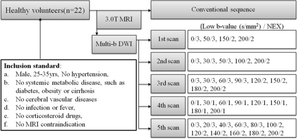
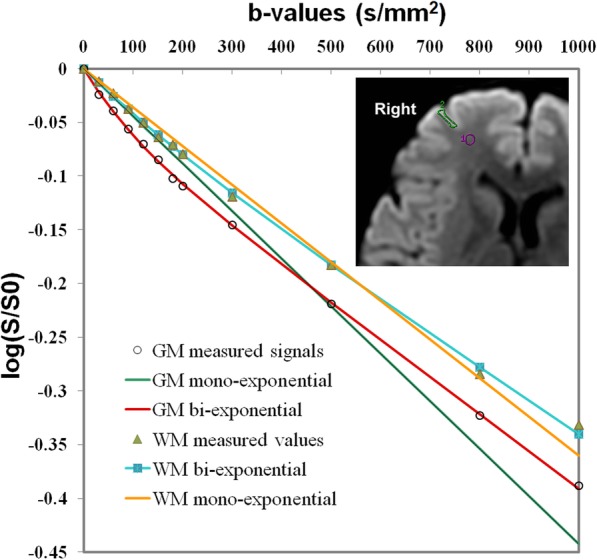
Methods: This prospective study was approved by the local Ethics Committee and informed consent was obtained from each participant. The five consecutive multi-b DWI with different b-value protocols (0-3500 s/mm2) were performed in 22 male healthy volunteers on a 3.0-T MRI system. The IVIM parameters from normal white matter (WM) and gray matter (GM) including slow diffusion coefficient (D), fast perfusion coefficient (D*) and perfusion fraction (f) were compared for differences among defined groups with different IVIM protocols by one-way ANOVA.
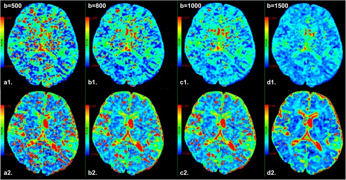
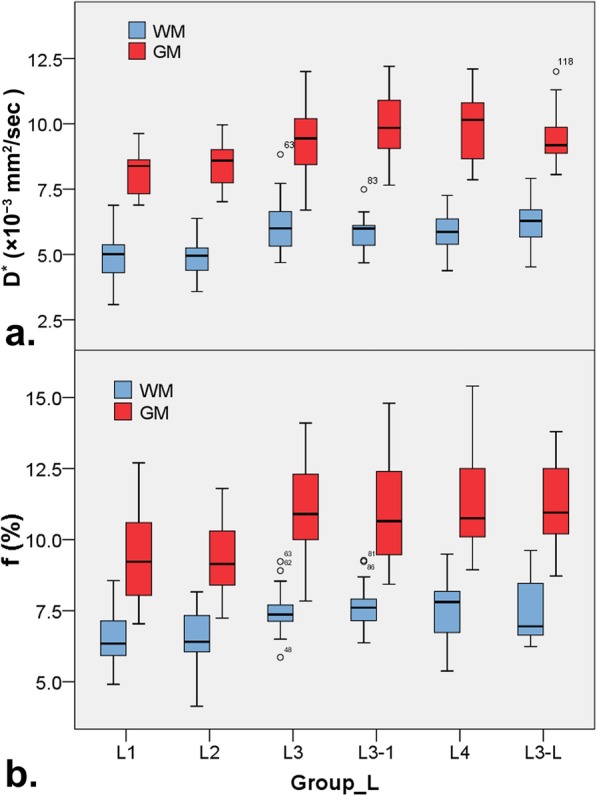
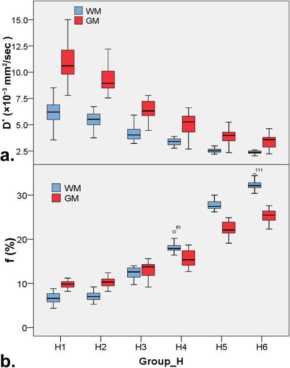
Results: The D* and f value of WM or GM in groups with less low b-values distribution (less than or equal to 5 b-values) were significantly lower than ones in any other group with more low b-values distribution (all P < 0.05), but no significant differences among groups with more low b-values distribution (P > 0.05). In addition, no significant differences in the D, D* and f value of WM or GM were found between group with one and more NEX of low b-values distribution (all P > 0.05). IVIM parameters in normal WM and GM strongly depended on the choice of the high b-value upper limit.
Conclusions: Metrics of IVIM parameters can be affected by low and high b value distribution. Eight low b-values distribution with high b-value upper limit of 800-1000 s/mm2 may be the relatively proper set when performing brain IVIM studies.
Keywords: B-value; Brain; Diffusion weighted imaging; Intravoxel incoherent motion; Number of excitation; Pseudodiffusion.
Conflict of interest statement
The authors declare that they have no competing interests.
Figures
Similar articles
-
Effect of intravoxel incoherent motion on diffusion parameters in normal brain.Neuroimage. 2020 Jan 1;204:116228. doi: 10.1016/j.neuroimage.2019.116228. Epub 2019 Sep 30. Neuroimage. 2020. PMID: 31580945 Free PMC article.
-
Fetal brain development at 25-39 weeks gestational age: A preliminary study using intravoxel incoherent motion diffusion-weighted imaging.J Magn Reson Imaging. 2019 Sep;50(3):899-909. doi: 10.1002/jmri.26667. Epub 2019 Jan 24. J Magn Reson Imaging. 2019. PMID: 30677192
-
Variability of non-Gaussian diffusion MRI and intravoxel incoherent motion (IVIM) measurements in the breast.PLoS One. 2018 Mar 1;13(3):e0193444. doi: 10.1371/journal.pone.0193444. eCollection 2018. PLoS One. 2018. PMID: 29494639 Free PMC article.
-
Deep learning reconstruction for brain diffusion-weighted imaging: efficacy for image quality improvement, apparent diffusion coefficient assessment, and intravoxel incoherent motion evaluation in in vitro and in vivo studies.Diagn Interv Radiol. 2023 Sep 5;29(5):664-673. doi: 10.4274/dir.2023.232149. Epub 2023 Aug 9. Diagn Interv Radiol. 2023. PMID: 37554957 Free PMC article.
-
Intravoxel Incoherent Motion MRI of Rectal Cancer: Correlation of Diffusion and Perfusion Characteristics With Prognostic Tumor Markers.AJR Am J Roentgenol. 2018 Apr;210(4):W139-W147. doi: 10.2214/AJR.17.18342. Epub 2018 Feb 15. AJR Am J Roentgenol. 2018. PMID: 29446674
Cited by
-
Signal to noise and b-value analysis for optimal intra-voxel incoherent motion imaging in the brain.PLoS One. 2021 Sep 23;16(9):e0257545. doi: 10.1371/journal.pone.0257545. eCollection 2021. PLoS One. 2021. PMID: 34555054 Free PMC article.
-
Exploring the Potential of Machine Learning Algorithms to Improve Diffusion Nuclear Magnetic Resonance Imaging Models Analysis.J Med Phys. 2024 Apr-Jun;49(2):189-202. doi: 10.4103/jmp.jmp_10_24. Epub 2024 Jun 25. J Med Phys. 2024. PMID: 39131437 Free PMC article.
-
High-fidelity intravoxel incoherent motion parameter mapping using locally low-rank and subspace modeling.Neuroimage. 2024 Apr 15;292:120601. doi: 10.1016/j.neuroimage.2024.120601. Epub 2024 Apr 7. Neuroimage. 2024. PMID: 38588832 Free PMC article.
-
Optimizing b-values schemes for diffusion MRI of the brain with segmented Intravoxel Incoherent Motion (IVIM) model.J Appl Clin Med Phys. 2023 Jun;24(6):e13986. doi: 10.1002/acm2.13986. Epub 2023 Apr 9. J Appl Clin Med Phys. 2023. PMID: 37031365 Free PMC article.
-
High-Grade Glioma Treatment Response Monitoring Biomarkers: A Position Statement on the Evidence Supporting the Use of Advanced MRI Techniques in the Clinic, and the Latest Bench-to-Bedside Developments. Part 1: Perfusion and Diffusion Techniques.Front Oncol. 2022 Mar 3;12:810263. doi: 10.3389/fonc.2022.810263. eCollection 2022. Front Oncol. 2022. PMID: 35359414 Free PMC article. Review.
References
-
- Lemke A, Laun FB, Klauss M, Re TJ, Simon D, Delorme S, Schad LR, Stieltjes B. Differentiation of pancreas carcinoma from healthy pancreatic tissue using multiple b-values: comparison of apparent diffusion coefficient and intravoxel incoherent motion derived parameters. Investig Radiol. 2009;44(12):769–775. doi: 10.1097/RLI.0b013e3181b62271. - DOI - PubMed
-
- Chandarana H, Kang SK, Wong S, Rusinek H, Zhang JL, Arizono S, Huang WC, Melamed J, Babb JS, Suan EF, et al. Diffusion-weighted intravoxel incoherent motion imaging of renal tumors with histopathologic correlation. Investig Radiol. 2012;47(12):688–696. doi: 10.1097/RLI.0b013e31826a0a49. - DOI - PubMed
Publication types
MeSH terms
LinkOut - more resources
Full Text Sources
