

The value of MRI in management of endometrial hyperplasia with atypia
- PMID: 32041614
- PMCID: PMC7011375
- DOI: 10.1186/s12957-020-1811-5
The value of MRI in management of endometrial hyperplasia with atypia
Abstract
Background: The value of the magnetic resonance imaging (MRI) in the assessment of women with endometrial hyperplasia and its role in diagnosis of myometrial invasion or coexistence of cancer is not known. This study aimed to evaluate the accuracy and usefulness of MRI in the management of patients diagnosed on endometrial biopsy with complex endometrial hyperplasia with atypia (CEHA).
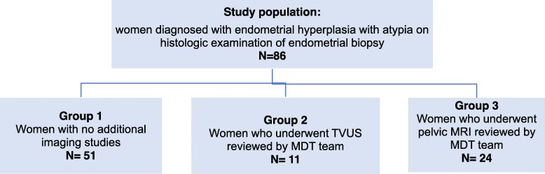
Methods: A retrospective study of 86 cases diagnosed with endometrial hyperplasia with atypia on the initial endometrial biopsy in a tertiary university teaching hospital between 2010 and 2015 was carried out. The MRI accuracy in predicting malignant changes and influence the clinical management was compared among women who had either pelvic MRI, transvaginal ultrasound (TVUS), or no additional imagistic studies.
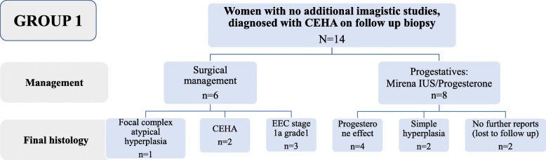
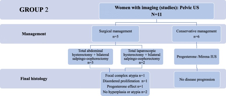
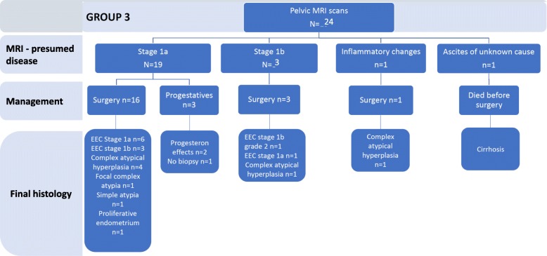
Results: MRI was performed in 24 (28%) and TVUS in 11 (13%)cases, while 51 (59%) women had no additional imagistic studies. In the group of women with no imaging studies, 26/51 (51%) were surgically treated and 8/26 (31%) were diagnosed with endometrial cancer (EEC) stage 1a. In the group of women who had TVUS, 5/11 (45%) were surgically treated and none was diagnosed with EEC. In the group of women who underwent an MRI examination, 20/24 (83%) were surgically treated. Among these, 11/20 (55%) were diagnosed with EEC, 7 had EEC stage 1a, and 4 had EEC stage 1b. Although MRI was able to identify malignant changes with a good sensitivity (91.7%), it had a low specificity in characterisation of malignant transformation (8%). MRI correctly identified 31% of the stage 1a and 33% of the stage 1b endometrial cancer.
Conclusion: In this study, we found a potential diagnostic value of MRI for identifying malignant transformation in patients with CEHA. However, pelvic MRI has a rather weak predictive value of myometrial invasion in women with CEHA and concurrent EEC. The diagnostic and therapeutic benefits of MRI assessment in patients with CEHA need further validation.
Keywords: Endometrial cancer; Endometrial hyperplasia; Magnetic resonance imaging; Myometrial invasion; Sensitivity and specificity; Ultrasonography.
Conflict of interest statement
The authors declare that they have no competing interests.
Figures
Similar articles
-
The value of ultrasonography in preoperative assessment of selected prognostic factors in endometrial cancer.Eur J Gynaecol Oncol. 2003;24(3-4):293-8. Eur J Gynaecol Oncol. 2003. PMID: 12807243
-
The value of magnetic resonance imaging in investigating complex atypical hyperplasia of the endometrium.Minerva Ginecol. 2016 Aug;68(4):400-4. Epub 2016 Mar 22. Minerva Ginecol. 2016. PMID: 27002383
-
Evaluation of the sensitivity, specificity, positive and negative predictive values of preoperative magnetic resonance imaging for staging endometrial cancer. A prospective study of 100 cases at the Dorset Cancer Centre.Eur J Obstet Gynecol Reprod Biol. 2008 Apr;137(2):232-5. doi: 10.1016/j.ejogrb.2007.02.029. Epub 2007 May 29. Eur J Obstet Gynecol Reprod Biol. 2008. PMID: 17537566
-
Clinical outcome of atypical endometrial hyperplasia diagnosed on an endometrial biopsy: institutional experience and review of literature.Am J Surg Pathol. 2012 Nov;36(11):1683-90. doi: 10.1097/PAS.0b013e31825dd4ff. Am J Surg Pathol. 2012. PMID: 23073327 Review.
-
Diagnostic accuracy of TVUS and MRI in the preoperative evaluation of myometrial infiltration in patients with endometrial cancer: A meta-analysis.Clin Radiol. 2025 Jun;85:106868. doi: 10.1016/j.crad.2025.106868. Epub 2025 Mar 12. Clin Radiol. 2025. PMID: 40215804
Cited by
-
Diagnostic value of the apparent diffusion coefficient in differentiating malignant from benign endometrial lesions.Front Oncol. 2023 Apr 14;13:1109495. doi: 10.3389/fonc.2023.1109495. eCollection 2023. Front Oncol. 2023. PMID: 37124536 Free PMC article.
-
Risk of More Advanced Lesions at Hysterectomy after Initial Diagnosis of Non-Atypical Endometrial Hyperplasia in Patients with Postmenopausal Bleeding and Oral Anticoagulant Treatment.Medicina (Kaunas). 2021 Sep 23;57(10):1003. doi: 10.3390/medicina57101003. Medicina (Kaunas). 2021. PMID: 34684040 Free PMC article.
-
A comparative study of mono-exponential and advanced diffusion-weighted imaging in differentiating stage IA endometrial carcinoma from benign endometrial lesions.J Cancer Res Clin Oncol. 2024 Mar 20;150(3):141. doi: 10.1007/s00432-024-05668-8. J Cancer Res Clin Oncol. 2024. PMID: 38504026 Free PMC article.
-
Multimodal MRI-Based Radiomics-Clinical Model for Preoperatively Differentiating Concurrent Endometrial Carcinoma From Atypical Endometrial Hyperplasia.Front Oncol. 2022 May 27;12:887546. doi: 10.3389/fonc.2022.887546. eCollection 2022. Front Oncol. 2022. PMID: 35692806 Free PMC article.
-
Evaluation and Monitoring of Endometrial Cancer Based on Magnetic Resonance Imaging Features of Deep Learning.Contrast Media Mol Imaging. 2022 Mar 18;2022:5198592. doi: 10.1155/2022/5198592. eCollection 2022. Contrast Media Mol Imaging. 2022. PMID: 35360265 Free PMC article.
References
MeSH terms
LinkOut - more resources
Full Text Sources
Medical
