. 2020 Apr;79(4):507-517.
doi: 10.1136/annrheumdis-2019-216542. Epub 2020 Feb 10.

Long non-coding RNA HOTAIR drives EZH2-dependent myofibroblast activation in systemic sclerosis through miRNA 34a-dependent activation of NOTCH

Affiliations

Long non-coding RNA HOTAIR drives EZH2-dependent myofibroblast activation in systemic sclerosis through miRNA 34a-dependent activation of NOTCH

Christopher W Wasson et al. Ann Rheum Dis. 2020 Apr.

Abstract

Background: Systemic sclerosis (SSc) is characterised by autoimmune activation, tissue and vascular fibrosis in the skin and internal organs. Tissue fibrosis is driven by myofibroblasts, that are known to maintain their phenotype in vitro, which is associated with epigenetically driven trimethylation of lysine 27 of histone 3 (H3K27me3).

Methods: Full-thickness skin biopsies were surgically obtained from the forearms of 12 adult patients with SSc of recent onset. Fibroblasts were isolated and cultured in monolayers and protein and RNA extracted. HOX transcript antisense RNA (HOTAIR) was expressed in healthy dermal fibroblasts by lentiviral induction employing a vector containing the specific sequence. Gamma secretase inhibitors were employed to block Notch signalling. Enhancer of zeste 2 (EZH2) was blocked with GSK126 inhibitor.

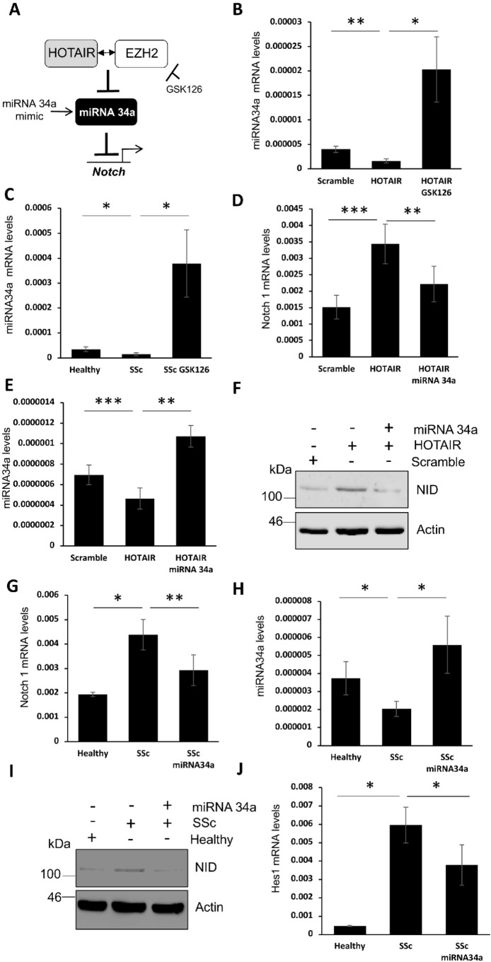
Results: SSc myofibroblasts in vitro and SSc skin biopsies in vivo display high levels of HOTAIR, a scaffold long non-coding RNA known to direct the histone methyltransferase EZH2 to induce H3K27me3 in specific target genes. Overexpression of HOTAIR in dermal fibroblasts induced EZH2-dependent increase in collagen and α-SMA expression in vitro, as well as repression of miRNA-34A expression and consequent NOTCH pathway activation. Consistent with these findings, we show that SSc dermal fibroblast display decreased levels of miRNA-34a in vitro. Further, EZH2 inhibition rescued miRNA-34a levels and mitigated the profibrotic phenotype of both SSc and HOTAIR overexpressing fibroblasts in vitro.

Conclusions: Our data indicate that the EZH2-dependent epigenetic phenotype of myofibroblasts is driven by HOTAIR and is linked to miRNA-34a repression-dependent activation of NOTCH signalling.

Keywords: autoimmune diseases; fibroblasts; systemic sclerosis.


Conflict of interest statement

Competing interests: H.Y.C. is a co-founder of Accent Therapeutics, Boundless Bio, and advisor to 10x Genomics, Arsenal Biosciences, and Spring Discovery.

Figures

Figure 1
Figure 1
lncRNA HOX transcript antisense RNA (HOTAIR) is upregulated in activated myofibroblasts. (A) Healthy and systemic sclerosis (SSc) fibroblasts were stained with a mouse alpha-smooth muscle actin (α-SMA) antibody and visualised with an alexa 594-conjugated mouse secondary antibody (red). Cells were counterstained with 4′,6-diamidino-2-phenylindole (DAPI) to stain the nuclei (blue). (B) α-SMA positive or negative single cells were dissected by laser capture microscopy on untreated SSc patient fibroblasts. RNA was extracted from α-SMA positive and negative cells. (C) α-SMA transcript levels were assessed by qPCR. Graph represents mRNA levels from three independent repeats. (D) HOX tiling array was performed on the RNA and the table represents the fold differences of each lncRNA within the HOX locus in α-SMA positive cells compared with negative. HOTAIR is labelled in red. (E) HOTAIR transcript levels were assessed from RNA extracted from α-SMA positive and negative cells by qPCR. Graph represents mRNA levels from three independent repeats. (F) RNA was extracted from telomerase reverse transcriptase (Human) (HTERT) immortalised fibroblasts that had been isolated from healthy and patient with SSc skin biopsies. HOTAIR transcript levels were assessed by qPCR. Graph represents HOTAIR mRNA levels from five healthy patients and five patient with diffuse SSc fibroblasts. (G) RNA was extracted from healthy and patient with diffuse SSc skin biopsies. HOTAIR transcript levels were assessed by qPCR. Graph represents HOTAIR mRNA levels from (n=3). (H) HOTAIR in situ hybridisation staining from healthy and patient with SSc skin. (H) Expanded panels represent areas of interest in the dermis regions and black boxes represent the region of the area of interest. (I) RNA was extracted from primary fibroblasts transfected with HOTAIR and scramble control antisense oligonucleotides. HOTAIR, α-SMA and connective tissue growth factor (CTGF) transcript levels were assessed by qPCR. Graph represents mRNA levels from three independent repeats. *p<0.05, **p<0.01, ***p<0.001.
Figure 2
Figure 2
HOX transcript antisense RNA (HOTAIR) drives profibrotic activation of dermal fibroblasts. (A) Representative image of healthy dermal fibroblasts infected with lentiviruses containing the scramble/HOTAIR vectors. Vectors contain a GFP reporter to determine infection efficiencies. (B) Histograms representing GFP cell sorting of fibroblasts-infected lentiviruses containing the scramble/HOTAIR vectors. GFP-positive fibroblasts were collected and cultured. RNA was extracted from fibroblasts stably expressing scramble and HOTAIR vectors. (C) HOTAIR, (D) collagen type 1A1, (E) 1A2, (F) alpha-smooth muscle actin (α-SMA) and (G) connective tissue growth factor (CTGF) transcript levels were assessed by qPCR. Graphs represents mRNA levels from three independent repeats. (H) Protein was extracted from fibroblasts stably expressing scramble and HOTAIR vectors. Lysates were probed with a pan collagen type 1 antibody and an α-SMA antibody by western blot. β-actin was probed for as a loading control. (I) Graph represents densitometry analysis of collagen type 1 and α-SMA western blots from three independent repeats. (J) α-SMA staining of scramble and HOTAIR expressing dermal fibroblasts. Fibroblasts were stained with a mouse α-SMA antibody and visualised with a mouse-specific alexa 594-conjugated secondary (red). Cells were counterstained with DAPI to visualise nuclei (blue). White lines represent 400 μM scale bar. Red lines represent 20 μM scale bar. Scramble and HOTAIR expressing fibroblasts were serum depleted for 16 hours prior to stimulation with TGF-β (10 ng/mL) for 48 hours. (K) RNA was extracted and α-SMA transcript levels were assessed by qPCR. Graphs represents mRNA levels from three independent repeats. *p<0.05, **p<0.01, ***p<0.001.
Figure 3
Figure 3
HOX transcript antisense RNA (HOTAIR) drives profibrotic activation through enhancer of zeste 2 (EZH2)-mediated H3K27me3 methylation. RNA was extracted from fibroblasts stably expressing scramble and HOTAIR vectors. (A) EZH2 transcript levels were assessed by qPCR. Graphs represents mRNA levels from three independent repeats. (B) Protein was extracted from fibroblasts stably expressing scramble and HOTAIR vectors. Lysates were probed with H3K27me3-specific antibody by western blot. β-actin was probed for as a loading control. (C) Graph represents densitometry analysis of H3K27me3 western blots from three independent repeats. RNA and protein were extracted from fibroblasts stably expressing the scramble and HOTAIR vectors, in addition to HOTAIR fibroblasts treated with the EZH2 inhibitor GSK126. (D) Collagen type 1A1, (E) collagen type 1A2, (F) alpha-smooth muscle actin (α-SMA) and (G) connective tissue growth factor (CTGF) transcript levels were assessed by qPCR. Graphs represents mRNA levels from three independent repeats. (H) Protein lysates were probed with pan collagen type 1, α-SMA and H3K27me3 antibodies by western blot. β-actin was probed for as a loading control. (I) α-SMA staining of scramble and HOTAIR expressing dermal fibroblasts in addition to HOTAIR fibroblasts treated with the EZH2 inhibitor GSK126. Fibroblasts were stained with a mouse α-SMA antibody and visualised with a mouse-specific alexa 594-conjugated secondary (red). Cells were counterstained with DAPI to visualise nuclei (blue). Red lines represent 20 μM scale bar. *p<0.05, **p<0.01, ***p<0.001. NS, not significant.
Figure 4
Figure 4
HOX transcript antisense RNA (HOTAIR) drives Notch 1 expression in dermal fibroblasts through enhancer of zeste 2 (EZH2). RNA was extracted from fibroblasts stably expressing scramble and HOTAIR vectors. (A) Notch1 and (D) Notch target gene Hes1 transcript levels were assessed by qPCR. Graph represents mRNA levels from three independent repeats. (B) Schematic of the Notch 1 receptor and the mechanism of activation. (C) Protein was extracted from fibroblasts stably expressing scramble and HOTAIR vectors. Lysates were probed with an antibody specific for the intracellular domain of Notch, Notch Intracellular Domain (NID) by western blot. β-actin was probed for as a loading control. (E) RNA and protein were extracted from fibroblasts stably expressing the scramble and HOTAIR vectors, in addition to HOTAIR fibroblasts treated with the EZH2 inhibitor GSK126. Notch 1 transcript levels were assessed by qPCR. Graphs represents mRNA levels from three independent repeats. (F) Protein lysates were probed with an NID and H3K27me3 antibodies by western blot. β-actin was probed for as a loading control. RNA was extracted from healthy and systemic sclerosis (SSc) fibroblasts, in addition to SSc fibroblasts treated with the EZH2 inhibitor GSK126. (G) Notch 1 transcript levels were assessed by qPCR. Graphs represents mRNA levels from three independent repeats. RNA and protein were extracted from fibroblasts stably expressing the scramble and HOTAIR vectors, in addition to HOTAIR fibroblasts treated with the gamma secretase inhibitor R04929097. (H) Hes1, (I) Col1A1 and (J) alpha-smooth muscle actin (α-SMA) transcript levels were assessed by qPCR. Graphs represents mRNA levels from three independent repeats. (K) Protein lysates were probed with a pan collagen type 1 antibody, an α-SMA antibody and an antibody specific for the intracellular domain of Notch 1 (NID) by western blot. β-actin was probed for as a loading control. (L) α-SMA staining of scramble and HOTAIR expressing dermal fibroblasts, in addition to HOTAIR fibroblasts treated with the gamma secretase inhibitor R04929097. Fibroblasts were stained with a mouse α-SMA antibody and visualised with a mouse-specific alexa 594-conjugated secondary (red). Cells were counterstained with DAPI to visualise nuclei (blue). Red lines represent 20 μM scale bar. *p<0.05, **p<0.01, ***p<0.001.
Figure 5
Figure 5
HOX transcript antisense RNA (HOTAIR) drives Notch 1 expression through the methylation of miRNA-34a. (A) Schematic of the mechanism HOTAIR employs to drive Notch transcription. (B) RNA was extracted from fibroblasts stably expressing the scramble and HOTAIR vectors, in addition to HOTAIR fibroblasts treated with the enhancer of zeste 2 (EZH2) inhibitor GSK126. MiRNA-34a transcript levels were assessed by qPCR. Graphs represents mRNA levels from three independent repeats. (C) RNA was extracted from healthy and SSc fibroblasts, in addition systemic sclerosis (SSc) fibroblasts treated with the EZH2 inhibitor GSK126. miRNA-34a transcript levels were assessed by qPCR. Graphs represents mRNA levels from three independent repeats. RNA and protein were extracted from scramble or HOTAIR expressing fibroblasts transfected with an miRNA-34a mimic or a negative control mimic. (D) Notch 1 and (E) miRNA-34a transcript levels were assessed by qPCR. Graphs represents mRNA levels from three independent repeats. (F) Protein lysates were probed with an antibody specific for the intracellular domain of Notch 1 (Notch Intracellular Domain (NID)) by western blot. β-actin was probed for as a loading control. RNA and protein were extracted from healthy and SSc fibroblasts transfected with an miRNA-34a mimic or a negative control mimic. Notch 1 (G) and miRNA-34a (H) transcript levels were assessed by qPCR. Graphs represents mRNA levels from three independent repeats. (I) Protein lysates were probed with an antibody specific for the intracellular domain of Notch 1 (NID) by western blot. β-actin was probed for as a loading control. (J) RNA was extracted from healthy and SSc fibroblasts transfected with an miRNA-34a mimic or a negative control mimic. Hes1 transcript levels were assessed by qPCR. Graphs represents mRNA levels from three independent repeats. *p<0.05, **p<0.01, ***p<0.001.
Figure 6
Figure 6
HOX transcript antisense RNA (HOTAIR)-mediated suppression of miRNA-34a is important for systemic sclerosis (SSc) fibrosis. RNA and protein were extracted from scramble or HOTAIR-expressing fibroblasts transfected with an miRNA-34a mimic or a negative control mimic. (A) Col1A1, (B) Col1A2 and (C) alpha-smooth muscle actin (α-SMA) transcript levels were assessed by qPCR. Graphs represents mRNA levels from three independent repeats. (D) Protein lysates were probed with a pan collagen type 1 and α-SMA antibody by western blot. β-actin was probed for as a loading control. (E) α-SMA staining of scramble and HOTAIR expressing dermal fibroblasts, in addition to HOTAIR fibroblasts were transfected with an miRNA-34a mimic or a negative control mimic. Fibroblasts were stained with a mouse α-SMA antibody and visualised with a mouse-specific alexa 594-conjugated secondary (red). Cells were counterstained with DAPI to visualise nuclei (blue). Red lines represent 20 μM scale bar. RNA and protein were extracted from healthy and SSc fibroblasts transfected with an miRNA-34a mimic or a negative control mimic. (F) Col1A1, (G) Col1A2 and (H) α-SMA transcript levels were assessed by qPCR. Graphs represents mRNA levels from three independent repeats. (I) Protein lysates were probed with an collagen type 1 antibody and an α-SMA antibody by western blot. β-actin was probed for as a loading control. (J) Schematic of the role HOTAIR plays in SSc-associated fibrosis. *p<0.05, **p<0.01, ***p<0.001.

Similar articles

Cited by

References

    1. Gabrielli A, Avvedimento EV, Krieg T. Scleroderma. N Engl J Med 2009;360:1989–2003. 10.1056/NEJMra0806188 - DOI - PubMed
    1. Denton CP, Khanna D. Systemic sclerosis. Lancet 2017;390:1685–99. 10.1016/S0140-6736(17)30933-9 - DOI - PubMed
    1. Lambova S, Batalov A, Dobrev H, et al. . Digital Ulcers in Systemic Sclerosis - How to Manage in 2013? Curr Rheumatol Rev 2013;9:274–8. 10.2174/157339710904140417125803 - DOI - PubMed
    1. Garrett SM, Baker Frost D, Feghali-Bostwick C. The mighty fibroblast and its utility in scleroderma research. J scleroderma relat Disord. May-Aug 2017;2:69–134. - PMC - PubMed
    1. Hinz B, Phan SH, Thannickal VJ, et al. . Recent developments in myofibroblast biology: paradigms for connective tissue remodeling. Am J Pathol 2012;180:1340–55. 10.1016/j.ajpath.2012.02.004 - DOI - PMC - PubMed

MeSH terms