. 2020 Apr 3;295(14):4696-4708.
doi: 10.1074/jbc.RA119.011280. Epub 2020 Feb 10.

The E3 ubiquitin ligase STUB1 attenuates cell senescence by promoting the ubiquitination and degradation of the core circadian regulator BMAL1

Affiliations

The E3 ubiquitin ligase STUB1 attenuates cell senescence by promoting the ubiquitination and degradation of the core circadian regulator BMAL1

Kifayat Ullah et al. J Biol Chem. .

Erratum in

Abstract

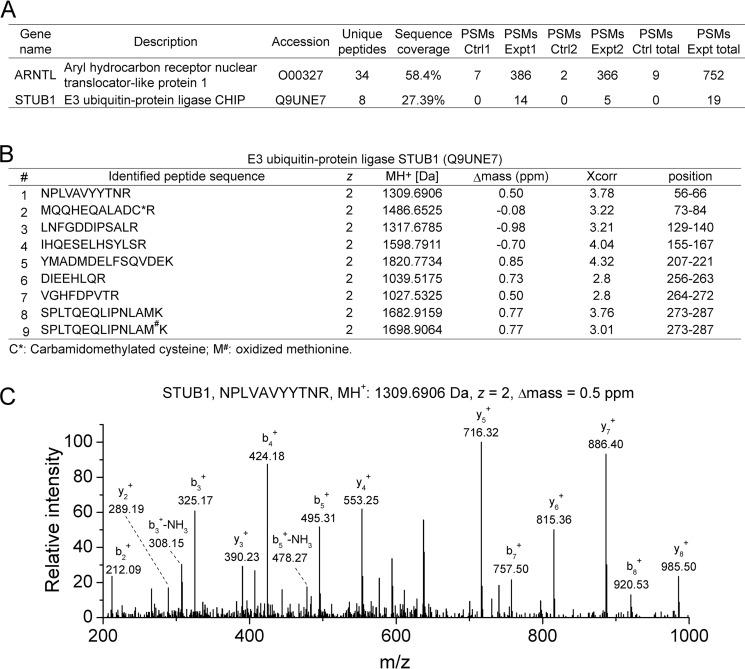
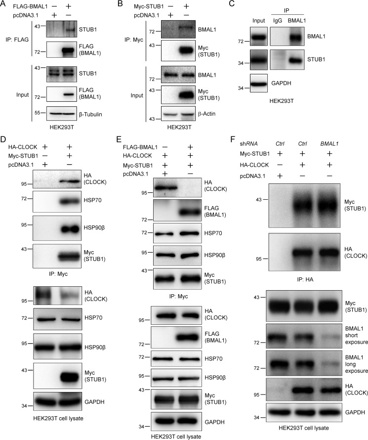
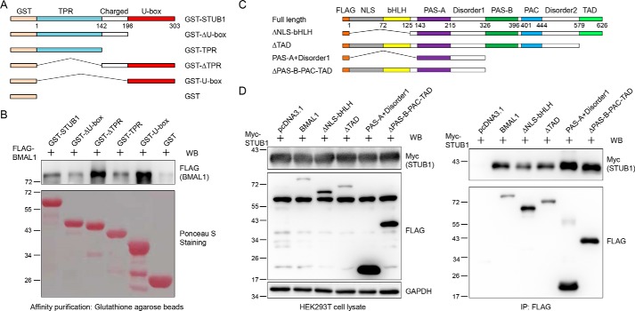
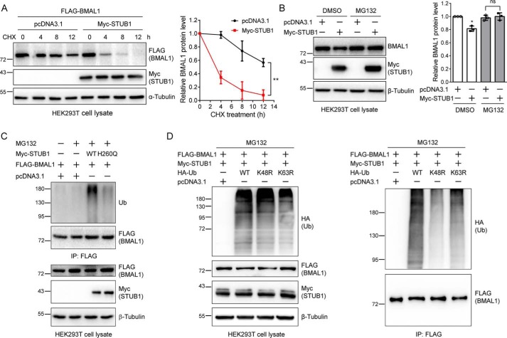
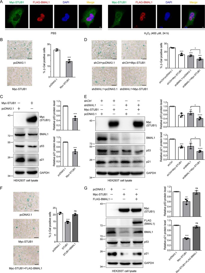
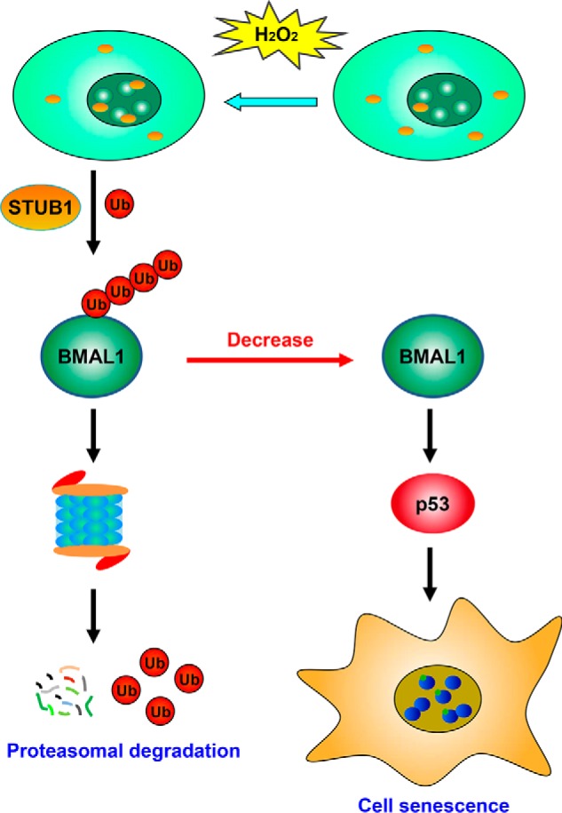
Cell senescence is one of the most important processes determining cell fate and is involved in many pathophysiological conditions, including cancer, neurodegenerative diseases, and other aging-associated diseases. It has recently been discovered that the E3 ubiquitin ligase STIP1 homology and U-box-containing protein 1 (STUB1 or CHIP) is up-regulated during the senescence of human fibroblasts and modulates cell senescence. However, the molecular mechanism underlying STUB1-controlled senescence is not clear. Here, using affinity purification and MS-based analysis, we discovered that STUB1 binds to brain and muscle ARNT-like 1 (BMAL1, also called aryl hydrocarbon receptor nuclear translocator-like protein 1 (ARNTL)). Through biochemical experiments, we confirmed the STUB1-BMAL1 interaction, identified their interaction domains, and revealed that STUB1 overexpression down-regulates BMAL1 protein levels through STUB1's enzymatic activity and that STUB1 knockdown increases BMAL1 levels. Further experiments disclosed that STUB1 enhances BMAL1 degradation, which is abolished upon proteasome inhibition. Moreover, we found that STUB1 promotes the formation of Lys-48-linked polyubiquitin chains on BMAL1, facilitating its proteasomal degradation. Interestingly, we also discovered that oxidative stress promotes STUB1 nuclear translocation and enhances its co-localization with BMAL1. STUB1 expression attenuates hydrogen peroxide-induced cell senescence, indicated by a reduced signal in senescence-associated β-gal staining and decreased protein levels of two cell senescence markers, p53 and p21. BMAL1 knockdown diminishes this effect, and BMAL1 overexpression abolishes STUB1's effect on cell senescence. In summary, the results of our work reveal that the E3 ubiquitin ligase STUB1 ubiquitinates and degrades its substrate BMAL1 and thereby alleviates hydrogen peroxide-induced cell senescence.

Keywords: E3 ubiquitin ligase; STIP1 homology and U-box-containing protein 1 (STUB1); brain and muscle ARNT-like 1 (BMAL1, ARNTL, MOP3); cell cycle regulation; circadian clock; hydrogen peroxide; proteasome; protein degradation; senescence; ubiquitylation (ubiquitination).


Conflict of interest statement

The authors declare that they have no conflicts of interest with the contents of this article

Figures

Figure 1.
Figure 1.
Immunoprecipitation and MS analysis identified STUB1 as an interacting partner of BMAL1. A, PSMs for BMAL1 and STUB1 identified from two biological replicates of MS analyses for the FLAG immunoprecipitates of cell lysates from HEK293T cells expressing control or FLAG-BMAL1 plasmid, respectively. The number of peptides, sequence coverage, and PSMs for BMAL1 and STUB1 were provided. B, information for the MS-identified tryptic peptides derived from STUB1 in two biological replicates. The peptide sequence, charge state (z), MH+, Δmass, Xcorr, and amino acid position of the peptides derived from STUB1 were provided. C, a representative annotated MS/MS spectrum of an identified STUB1 tryptic peptide. The amino acid sequence, MH+, charge state (z), and Δmass of the identified peptide were also shown along with the annotated b- and y-ions.
Figure 2.
Figure 2.
STUB1 preferably interacts with BMAL1 over CLOCK. A and B, STUB1 co-immunoprecipitates BMAL1 (A) and BMAL1 co-immunoprecipitates STUB1 (B). HEK293T cells were transfected with pcDNA3.1, Myc-STUB1, or FLAG-BMAL1 plasmid using PEI transfection reagent for 48 h. STUB1 or BMAL1 was immunoprecipitated (IP) with Myc or FLAG antibodies, respectively. C, STUB1 interacts with BMAL1 endogenously. Endogenous BMAL1 in cell lysate from HEK293T cells was immunoprecipitated with an anti-BMAL1 antibody. IgG was used as a control. D, STUB1 interacts with CLOCK. HEK293T cells were transfected with HA-CLOCK plasmid and then split into two plates for the transfection of control or Myc-STUB1 plasmid for 48 h, respectively. STUB1 was immunoprecipitated with an anti-Myc antibody. HSP70 and HSP90β were used as positive controls for their interaction with STUB1. E, STUB1 preferably interacts with BMAL1 over CLOCK. HEK293T cells were co-transfected with HA-CLOCK and Myc-STUB1 plasmids and then split into two plates for the transfection of control or FLAG-BMAL1 plasmid, respectively. STUB1 was immunoprecipitated with anti-Myc magnetic beads. F, BMAL1 knockdown enhances the interaction between STUB1 and CLOCK. HEK293T cells were co-transfected with HA-CLOCK and Myc-STUB1 plasmids and then split into two plates for the transfection of control or shBMAL1 plasmid, respectively. CLOCK was immunoprecipitated with anti-HA magnetic beads. The cell lysates and immunoprecipitates in A–F were immunoblotted with the indicated antibodies.
Figure 3.
Figure 3.
BMAL1 interacts with the STUB1 U-box domain and STUB1 interacts with the BMAL1 middle domain. A, illustration of domains for the GST-tagged full-length STUB1 and its truncations. B, BMAL1 preferably interacts with the STUB1 U-box domain. GST-tagged STUB1 and its truncations were expressed in E. coli, purified with GSH-agarose beads, incubated with cell lysate from HEK293T cells expressing FLAG-BMAL1, washed, and eluted by heating with 2× SDS loading buffer. The eluates were stained with Ponceau S or immunoblotted (WB) with an anti-FLAG antibody. C, illustration of domains for the FLAG-tagged full-length BMAL1 and its truncations. D, STUB1 interacts with the BMAL1 middle domain. FLAG-tagged full-length BMAL1 and its truncations were co-transfected with Myc-STUB1 into HEK293T cells. Cells were lysed, and BMAL1 and its truncations were immunoprecipitated with an anti-FLAG antibody. Cell lysates and immunoprecipitates were immunoblotted with the indicated antibodies.
Figure 4.
Figure 4.
The E3 ligase activity is required for the STUB1-mediated BMAL1 reduction. A, STUB1 expression down-regulates BMAL1 protein. HEK293T cells were transfected with the control or Myc-STUB1 plasmid, respectively. Cell lysates were immunoblotted for endogenous BMAL1, Myc-STUB1, and β-tubulin. The experiments were carried out three times, and means ± S.D. (error bars) were plotted with GraphPad Prism. p value was calculated using Student's t test. **, p < 0.01. B, gradient-increased transfection of STUB1 gradually reduces BMAL1 protein level. HEK293T cells were transfected with different amounts of the control or Myc-STUB1 plasmid in 6-well plates. Cell lysates were immunoblotted for endogenous BMAL1, Myc-STUB1, and β-tubulin. The experiments were carried out three times, and means ± S.D. were plotted with GraphPad Prism. Student's t test was used: *, p < 0.05; ns, not significant. C, the regulation of BMAL1 by STUB1 requires its E3 ligase activity. HEK293T cells were transfected with the control, WT STUB1, or H260Q STUB1 (inactive mutant) plasmid, and cell lysates were immunoblotted. Student's t test was performed for the data from triplicates, and means ± S.D. were plotted. ***, p < 0.001; ns, not significant. D, STUB1 knockdown increases BMAL1 protein level. HEK293T cells were transfected with control or STUB1 specific siRNAs, respectively, for 48 h using Lipofectamine 2000 transfection reagent. Cell lysates were immunoblotted with the indicated antibodies. The experiments were performed in triplicates, and means ± S.D. were plotted. Student's t test was used: ***, p < 0.001.
Figure 5.
Figure 5.
STUB1 mediates the BMAL1 protein level through the ubiquitin-proteasome system. A, STUB1 enhances BMAL1 degradation. HEK293T cells were transfected with FLAG-BMAL1 plasmid and the control or Myc-STUB1 plasmid. Cells were split into 6-well plates, and at 48 h post-transfection, cells were treated with CHX (200 μg/ml) for the indicated time. Cell lysates were immunoblotted with the indicated antibodies. Experiments were repeated three times, and means ± S.D. (error bars) were plotted. Two-way analysis of variance was used: **, p < 0.01. Note that the amount of plasmid used for transfection was slightly adjusted to ensure that the FLAG-BMAL1 protein level at the zero time point was similar in the absence or presence of Myc-STUB1. B, MG132 eliminates the effect of STUB1 on BMAL1 protein level. HEK293T cells were transfected with a control or Myc-STUB1 plasmid, respectively, and at 48 h after transfection, cells were treated with DMSO or MG132 (10 μm) for 12 h. The resulting cell lysates were immunoblotted. The experiments were repeated three times, and means ± S.D. were plotted. Student's t test was used: *, p < 0.05; ns, not significant. C, STUB1 increases BMAL1 ubiquitination. HEK293T cells were first transfected with FLAG-BMAL1 plasmid and divided into four plates 6 h after transfection. The cells were again transfected with a control, WT, or H260Q STUB1 plasmid for 36 h. Cells were treated with DMSO or MG132 (20 μm) for 6 h. FLAG-BMAL1 was immunoprecipitated with FLAG M2 affinity gel. The cell lysates and immunoprecipitates were immunoblotted. D, STUB1 promotes the formation of the Lys-48–linked polyubiquitin chains on BMAL1. HEK293T cells were first transfected with FLAG-BMAL1 and Myc-STUB1 and then split into four plates 6 h after transfection. Cells were again transfected with a control, WT, K48R, or K63R ubiquitin (Ub) plasmid, and at 36 h after the second transfection, cells were treated with MG132 (10 μm) for 12 h. The cell lysates and FLAG immunoprecipitates were immunoblotted with the indicated antibodies.
Figure 6.
Figure 6.
Hydrogen peroxide enhances STUB1 nuclear localization, and STUB1 reduces cell senescence by down-regulating BMAL1. A, H2O2 elevates STUB1 nuclear translocation and promotes the co-localization between STUB1 and BMAL1. HEK293 cells were first transfected with Myc-STUB1 and FLAG-BMAL1, split into two plates, and treated with PBS or 400 μm H2O2 for 24 h. Cells were fixed and incubated with anti-Myc and anti-FLAG antibodies for immunofluorescence measurement. Scale bar, 5 μm. B and C, STUB1 attenuates H2O2-induced cell senescence. HEK293T cells were first transfected with the control or Myc-STUB1 plasmid and then treated with 400 μm H2O2 for 24 h. Half of the cells were stained with an SA-β-Gal staining kit (B), and the other half were used for immunoblotting (C). Images were taken under a microscope, and the senescent cells (blue cells) were counted. Three images were taken for quantification (means ± S.D. (error bars)). Scale bar, 50 μm. Student's t test was used: ***, p < 0.001. The experiments were repeated in triplicates, and similar results were obtained. D and E, BMAL1 knockdown almost completely eliminates the effect of STUB1 on hydrogen peroxide–induced cell senescence. HEK293T cells were first transfected with the control or BMAL1-specific shRNA, and then each was split into two plates for the transfection of the control or Myc-STUB1 plasmid. Cells were treated and processed as described in B and C. Scale bar, 50 μm. Student's t test was used: *, p < 0.05; **, p < 0.01; ***, p < 0.001. F and G, BMAL1 expression abolishes the effect of STUB1 on cell senescence. HEK293T cells were transfected with the control or Myc-STUB1 plasmid with or without FLAG-BMAL1 plasmid. Cells were treated and processed as described in B and C. Scale bar, 50 μm. Student's t test was used: **, p < 0.01; ***, p < 0.001; ns, not significant.
Figure 7.
Figure 7.
Proposed model for the regulation of cell senescence by STUB1. Upon hydrogen peroxide treatment, STUB1 is translocated to the nucleus, promotes the ubiquitination and proteasomal degradation of BMAL1, decreases the BMAL1 protein level, and further reduces the expression of two cell senescence marker proteins, p53 and p21, leading to the attenuated cell senescence induced by hydrogen peroxide.

Similar articles

Cited by

References

    1. Hernandez-Segura A., Nehme J., and Demaria M. (2018) Hallmarks of cellular senescence. Trends Cell Biol. 28, 436–453 10.1016/j.tcb.2018.02.001 - DOI - PubMed
    1. Chen Q. M., Bartholomew J. C., Campisi J., Acosta M., Reagan J. D., and Ames B. N. (1998) Molecular analysis of H2O2-induced senescent-like growth arrest in normal human fibroblasts: p53 and Rb control G1 arrest but not cell replication. Biochem. J. 332, 43–50 10.1042/bj3320043 - DOI - PMC - PubMed
    1. Dimri G. P., Lee X., Basile G., Acosta M., Scott G., Roskelley C., Medrano E. E., Linskens M., Rubelj I., and Pereira-Smith O. (1995) A biomarker that identifies senescent human cells in culture and in aging skin in vivo. Proc. Natl. Acad. Sci. U.S.A. 92, 9363–9367 10.1073/pnas.92.20.9363 - DOI - PMC - PubMed
    1. Shay J. W., Pereira-Smith O. M., and Wright W. E. (1991) A role for both RB and p53 in the regulation of human cellular senescence. Exp. Cell Res. 196, 33–39 10.1016/0014-4827(91)90453-2 - DOI - PubMed
    1. Mavrogonatou E., Pratsinis H., and Kletsas D. (2019) The role of senescence in cancer development. Semin. Cancer Biol. 10.1016/j.semcancer.2019.06.018 - DOI - PubMed

Publication types

MeSH terms