. 2020 Apr 13;40(9):e00429-19.
doi: 10.1128/MCB.00429-19. Print 2020 Apr 13.

TRAF2 Ser-11 Phosphorylation Promotes Cytosolic Translocation of the CD40 Complex To Regulate Downstream Signaling Pathways

Affiliations

TRAF2 Ser-11 Phosphorylation Promotes Cytosolic Translocation of the CD40 Complex To Regulate Downstream Signaling Pathways

Lauren M Workman et al. Mol Cell Biol. .

Abstract

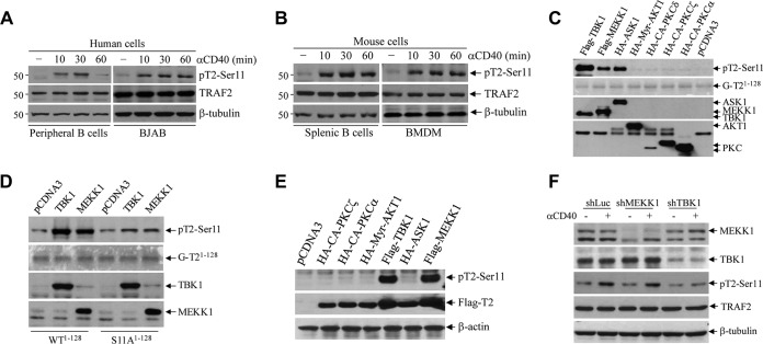
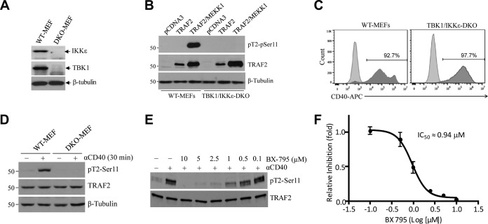
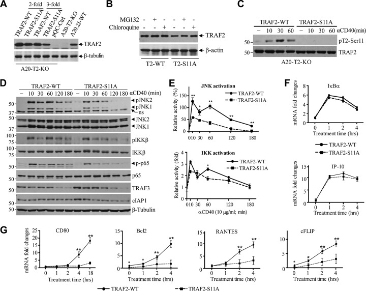
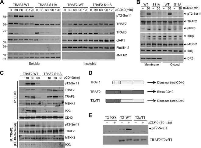
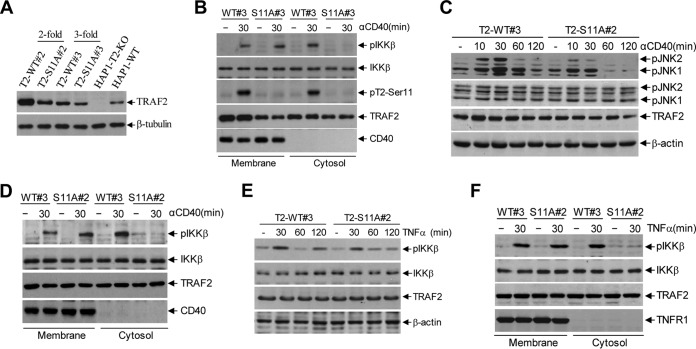
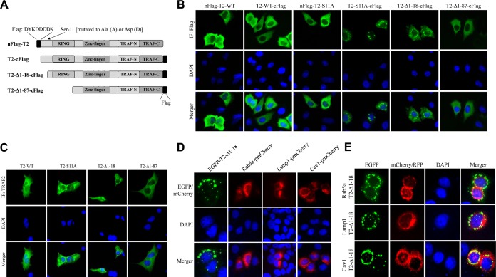
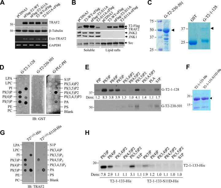
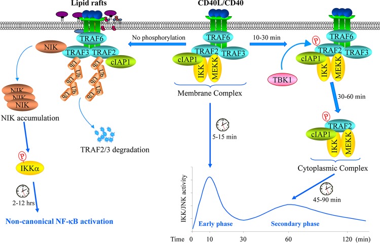
CD40 plays an important role in immune responses by activating the c-Jun N-terminal protein kinase (JNK) and NF-κB pathways; however, the precise mechanisms governing the spatiotemporal activation of these two signaling pathways are not fully understood. Here, using four different TRAF2-deficient cell lines (A20.2J, CH12.LX, HAP1, and mouse embryonic fibroblasts [MEFs]) reconstituted with wild-type or phosphorylation mutant forms of TRAF2, along with immunoprecipitation, immunoblotting, gene expression, and immunofluorescence analyses, we report that CD40 ligation elicits TANK-binding kinase 1 (TBK1)-mediated phosphorylation of TRAF2 at Ser-11. This phosphorylation interfered with the interaction between TRAF2's RING domain and membrane phospholipids and enabled translocation of the TRAF2 complex from CD40 to the cytoplasm. We also observed that this cytoplasmic translocation is required for full activation of the JNK pathway and the secondary phase of the NF-κB pathway. Moreover, we found that in the absence of Ser-11 phosphorylation, the TRAF2 RING domain interacts with phospholipids, leading to the translocation of the TRAF2 complex to lipid rafts, resulting in its degradation and activation of the noncanonical NF-κB pathway. Thus, our results provide new insights into the CD40 signaling mechanisms whereby Ser-11 phosphorylation controls RING domain-dependent subcellular localization of TRAF2 to modulate the spatiotemporal activation of the JNK and NF-κB pathways.

Keywords: CD40; JNK; NF-κB; TRAF2; phosphorylation; signaling mechanisms.


Figures

FIG 1
FIG 1
CD40 ligation elicits TBK1-mediated TRAF2 phosphorylation on Ser-11. (A and B) Human peripheral resting B cells (CD19+ CD43) and BJAB B cell lymphoma cells (A) and mouse splenic B cells and bone marrow-derived macrophages (BMDMs) (B) were stimulated with anti-human CD40 (G28.5, 10 μg/ml) or anti-mouse CD40 (1C10, 5 μg/ml) agonistic antibodies (αCD40) for the indicated times. TRAF2 Ser-11 phosphorylation (pT2-Ser11) was then monitored by Western blotting with a site-specific phosphoantibody. (C) Kinases previously implicated in receptor-mediated NF-κB activation were expressed and immunopurified from 293T cells and then subjected to in vitro phosphorylation assays with GST-TRAF21–128 as the substrate. The reaction mixtures were separated by SDS-PAGE and transferred to nitrocellulose membrane, and TRAF2 phosphorylation was then assessed by autoradiography. The same membrane was stained with Ponceau S to visualize the substrate (GST-TRAF21–128) and then probed with anti-HA and anti-Flag antibodies to monitor the expression of kinases. (D) TBK1 and MEKK1 were immunopurified from 293T cells and subjected to in vitro phosphorylation assays using GST-TRAF2-WT1–128 and GST-TRAF2-S11A1–128 as substrates as described in the legend to panel C. (E) 293T cells were cotransfected with TRAF2 and the indicated kinases, and 24 h after transfection, TRAF2 phosphorylation and expression of kinases were examined by Western blotting. (F) A20.2J TRAF2-WT cells stably transduced with shRNAs targeting MEKK1, TBK1, or firefly luciferase (Luc) were stimulated with 1C10 (αCD40; 5 μg/ml) for 30 min, and TRAF2 Ser-11 phosphorylation was monitored by Western blotting.
FIG 2
FIG 2
TBK1 directly phosphorylates TRAF2 at Ser-11. (A) Expression of TBK1 and IKKε in wild-type (WT) and TBK1/IKKε double-knockout (DKO) MEFs was analyzed by Western blotting. (B) TRAF2 Ser-11 phosphorylation was monitored in WT and TBK1/IKKε DKO MEFs following transient overexpression of TRAF2 with or without MEKK1. (C) WT and TBK1/IKKε DKO MEFs were stably transduced with pQCXIP-hCD40 (human CD40), and surface expression of hCD40 was then analyzed by FACS. (D) WT and TBK1/IKKε DKO MEFs stably expressing hCD40 were stimulated with G28.5 (αCD40; 10 μg/ml), and TRAF2 Ser-11 phosphorylation was examined by Western blotting. (E) A20.2J cells were pretreated with a TBK1 inhibitor (BX-795) as indicated before being stimulated with 1C10 (αCD40; 5 μg/ml) for 20 min, and then TRAF2 Ser-11 phosphorylation was monitored by Western blotting. (F) The TRAF2 Ser-11 phosphorylated-protein bands were quantified by densitometry, and the average levels of TRAF2 phosphorylation from three independent experiments were plotted in a log scale graph to measure dose response and 50% inhibitory concentration (IC50) values. Data represent the mean values ± SD.
FIG 3
FIG 3
TRAF2 Ser-11 phosphorylation regulates full activation of JNK and the secondary phase of the canonical NF-κB pathway following CD40 ligation. (A) Retroviral supernatants of pBabe-TRAF2-WT and -S11A were diluted 2- and 3-fold before being used for infection of A20.2J-TRAF2-KO (A20-T2-KO) cells. After puromycin selection, exogenous TRAF2 expression in A20-TRAF2-WT and -S11A cells was analyzed in parallel with that of endogenous TRAF2 in parental A20.2J-WT cells by Western blotting. (B) A20-T2-KO cells reconstituted with TRAF2-WT or -S11A were treated with proteasome inhibitor MG132 (20 μM) or lysosome inhibitor chloroquine (100 μM) for 6 h, and then TRAF2 expression was monitored by Western blotting. (C) A20-TRAF2-WT and -S11A cells were stimulated with 1C10 (5 μg/ml) as indicated, and TRAF2 Ser-11 phosphorylation was examined by Western blotting. (D and E) A20-TRAF2-WT or -S11A cells were treated with 1C10 (5 μg/ml) as indicated, and phosphorylation of JNK, IKK, and p65, as well as the degradation of TRAF3 and cIAP1, were examined by Western blotting; JNK and IKK phosphorylation from three independent experiments was then quantified by densitometry, and the results (mean values ± SD) were plotted in the graphs. *, P < 0.05; **, P < 0.01. (F and G) A20-TRAF2-WT or -S11A cells were stimulated with MegaCD40L, and the expression of indicated gene products was analyzed by quantitative RT-PCR. Data represent the mean values ± SD from three independent experiments. *, P < 0.05; **, P < 0.01.
FIG 4
FIG 4
TRAF2 Ser-11 phosphorylation regulates the cytoplasmic translocation of the signaling complex. (A) Nonionic detergent soluble and insoluble fractions were prepared from A20-TRAF2-WT or -S11A cells treated with 1C10 (5 μg/ml) as indicated, and the localization of phosphorylated and nonphosphorylated TRAF2, TRAF3, and cIAP1 in these fractions was monitored by Western blotting. (B) Membrane and cytosolic fractions were isolated from A20-TRAF2-WT and -S11A cells following CD40 stimulation, and the localization of indicated phosphorylated and nonphosphorylated proteins was monitored by Western blotting. (C) A20-TRAF2-WT and -S11A cells were treated with 1C10 (5 μg/ml) as indicated, and the CD40-associated membrane complex was immunoprecipitated first; after that, the TRAF2-associated cytosolic complex was immunoprecipitated from the CD40-depleted lysates. Recruitment of the indicated proteins to the complexes was then assessed by Western blotting. (D) A diagram showing schematically the domains of TRAF1, TRAF2, and TRAF1-TRAF2 fusion (T2zfT1) proteins. (E) A20-T2-KO cells reconstituted with TRAF2-WT or T2zfT1 were treated with 1C10 (5 μg/ml) as indicated, and TRAF2 phosphorylation was then monitored by Western blotting.
FIG 5
FIG 5
TRAF2 Ser-11 phosphorylation inhibits noncanonical NF-κB activation. (A) Expression of p100 and p52 in indicated cells was monitored by Western blotting. (B) Basal TRAF2 and TRAF3 expression in CH12-T2-KO cells reconstituted with TRAF2-WT or -S11A was examined by Western blotting, in parallel with their expression in parental CH12.LX and CH12-T2-KO cells. (C and D) Exogenous TRAF2 mRNA expression in CH12-T2-WT and -S11A cells was examined by real-time and conventional RT-PCR. Data in panel C represent the mean values ± SD. (E) CH12-T2-WT and -S11A cells were treated with MG132 (20 μM) or chloroquine (100 μM) for 6 h, and then TRAF2 expression was monitored by Western blotting. (F and G) CH12-T2-WT or -S11A cells were treated with 1C10 (5 μg/ml) as indicated, and TRAF2 phosphorylation, p100 processing to p52, and degradation of TRAF2, TRAF3, and cIAP1/2 were assessed by Western blotting. (H) p100 processing to p52 in WT and TBK1/IKKε DKO MEFs stably expressing hCD40 was monitored by Western blotting. (I) WT MEFs expressing hCD40 were stably transduced with TRAF2-WT and -S11A, and basal and CD40-induced p100 processing to p52 and degradation of TRAF2, TRAF3, and cIAP1 were examined by Western blotting.
FIG 6
FIG 6
TRAF2 Ser-11 phosphorylation regulates full activation of JNK and the secondary phase of the canonical NF-κB pathway in human cells following CD40 and TNFR1 ligation. (A) Retroviral supernatants of pBabe-TRAF2-WT and -S11A were diluted 2- and 3-fold before being used for infection of human HAP1-T2-KO cells. After puromycin selection, exogenous TRAF2 expression was analyzed in parallel with endogenous TRAF2 expression in parental cells by Western blotting. (B) Membrane and cytosolic fractions were isolated from HAP1-T2-WT#3 and -S11A#3 cells following G28.5 (10 μg/ml) stimulation, and the localization of indicated phosphorylated and nonphosphorylated proteins was monitored by Western blotting. (C) HAP1-T2-WT#3 and -S11A#2 cells were treated with G28.5 (10 μg/ml), and phosphorylation of JNK was examined by Western blotting. (D) Membrane and cytosolic fractions were isolated from HAP1-T2-WT#3 and -S11A#2 cells following CD40 ligation with G28.5 (10 μg/ml), and the localization of indicated phosphorylated and nonphosphorylated proteins was monitored by Western blotting. (E) HAP1-T2-WT#3 and -S11A#2 cells were treated with TNF-α (20 ng/ml), and phosphorylation of IKK was examined by Western blotting. (F) Membrane and cytosolic fractions were isolated from HAP1-T2-WT#3 and -S11A#2 cells following TNF-α (20 ng/ml) stimulation, and the localization of indicated phosphorylated and nonphosphorylated proteins was monitored by Western blotting.
FIG 7
FIG 7
TRAF2 Ser-11 phosphorylation regulates its subcellular localization. (A) A diagram showing schematically the full-length and truncated forms of TRAF2 constructs, as well as their N- or C-terminal Flag tags. (B) HeLa cells cultured on glass coverslips were transfected with the indicated Flag-tagged TRAF2 plasmids, and 36 h after transfection, the cells were stained with fluorescein isothiocyanate (FITC)-labeled anti-FLAG antibody and DAPI to visualize TRAF2 localization and nuclei under an immunofluorescence (IF) microscope. (C) TRAF2/5 DKO MEFs cultured on glass coverslips were transfected with the indicated nontagged TRAF2 plasmids, and 36 h after transfection, the cells were stained with anti-TRAF2 antibody and DAPI to visualize TRAF2 localization. (D) HeLa cells cultured on glass coverslips were transfected with the indicated plasmids, and 36 h after transfection, the cells were fixed and stained with DAPI. The localization of corresponding proteins was then visualized by IF microscope. (E) EGFP-TRAF2-Δ1-18 was cotransfected with Rab5a-pmCherryC1, Lamp1-RFP, or Cav1-mRFP to HeLa cells, and their localizations in the cells were observed under IF microscope.
FIG 8
FIG 8
TRAF2 Ser-11 phosphorylation regulates its phospholipid binding. (A) The indicated TRAF2 constructs with N- or C-terminal Flag tags were transiently expressed in 293T cells, and 36 h after transfection, exogenous TRAF2 protein and mRNA expression were monitored by Western blotting and RT-PCR. (B) The indicated TRAF2 constructs were transiently expressed in 293T cells, and the presence of TRAF2 in the soluble and lipid raft fractions was monitored by Western blotting. (C) GST-tagged TRAF2 N-terminal (G-T2-1-128) and C-terminal (G-T2-230-501) domains were expressed in BL21 E. coli cells and purified with glutathione (GSH) beads. (D) Phospholipid binding abilities of G-T2-1-128 and G-T2-230-501 were examined by lipid-protein overlay assays using purified proteins and membrane-immobilized phospholipids, with G-PLC-PH as a control for PI(4,5)P binding. (E) Purified G-T2-1-128 and G-T2-230-501were incubated with various lipid affinity matrices for 60 min at 4°C; after that, the beads were washed extensively, and protein-lipid binding was assessed by Western blotting using anti-GST Ab. The intensities of bands were quantified by densitometry; the weakest band [PI(4,5)P2] was set as 1.0, and the rest were normalized to the PI(4,5)P2 signal. (F) C-terminally His-tagged TRAF2-1-133 (T2-1-133-His) and T2-1-133-S11D-His were expressed in BL21 E. coli cells and purified with Ni-NTA beads. (G) Phospholipid binding abilities of T2-1-133-His and T2-1-133-S11D-His were examined as described in the legend to panel D, using anti-TRAF2 Ab. (H) Purified T2-1-133-His and T2-1-133-S11D-His were incubated with indicated lipid affinity matrices for 60 min at 4°C, and then the beads were washed extensively before being assessed for protein-lipid binding by Western blotting. The intensities of bands were quantified as described in the legend to panel E.
FIG 9
FIG 9
A possible model for the CD40 signaling mechanisms. Upon ligation, CD40 recruits TRAF6, TRAF3, TRAF2, cIAP1, IKK, and MEKK to initiate the immediate phases of IKK and JNK activation within 10 min. Thereafter, if TRAF2 is phosphorylated on Ser-11 by TBK1, the TRAF2-cIAP1-IKK-MEKK complex dissociates from CD40 and translocates to the cytoplasm to initiate the secondary/prolonged phases of IKK and JNK activation within 45 to 90 min; if TRAF2 is not phosphorylated, the TRAF2-associated complex then translocates to lipid rafts where TRAF2 and TRAF3 are degraded, resulting in NIK protein accumulation and NIK-dependent activation of the noncanonical NF-κB pathway within 2 to 12 h after ligation.

Similar articles

Cited by

References

    1. Bishop GA. 2004. The multifaceted roles of TRAFs in the regulation of B-cell function. Nat Rev Immunol 4:775–786. doi:10.1038/nri1462. - DOI - PubMed
    1. Graham JP, Arcipowski KM, Bishop GA. 2010. Differential B-lymphocyte regulation by CD40 and its viral mimic, latent membrane protein 1. Immunol Rev 237:226–248. doi:10.1111/j.1600-065X.2010.00932.x. - DOI - PubMed
    1. Ha H, Han D, Choi Y. 2009. TRAF-mediated TNFR-family signaling. Curr Protoc Immunol 11:Unit 11.19D. doi:10.1002/0471142735.im1109ds87. - DOI - PubMed
    1. Chen ZJ, Sun LJ. 2009. Nonproteolytic functions of ubiquitin in cell signaling. Mol Cell 33:275–286. doi:10.1016/j.molcel.2009.01.014. - DOI - PubMed
    1. Alvarez SE, Harikumar KB, Hait NC, Allegood J, Strub GM, Kim E, Maceyka M, Jiang H, Luo C, Kordula T, Milstien S, Spiegel S. 2010. Sphingosine-1-phosphate: a missing cofactor for the E3 ubiquitin ligase TRAF2. Nature 465:1084–1088. doi:10.1038/nature09128. - DOI - PMC - PubMed

Publication types

MeSH terms