. 2020 Feb 25;117(8):4400-4410.
doi: 10.1073/pnas.1912513117. Epub 2020 Feb 10.

Multistep peripherin-2/rds self-assembly drives membrane curvature for outer segment disk architecture and photoreceptor viability

Affiliations

Multistep peripherin-2/rds self-assembly drives membrane curvature for outer segment disk architecture and photoreceptor viability

Michelle L Milstein et al. Proc Natl Acad Sci U S A. .

Abstract

Rod and cone photoreceptor outer segment (OS) structural integrity is essential for normal vision; disruptions contribute to a broad variety of retinal ciliopathies. OSs possess many hundreds of stacked membranous disks, which capture photons and scaffold the phototransduction cascade. Although the molecular basis of OS structure remains unresolved, recent studies suggest that the photoreceptor-specific tetraspanin, peripherin-2/rds (P/rds), may contribute to the highly curved rim domains at disk edges. Here, we demonstrate that tetrameric P/rds self-assembly is required for generating high-curvature membranes in cellulo, implicating the noncovalent tetramer as a minimal unit of function. P/rds activity was promoted by disulfide-mediated tetramer polymerization, which transformed localized regions of curvature into high-curvature tubules of extended lengths. Transmission electron microscopy visualization of P/rds purified from OS membranes revealed disulfide-linked tetramer chains up to 100 nm long, suggesting that chains maintain membrane curvature continuity over extended distances. We tested this idea in Xenopus laevis photoreceptors, and found that transgenic expression of nonchain-forming P/rds generated abundant high-curvature OS membranes, which were improperly but specifically organized as ectopic incisures and disk rims. These striking phenotypes demonstrate the importance of P/rds tetramer chain formation for the continuity of rim formation during disk morphogenesis. Overall, this study advances understanding of the normal structure and function of P/rds for OS architecture and biogenesis, and clarifies how pathogenic loss-of-function mutations in P/rds cause photoreceptor structural defects to trigger progressive retinal degenerations. It also introduces the possibility that other tetraspanins may generate or sense membrane curvature in support of diverse biological functions.

Keywords: cilium; digenic retinitis pigmentosa; membrane curvature; photoreceptor outer segment; tetraspanin.


Conflict of interest statement

The authors declare no competing interest.

Figures

Fig. 1.
Fig. 1.
TEM imaging of purified P/rds reveals linear chains of polymerized tetramers. (A) Western blot analysis of P/rds immunoaffinity purification; CHAPS-lysed membrane fraction (LYS), particulate fraction (PEL), soluble fraction (SOL), unbound fraction (UB), and eluted fraction (ELU). Samples were boiled prior to analysis to aggregate rhodopsin, since its monomeric form (39 kDa) overlaps that of P/rds and impedes detection. The results demonstrate effective P/rds solubilization, binding, and elution. (B) Aliquots of identical fractions assayed by Coomassie-stained SDS/PAGE. (Left) Large quantities of aggregated rhodopsin are visible in the LYS, SOL, and UB fractions. A single band of the expected molecular weight for P/rds is visible in the ELU fraction (arrowhead). (Right) A more amplified scan of the UB and ELU fractions illustrates that no other major bands are present in the purified sample. Saturated pixels (due to aggregated rhodopsin) appear blue in the UB fraction. (C) Negative-staining TEM of purified P/rds reveals linear chains of heterogeneous lengths. Boxed area is enlarged in D. Single-particle dimensions (arrowheads show several examples) are comparable to those of noncovalent tetramers (26). Tetramer chains of varied lengths are evident. (E) Histogram illustrating tetramer chain lengths extracted from OS membranes under nondenaturing nonreducing conditions. (F) Reduction of purified P/rds dissociated tetramer chains into largely individual tetramers. Boxed area is enlarged in G; arrowheads show several individual tetramers. (H) Histogram illustrating the dissociation of chains into individual tetramers. nm, nanometers. (I) Depiction of individual tetramers and disulfide-mediated tetramer chains found in OS disk rim membranes.
Fig. 2.
Fig. 2.
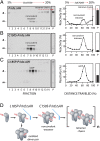
Genetic blockade of activated P/rds self-assembly. Detergent extracts from transiently transfected HEK239 cells were subjected to velocity sedimentation under nonreducing conditions on 5 to 20% sucrose gradients. (Left) Gradient (numbered) and particulate (P) fractions were analyzed for P/rds by nonreducing SDS/PAGE and Western blotting. SDS-denatured species are indicated: monomeric (m); disulfide-linked dimeric (d). (Right) Sedimentation profiles display P/rds variant distributions between the gradients and particulate (P) fractions, and report on the stoichiometry of self-assembled forms. (A) P/rdsΔAH shows normal self-assembly (23), which includes noncovalent tetramers and an abundance of disulfide-linked tetramer chains. (B) C150S-P/rdsΔAH assembled into noncovalent tetramers, but was unable to form disulfide-linked tetramer chains. (C) L185P-P/rdsΔAH was unable to form normal noncovalent tetramers or disulfide-linked tetramer chains, and instead generated oxidized dimer pairs (oxDPs). (D) The cartoon illustrates how the point mutations utilized here trap activated P/rds intermediates along the self-assembly pathway. Free thiols (-SH, HS-) and disulfide bonds (SS) are illustrated. Tetramer chains form varied (n) lengths.
Fig. 3.
Fig. 3.
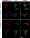
ICC analysis of P/rds variant distributions in transfected HEK239 cells by LSCM. P/rds variants (red), the ER marker KDEL (green), and cell nuclei (blue) are shown in single optical sections from cells with moderate protein expression levels and typical protein distributions. Examples shown are representative of two to three independent experiments (transfections). P/rdsΔAH accumulated in large oblate perinuclear accumulations (Top panels), a distribution documented previously for this variant (23). C150S-P/rdsΔAH localization showed that it was distributed similarly to P/rdsΔAH. In contrast, L185P-P/rdsΔAH was more diffusely distributed and included small puncta. The latter pattern resembled that of WT P/rds (Bottom panels), which does not produce large oblate perinuclear accumulations (60). In every case, P/rds was efficiently released from the ER. (Scale bar applies to all images.) Additional examples are provided in SI Appendix, Fig. S3.
Fig. 4.
Fig. 4.
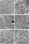
P/rds self-assembly drives membrane curvature and morphology. TEM images of P/rds-induced membrane structures in transfected HEK293 cells. (A, Left) Expression of P/rdsΔAH generates stereotypical tubulovesicular networks of interconnected high-curvature tubules. (Scale bar applies to AC, Left.) (Right) A higher-magnification view shows membrane structure details, including high-curvature tubules of extended lengths and constant diameters. Densely stained circular profiles (arrows) derive from tubule cross-sections and directly demonstrate the induction of high curvature; 25-nm OD circle (red) is provided as a size reference. (Inset) A longitudinal TEM section through a X. laevis rod OS shows disk rims aligned along the plasma membrane. A 25-nm OD circle (red) is provided as a size reference for disk rim (arrowhead) diameter. (Scale bar applies to AC, Right.) (B, Left) Expression of C150S-P/rdsΔAH, which self-assembles as individual tetramers only, produces tubulovesicular networks with a distinct ultrastructure. M, mitochondrion. (Right) A higher-magnification view shows membrane structure details, including abundant high-curvature tubules of reduced length. Densely stained circular profiles (arrows) derive from tubule cross-sections and directly demonstrate the induction of high curvature; 25-nm OD circles (red) are provided as size references. (C, Left) Expression of L185P-P/rdsΔAH, which cannot form normal tetramers (or polymerized tetramer chains), did not induce any distinctive membrane structures; biosynthetic membranes of normal appearance are apparent. (Right) A higher-magnification view shows typical ER with 50- to 60-nm diameters. The findings illustrate that, although individual tetramers produce high-curvature membranes, disulfide-mediated tetramer polymerization is required to organize that curvature over extended distances.
Fig. 5.
Fig. 5.
Three-dimensional renderings of induced membrane structures imaged by ET. (A) Tubulovesicular networks generated by P/rdsΔAH included extended length tubules of constant diameter and high curvature (arrowheads). Movie S1 presents a volume reconstruction that emphasizes the geometry of P/rdsΔAH-shaped high-curvature tubules. (B) Tubulovesicular networks generated by C150S-P/rdsΔAH showed an abundance of high-curvature membranes; however, no extended-length tubules were observed. Movie S2 presents a volume reconstruction that emphasizes the lack of extended tubules induced by C150S-P/rdsΔAH.
Fig. 6.
Fig. 6.
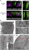
P/rds localization in high-curvature OS membrane domains is correlated with its functional activity for membrane curvature generation. IHC/LSCM analyses of X. laevis tadpole eye cryosections compare the distributions of transgenic GFP-fusion proteins (green), with endogenous P/rds (magenta) localized in OS disk rims (high-curvature membranes). X. laevis disks possess numerous incisures, which produce a striated appearance in longitudinal sections, and a flower-petal appearance in cross-sections (61). (A and B) Projection views of longitudinal image fields span photoreceptor nuclei (white), inner segments (ISs), and OSs. (A) WT-P/rds-GFP (green) showed localization predominately at rims/incisures, as compared to endogenous P/rds labeling (magenta); some transgenic protein mislocalization was also evident. Overlap in transgenic-endogenous P/rds distributions (arrowheads show examples) is shown in white, as are cell nuclei. (B) The L185P-P/rds-GFP mutant (green), which lacks activity for membrane curvature generation, was more diffusely distributed, although it was occasionally aligned along incisures (arrowheads). (Scale bars, 10 μm.) (C and D) Transverse views obtained by optical sectioning through reconstructed volumes. (C) WT-P/rds-GFP (green) showed localization mainly at rims/incisures, as identified by endogenous P/rds labeling (magenta). Overlap in transgenic-endogenous P/rds distributions show as white. (D) In addition to a presence at rims/incisures, L185P-P/rds-GFP (green) partially mislocalized to central disk regions (arrowheads). (Scale bars, 5 μm.) Movies S3 and S4 present 3D views from reconstructed volumes.
Fig. 7.
Fig. 7.
OS incisure alignment and rim formation is specifically affected by nonpolymerized P/rds tetramers. (A) IHC/LSCM analyses (projection views) of X. laevis tadpole eye cryosections compare the distributions of transgenic GFP-fusion proteins (green), with endogenous P/rds (magenta). (Scale bars: Left, 10 μm; Right, 5 μm.) Longitudinal cryosections span photoreceptor nuclei (white), ISs, and OSs. (Upper) WT-P/rds-GFP (green) showed localization predominately at disk rims/incisures, in some cases displacing endogenous P/rds labeling, but retaining striation (incisure) integrity (magenta). (Lower) The C150S-P/rds-GFP mutant (green), which retains activity for generating membrane curvature, but is unable to polymerize into linear chains, was mainly distributed in irregular patterns. Normal incisures were present in OS regions containing low transgenic protein expression, with clear transition zones between normal and disrupted incisures (arrowheads). Movie S5 presents 3D views from a reconstructed volume. (B) TEM image illustrating the transition (arrowhead) between normal and disrupted incisure patterns documented by IHC/LSCM. High-magnification views of each boxed region (Right) illustrate the specific changes that accompany C150S-P/rds-GFP expression. (C) An abnormally short incisure that has bifurcated (arrowheads) and includes a double row of high-curvature “vesicular profiles” adjacent to the disk rims (arrow). (D) An abnormally short incisure that includes numerous vesicular profiles adjacent to the disk rims. (E) Vesicular profiles are present adjacent to disk rims and the OS plasma membrane (PM). (F) Transverse view of a C150S-P/rds-GFP expressing OS disk. Ectopic disk rims (arrows) are present at each of the two incisures (asterisks), and fill the space between the OS plasma membrane and the disk rim. These types of images reveal that the numerous vesicular profiles observed in longitudinal views are ectopic disk rims, rims lacking lamellar regions.
Fig. 8.
Fig. 8.
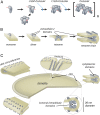
Mechanistic model for P/rds molecular function. (A) Noncovalent P/rds tetramers are the minimal unit of function for membrane curvature generation, but disulfide-linked chains of polymerized tetramers are required to organize curvature for normal rim formation and disk morphogenesis. The L185P mutation leads to oxDP and loss-of-function, which in combination with a heterozygous loss of rom1, compromises disk morphogenesis, OS structure, and photoreceptor viability. (B) Working model for P/rds curvature induction via bilayer deformation and higher-order self-assembly. Dimerization of dimers, driven by EC2 domains, create V-shaped tetramers that generate localized curvature. Lateral organization, driven by disulfide-mediated tetramer polymerization, creates extended rims of high-curvature with internalized EC2 domains. (C) Integrated model of P/rds organization and function for rim structure and mature disk morphology. P/rds tetramers and tetramer chains organized around the disk circumference function to shape the high curvature of OS disk rims, which are present both at disk peripheries and at disk incisures. A single incisure, which is typical of mammalian disks, is illustrated.

Similar articles

Cited by

References

    1. Molday R. S., Moritz O. L., Photoreceptors at a glance. J. Cell Sci. 128, 4039–4045 (2015). - PMC - PubMed
    1. Bujakowska K. M., Liu Q., Pierce E. A., Photoreceptor cilia and retinal ciliopathies. Cold Spring Harb. Perspect. Biol. 9, a028274 (2017). - PMC - PubMed
    1. Arshavsky V. Y., Burns M. E., Photoreceptor signaling: Supporting vision across a wide range of light intensities. J. Biol. Chem. 287, 1620–1626 (2012). - PMC - PubMed
    1. Palczewski K., Chemistry and biology of vision. J. Biol. Chem. 287, 1612–1619 (2012). - PMC - PubMed
    1. Goldberg A. F., Moritz O. L., Williams D. S., Molecular basis for photoreceptor outer segment architecture. Prog. Retin. Eye Res. 55, 52–81 (2016). - PMC - PubMed

Publication types

MeSH terms