. 2020 Feb 25;117(8):4252-4261.
doi: 10.1073/pnas.1908975117. Epub 2020 Feb 10.

A hemipteran insect reveals new genetic mechanisms and evolutionary insights into tracheal system development

Affiliations

A hemipteran insect reveals new genetic mechanisms and evolutionary insights into tracheal system development

Lisa Hanna et al. Proc Natl Acad Sci U S A. .

Abstract

The diversity in the organization of the tracheal system is one of the drivers of insect evolutionary success; however, the genetic mechanisms responsible are yet to be elucidated. Here, we highlight the advantages of utilizing hemimetabolous insects, such as the milkweed bug Oncopeltus fasciatus, in which the final adult tracheal patterning can be directly inferred by examining its blueprint in embryos. By reporting the expression patterns, functions, and Hox gene regulation of trachealess (trh), ventral veinless (vvl), and cut (ct), key genes involved in tracheal development, this study provides important insights. First, Hox genes function as activators, modifiers, and suppressors of trh expression, which in turn results in a difference between the thoracic and abdominal tracheal organization. Second, spiracle morphogenesis requires the input of both trh and ct, where ct is positively regulated by trh As Hox genes regulate trh, we can now mechanistically explain the previous observations of their effects on spiracle formation. Third, the default state of vvl expression in the thorax, in the absence of Hox gene expression, features three lateral cell clusters connected to ducts. Fourth, the exocrine scent glands express vvl and are regulated by Hox genes. These results extend previous findings [Sánchez-Higueras et al., 2014], suggesting that the exocrine glands, similar to the endocrine, develop from the same primordia that give rise to the trachea. The presence of such versatile primordia in the miracrustacean ancestor could account for the similar gene networks found in the glandular and respiratory organs of both insects and crustaceans.

Keywords: Oncopeltus; cut; tracheal system; trachealess; ventral veinless.


Conflict of interest statement

The authors declare no competing interest.

Figures

Fig. 1.
Fig. 1.
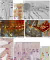
Spiracle location and trachea organization in Oncopeltus. (AC) Location and number of spiracles are conserved from first nymph to adult. The spiracle of the second thoracic segment (T2) is pseudocolored in magenta, while the A1 segment is pseudocolored in yellow. (DF) Tracheal organization (pseudocolored in blue) is also conserved throughout postembryonic development. Anterior is Left in all panels.
Fig. 2.
Fig. 2.
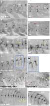
trh mRNA expression during Oncopeltus embryogenesis. (A and B) The formation of tracheal placodes (Tr) begins in T2–A1 segments, followed by the formation of abdominal placodes in A2–A8. (C) Head branch (orange arrowhead) and leg branches (green arrowheads) migrate from Tr1 and Tr2 placodes. (C, Inset) Tr1 placode extends further into the posterior T1 segment. (A′–B′) Double labeling of trh and en used to precisely determine the location of placode formation. Arrowhead in B′ points to expansion of Tr1 into en domain. (C′ and C″) While thoracic placodes extend anteriorly (arrowheads), abdominal placodes extend only laterally. (D) DBs, VBs, and DTs form in the abdomen, except for Tr3 which lacks a VB (asterisk). Also, the signal appears in the ScGs. The head branch continues to extend anteriorly (orange arrowhead). Thoracic placodes form large branches extending dorsoanteriorly (red arrowheads). (E) Thoracic branches continue to extend (red arrowheads). (E, Inset) A fainter trh signal appears posterior to the abdominal primary placodes (blue arrowhead). (F) Diagram summarizing the progression of tracheal branching at 40, 50, and 75% embryonic development. Anterior is Top in AC″. Anterior is Left in DF. Combined images are used for A, B, and C′.
Fig. 3.
Fig. 3.
Hox gene regulation of trh expression. (A and A′) Compared to WT, trh expression is absent in Tr1 and Tr2 placodes (red asterisks) and ectopic branches arise from Tr3 (arrowheads) in Antp RNAi. (B and B′) Compared to WT, Scr RNAi embryos show loss of signal in SGs (red asterisk). (C and C′) Compared to WT, trh expression in the Tr3 placode becomes thoracic-like (yellow arrowhead). (D and D′) Ubx/abd-A RNAi embryos show an increase of the size of Tr3–Tr5 as well as the appearance of ectopic leg branches (yellow arrowheads). (E and F) Antp/Ubx/abd-A RNAi and Antp/nub RNAi embryos show consistent loss of signal in the thorax (red asterisks) and its continued presence in the abdomen. The trend toward the formation of ectopic abdominal leg branches (yellow arrowheads) is more prominent in Antp/nub RNAi embryos. In both panels, white arrowheads point to ectopic abdominal legs. (G and G′) Compared to WT, Abd-B RNAi results in the formation of two ectopic tracheal placodes (Tr11 and Tr12). All panels show embryos between 65 and 75% embryonic development. Anterior is Left in all panels.
Fig. 4.
Fig. 4.
Expression and function of ct in Oncopeltus. (A and B) ct is expressed in the Sp from posterior T1/anterior T2 (arrowhead) to A8 and continues to late embryogenesis. (A′) Double labeling of ct and en shows that signal in Sp1 extends into the en domain of the T1 segment (arrowhead). The arrows in A and B points to ct expression in the A1 segment where a spiracle does not form. (C) In WT first nymphs, there are two thoracic spiracles located in posterior T1 and posterior T2 (arrowheads), and seven abdominal spiracles (only three shown) located anteriorly (arrows). Note that T3 and A1 lack a spiracle. (C′) All spiracles are lost in ct RNAi nymphs (asterisks). Differences in melanization levels between C and C′ are due to the lethality of ct knockdown and death of nymphs prior to full melanization. (D) ct is colocalized with trh in thoracic and abdominal placodes. (E and E′) Finer characterization of ct in the abdomen, showing that the signal is colocalized with trh weak expression posterior to the primary placodes (arrows). Anterior is Top in A and A′ and D. Anterior is Left in B, C, C′, E, and E′. Combined images are used for C and E.
Fig. 5.
Fig. 5.
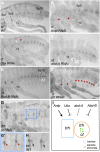
Regulation of ct expression by Hox genes and trh. (A) Expression of ct in WT embryos. (B) Antp RNAi embryos display loss of ct signal in the thorax (asterisks). (C) Ubx RNAi embryos showing ectopic expression of ct in posterior T3 (arrow) and loss of ct expression in A1 (asterisk). Arrowhead points to ectopic leg formation in A1. (D) ct expression is unaltered in abd-A RNAi; arrowheads point to ectopic legs in the abdomen. (E) Abd-B RNAi embryos showing ectopic expression of ct in the last two abdominal segments (arrows). (F) All ct expression is lost in trh RNAi (asterisks). (G) trh expression in the tracheal placodes and branches is unaltered in ct RNAi. (G′) Magnified detail from G showing absence of trh in the spiracle primordia (asterisks). (H) trh expression in spiracle primordia in WT (arrows). (I) Diagram of ct regulation by trh and Hox genes. Hox genes regulate trh in the trachea (blue rectangle) and in the spiracle primordia (orange circle); trh and ct cross-regulate each other in the spiracle primordia only (green arrows). The dotted line indicates a lack of effect of abd-A on trh and ct. All panels show embryos between 65 and 75% embryonic development. Anterior is Left in all panels.
Fig. 6.
Fig. 6.
vvl mRNA expression during Oncopeltus embryogenesis. (A and B) vvl expression starts in the appendages and CNS, followed by appearance in PGs. (C) New expression appears in the lateral thorax (arrowheads), while the signal in PGs expands. (D) There is an additional expression in SGs, and further modulation of vvl signal in the thorax (Inset, arrowheads) and abdomen (arrows). (E) vvl is colocalized with trh in the SG, Tr1–Tr2 (arrowheads), and abdomen lateral branches (arrows). (F) At this stage, PG moves into T1, and vvl becomes visible in the excretory cells (black arrows) and duct cells (white arrows) of ScG. (GG″) While vvl is colocalized with trh in the SG, trachea, and duct cells of the ScG (see G″, arrows), it is solely expressed in the abdominal placodes and not in the spiracle primordia (see G′, arrows). Mandibles, Mn; maxillary, Mx; labial, Lb; pr sali. Anterior is Top in AE. Anterior is Left in FG″. Combined images are used for A.
Fig. 7.
Fig. 7.
trh expression in vvl RNAi embryos. (A and B) Compared to WT, trh expression is unaltered, except for reduced expression in the Tr3 placode (red arrow). (C and D) At a later stage, trh is lost in Tr3 (red asterisk), the VBs (blue asterisks), and in the duct cells of ScGs (green asterisks). (C′) A vvl RNAi embryo with a mild phenotype showing presence of Tr3 placode (red arrow), but the lack of dorsal branch formation (red asterisk). Anterior is Top in A and B. Anterior is Left in C′ and D.
Fig. 8.
Fig. 8.
Hox gene regulation of gland organogenesis. (A and A′) vvl expression in the PG is lost in Scr RNAi (asterisks). (B and B′) phm expression in the PG is also lost in vvl RNAi (asterisk). (CE) While vvl is only lost from the excretory cells of the ScG (red asterisks) in abd-A RNAi embryos, it is lost from both the excretory cells (red asterisks) and the duct cells (white asterisks) in Ubx/abd-A RNAi embryos. White arrowheads in C and E point to the duct cells; red arrowheads in E point to the excretory cells. (F and F′) Compared to WT, the vvl signal is lost from the tracheal placodes and transformed into three cell clusters in the thorax (purple arrows) in Antp RNAi embryos. (F″) Magnification (40×) of the vvl-expressing cell clusters showing connective ducts (purple arrowheads). A and A′ show embryos at ∼30% embryogenesis. All remaining panels show embryos between 65 and 75% embryogenesis. Anterior is Top in A and A′. Anterior is Left in all remaining panels.

Similar articles

Cited by

References

    1. Manning G., Krasnow M. A., “Development of the Drosophila tracheal system” in The development of Drosophila Melanogaster, Bate M., Arias A. M., Eds. (Cold Spring Harbor Laboratory Press, Cold Spring Harbor, New York, 1993), pp. 609–685.
    1. Isaac D. D., Andrew D. J., Tubulogenesis in Drosophila: A requirement for the trachealess gene product. Genes Dev. 10, 103–117 (1996). - PubMed
    1. Anderson M. G., Perkins G. L., Chittick P., Shrigley R. J., Johnson W. A., drifter, a Drosophila POU-domain transcription factor, is required for correct differentiation and migration of tracheal cells and midline glia. Genes Dev. 9, 123–137 (1995). - PubMed
    1. de Celis J. F., Llimargas M., Casanova J., Ventral veinless, the gene encoding the Cf1a transcription factor, links positional information and cell differentiation during embryonic and imaginal development in Drosophila melanogaster. Development 121, 3405–3416 (1995). - PubMed
    1. Hayashi S., Kondo T., Development and function of the Drosophila tracheal system. Genetics 209, 367–380 (2018). - PMC - PubMed

Publication types

Substances

Associated data