. 2020 Mar 19;133(6):jcs236158.
doi: 10.1242/jcs.236158.

The Sts1 nuclear import adapter uses a non-canonical bipartite nuclear localization signal and is directly degraded by the proteasome

Affiliations

The Sts1 nuclear import adapter uses a non-canonical bipartite nuclear localization signal and is directly degraded by the proteasome

Lauren Budenholzer et al. J Cell Sci. .

Abstract

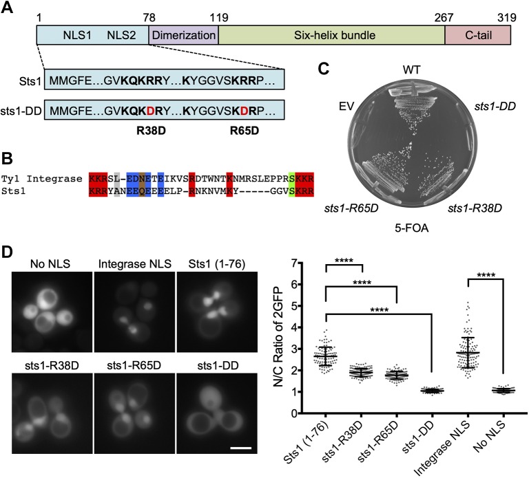
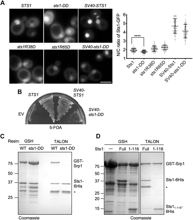
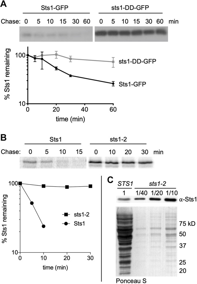
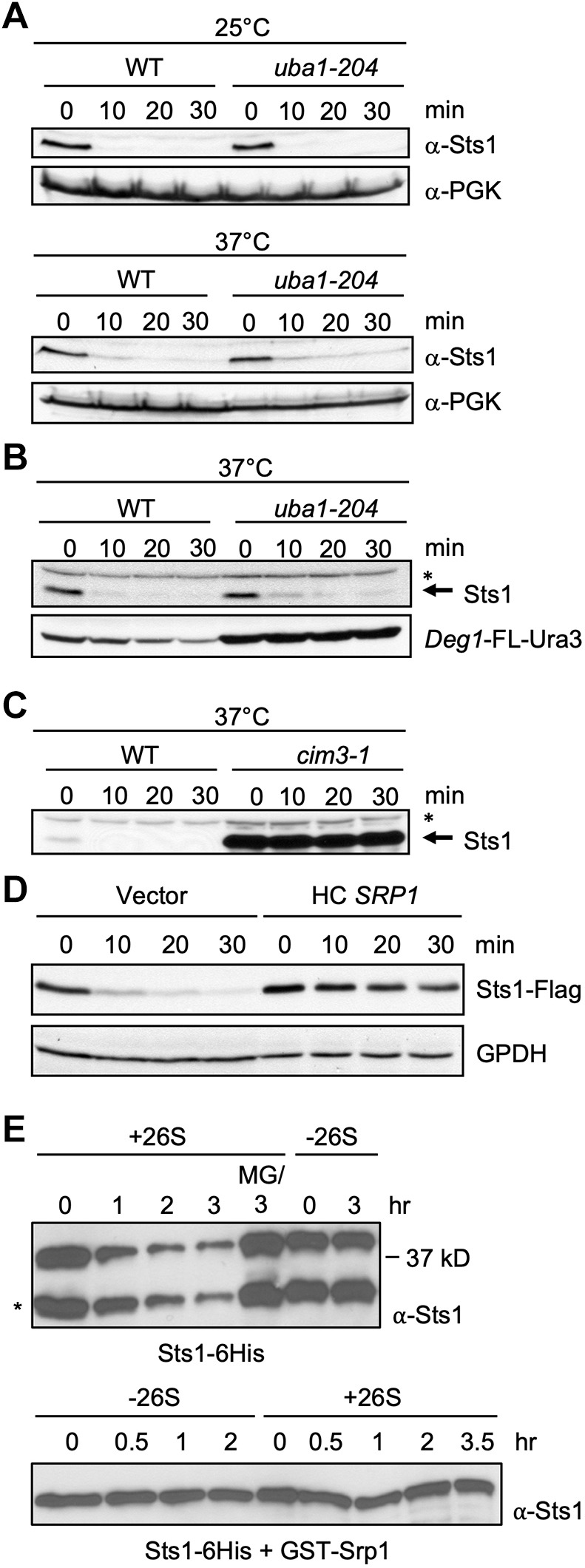
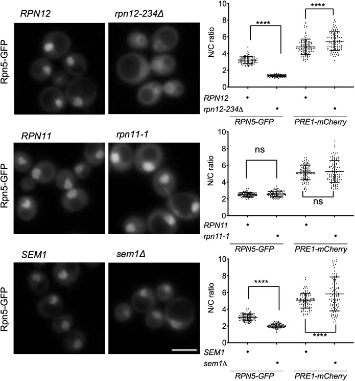
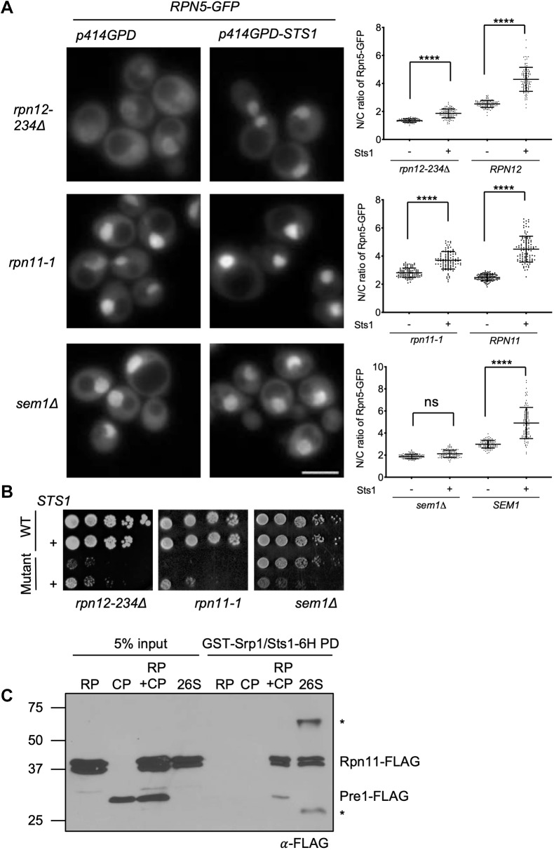
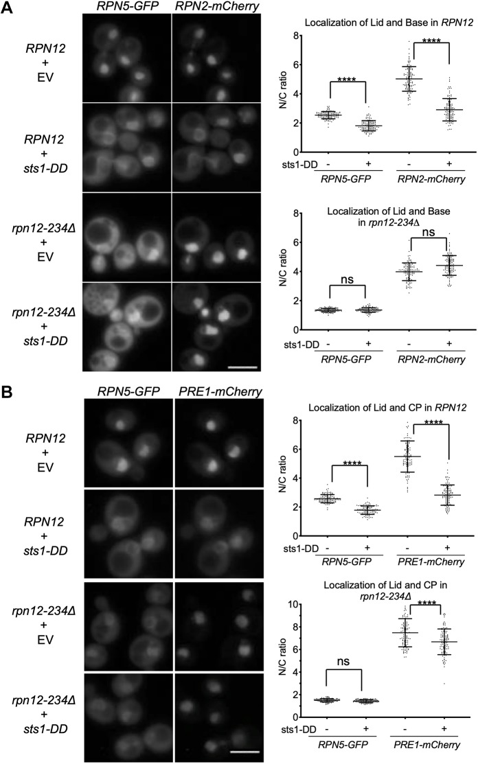
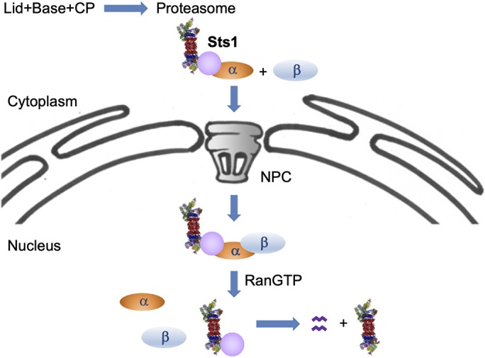
The proteasome is an essential regulator of protein homeostasis. In yeast and many mammalian cells, proteasomes strongly concentrate in the nucleus. Sts1 from the yeast Saccharomyces cerevisiae is an essential protein linked to proteasome nuclear localization. Here, we show that Sts1 contains a non-canonical bipartite nuclear localization signal (NLS) important for both nuclear localization of Sts1 itself and the proteasome. Sts1 binds the karyopherin-α import receptor (Srp1) stoichiometrically, and this requires the NLS. The NLS is essential for viability, and over-expressed Sts1 with an inactive NLS interferes with 26S proteasome import. The Sts1-Srp1 complex binds preferentially to fully assembled 26S proteasomes in vitro Sts1 is itself a rapidly degraded 26S proteasome substrate; notably, this degradation is ubiquitin independent in cells and in vitro and is inhibited by Srp1 binding. Mutants of Sts1 are stabilized, suggesting that its degradation is tightly linked to its role in localizing proteasomes to the nucleus. We propose that Sts1 normally promotes nuclear import of fully assembled proteasomes and is directly degraded by proteasomes without prior ubiquitylation following karyopherin-α release in the nucleus.

Keywords: Cut8; Karyopherin; NLS; Proteasome; Sts1; Yeast.


Conflict of interest statement

Competing interestsThe authors declare no competing or financial interests.

Figures

Fig. 1.
Fig. 1.
Sts1 contains an apparent bipartite NLS essential for cell viability. (A) Predicted domain architecture and selected functional elements of Sts1 based on sequence analysis and comparison with the crystal structure of the S. pombe homolog Cut8. The suggested NLS elements are indicated with the sequences shown below; the two mutations in the sts1-DD mutant are also shown. (B) Sts1 lacks a strong match to canonical NLS, but has substantial similarity to a confirmed bipartite NLS in the Ty1 integrase; in both cases the linker separating the two basic elements is unusually long. (C) Viability assay of Sts1 NLS mutants. The noted sts1 alleles, expressed under the endogenous STS1 promoter and terminator from pRS314-based plasmids, were transformed into MHY9580 yeast, in which the chromosomal sts1Δ allele is covered by pRS316-STS1. Transformed cells were struck on 5-FOA plates to evict the cover plasmid. EV, empty vector; WT, wild-type. (D) WT MHY500 yeast transformed with MET25 promoter-based plasmids expressing the indicated NLS sequences fused to 2GFP. Sts1 constructs expressed Sts1 residues 1–76 appended to the N-terminus of 2GFP. The NLS sequence from Ty1 integrase N-terminally tagged with 2GFP was used as a positive control for a bipartite NLS, and 2GFP without an NLS (‘No NLS’) was used as a negative control (both obtained from Anita Corbett). Transformants were grown to mid-log phase at 30°C prior to fluorescence imaging. Three biological replicates of at least 100 cells each were counted (right panel). A t-test was used to determine statistical significance of localization differences (****P<0.0001). Scale bar: 5 μm.
Fig. 2.
Fig. 2.
Nuclear localization is essential for Sts1 function. (A) Wild-type MHY500 yeast was transformed with p415GPD-based plasmids expressing the indicated sts1-GFP fusion alleles. ‘SV40’ denotes the cNLS from SV40T antigen fused to the N-terminus of the test protein. The transformants were grown at room temperature prior to imaging by fluorescence microscopy in mid-log phase. Three replicates of at least 100 cells were counted (right panel). A t-test was used to determine statistical significance of differences in localization (****P<0.0001). Scale bar: 5 μm. (B) Viability assay. MHY9580 cells (sts1Δ, pRS316-STS1) were transformed with the p415GPD-based alleles noted and grown at room temperature on 5-FOA to select against the original cover plasmid. (C) Sts1–Srp1 complex formation depends on the Sts1 bipartite NLS. The indicated recombinant proteins were co-expressed in E. coli, and binding was determined based on co-purification on a GST-binding glutathione resin or polyHis-binding TALON resin followed by SDS-PAGE. (D) The indicated recombinant proteins were co-expressed in E. coli, and binding was determined based on co-purification on either GSH beads or TALON beads. *Proteolytic fragments derived from Sts1–6His.
Fig. 3.
Fig. 3.
Degradation of wild-type and mutant forms of Sts1 in cells. (A) Cycloheximide-chase analysis was performed to determine the degradation rates of Sts1-GFP and sts1-DD-GFP using anti-GFP immunoblotting. MHY500 cells carried either p415MET25-STS1-GFP or p415MET25-sts1-DD-GFP and were grown at 30°C; cycloheximide was added at time 0 to block further protein synthesis. For quantitation, Sts1-GFP and sts1-DD-GFP levels were normalized to a PGK loading control. (B) Radioactive pulse-chase analysis of Sts1 degradation in MHY9692 (STS1) and MHY9693 (sts1-2) cells at 30°C. Proteins were immunoprecipitated using affinity-purified anti-Sts1 antibodies. Bottom panel shows the phosphorimager quantification of degradation rates. (C) Cell extracts from MHY9692 and MHY9693 were separated by SDS-PAGE and immunoblotted with anti-Sts1 antibodies. Extracts from MHY9693 were diluted to the indicated fraction of extract loaded for MHY9692. Ponceau S-stained membrane shows relative amounts of total protein extract.
Fig. 4.
Fig. 4.
Sts1 is degraded by the proteasome in a ubiquitin-independent manner. (A) Cycloheximide-chase analysis to determine the degradation rates of endogenous Sts1 in the indicated strains at both the permissive (25°C) and restrictive (37°C) temperatures for the uba1-204 strain (RJD3269). Immunoblot for PGK serves as a loading control. (B) Cycloheximide-chase analysis of endogenous Sts1 and plasmid-expressed Deg1-FLAG-Ura3 in the indicated strains at the restrictive temperature (37°C) for the uba1-204 strain. A cross-reactive band in the immunoblot for Sts1, indicated by an asterisk, shows unchanging levels of a protein that is not degraded over the 30 min chase. (C) Cycloheximide-chase analysis of endogenous Sts1 in the indicated strains at the restrictive temperature (37°C) for the cim3-1 strain (MHY4464, which carries a mutation in the Rpt6 subunit of the proteasome). (D) Cycloheximide-chase analysis of Flag-tagged Sts1 in WT cells carrying either an empty high-copy (HC) vector or the same plasmid with SRP1. GPDH served as a loading control. Threefold less protein was loaded for the latter extracts to achieve roughly equal Sts1-Flag levels for the zero-minute samples. The endogenous STS1 locus was 3′-tagged with 6xGly-3xFLAG. (E) In vitro degradation of purified recombinant Sts1–6His by 26S proteasomes purified from yeast. For the ‘MG/3 hr’ sample, proteasomes were treated with 50 µM MG132 inhibitor for 10 min prior to addition of Sts1–6His. Degradation was measured at room temperature. *Sts1 fragment that is often generated during purification from E. coli when Sts1 is not co-expressed with Srp1 (see Fig. 2C,D) and is also a substrate for the proteasome in vitro. In the lower samples, recombinant Sts1–6His was co-purified from bacteria with GST-Srp1 and used as substrate.
Fig. 5.
Fig. 5.
Select lid mutants alter proteasome subparticle localization. Lid mutants were grown in minimal medium to log phase and imaged by fluorescence microscopy; three replicates of at least 100 cells were counted. Two-way ANOVA was used to determine statistical significance of differences in localization (****P<0.0001; ns, not significant). Scale bar: 5 μm. Yeast strains used: MHY6956, MHY6954, MHY6377, MHY9583, MHY6938, MHY9581 and MHY9172. Pre1-mCherry (quantified here) and Rpn2-mCherry images are shown in Fig. S4.
Fig. 6.
Fig. 6.
High levels of Sts1 rescue certain lid mutant mislocalization and growth defects. (A) Yeast was grown in SD–Trp and imaged by fluorescence microscopy; three replicates of at least 100 cells were counted. Two-ANOVA was used to determine statistical significance of differences in localization (****P<0.0001; ns, not significant). Scale bar: 5 μm. The indicated mutant yeast expressed Rpn5-GFP and was transformed with either p414GPD (empty vector) or p414GPD-STS1. Strains used: MHY6964, MHY9583 and MHY9174. (B) Serial dilution growth assays. Matched wild-type (WT) and mutant transformants were grown as follows, from left to right: SD –Trp –Arg+1 µM canavanine, 34°C (5 days); SD–Trp, 30°C (5 days); and SD–Trp, 34°C (2 days). Strains used: MHY500, MHY5748, MHY9509 and MHY9134. (C) Recombinant GST-Srp1–Sts1-6His complex (1 μM) was immobilized on glutathione (GSH) resin and incubated with 1 μM purified CP, RP, 26S proteasome or 26S proteasomes reconstituted from RP and CP (‘RP+CP’) to detect interactions. All input complexes were isolated from yeast using anti-Flag affinity purifications. Proteins from the GST pulldowns (PD) were examined by anti-Flag immunoblotting.
Fig. 7.
Fig. 7.
The sts1-DD mutant induces 26S proteasome mislocalization in vivo. Microscopy was performed as in Fig. 5A. Yeast was transformed with either empty vector (p414GPD) or p414GPD-sts1-DD. Lid and base were examined in panel A, while lid and CP were imaged in panel B. Two-ANOVA was used to determine statistical significance of differences (****P<0.0001; ns, not significant). Scale bars: 5 μm. Strains used were MHY6966, MHY6964, MHY6956 and MHY6954.
Fig. 8.
Fig. 8.
Model of Sts1 degradation and function in proteasome nuclear import. Cytoplasmic 26S proteasomes are transported into the nucleus via Sts1. The bipartite NLS in Sts1 binds Srp1 (karyopherin α), and the complex of proteasome–Sts1–Srp1 can then interact with karyopherin β (Kap95), which promotes import of the complex through the NPC. Sts1, in complex with Srp1, appears to bind more efficiently to fully assembled 26S proteasomes than to proteasomal subparticles, although Sts1 can also promote lid import when Sts1 is overproduced. In the nucleus, RanGTP binding breaks up the import complex. It is possible that this is insufficient for Sts1 release from Srp1, requiring the Cdc48Ubx4 segregase (Chien and Chen, 2013) or other factors to fully dissociate it. When Srp1 is removed from the disordered Sts1 N-terminal domain, Sts1 is degraded by the proteasome to which it is bound.

Similar articles

Cited by

References

    1. Amrani N., Dufour M. E., Bonneaud N. and Lacroute F. (1996). Mutations in STS1 suppress the defect in 3′ mRNA processing caused by the rna15-2 mutation in Saccharomyces cerevisiae. Mol. Gen. Genet. 252, 552-562. 10.1007/BF02172401 - DOI - PubMed
    1. Bayliss R., Littlewood T. and Stewart M. (2000). Structural basis for the interaction between FxFG nucleoporin repeats and importin-beta in nuclear trafficking. Cell 102, 99-108. 10.1016/S0092-8674(00)00014-3 - DOI - PubMed
    1. Budenholzer L., Cheng C. L., Li Y. and Hochstrasser M. (2017). Proteasome structure and assembly. J. Mol. Bio. 429, 3500-3524. 10.1016/j.jmb.2017.05.027 - DOI - PMC - PubMed
    1. Chen L. and Madura K. (2014). Degradation of specific nuclear proteins occurs in the cytoplasm in Saccharomyces cerevisiae. Genetics 197, 193-197. 10.1534/genetics.114.163824 - DOI - PMC - PubMed
    1. Chen L., Romero L., Chuang S.-M., Tournier V., Joshi K. K., Lee J. A., Kovvali G. and Madura K. (2011). Sts1 plays a key role in targeting proteasomes to the nucleus. J. Biol. Chem. 286, 3104-3118. 10.1074/jbc.M110.135863 - DOI - PMC - PubMed

Publication types

MeSH terms

Substances