. 2020 Feb 10;12(3):3010-3024.
doi: 10.18632/aging.102795. Epub 2020 Feb 10.

microRNA-1203 targets and silences cyclophilin D to protect human endometrial cells from oxygen and glucose deprivation-re-oxygenation

Affiliations

microRNA-1203 targets and silences cyclophilin D to protect human endometrial cells from oxygen and glucose deprivation-re-oxygenation

Hong-Bin Xu et al. Aging (Albany NY). .

Abstract

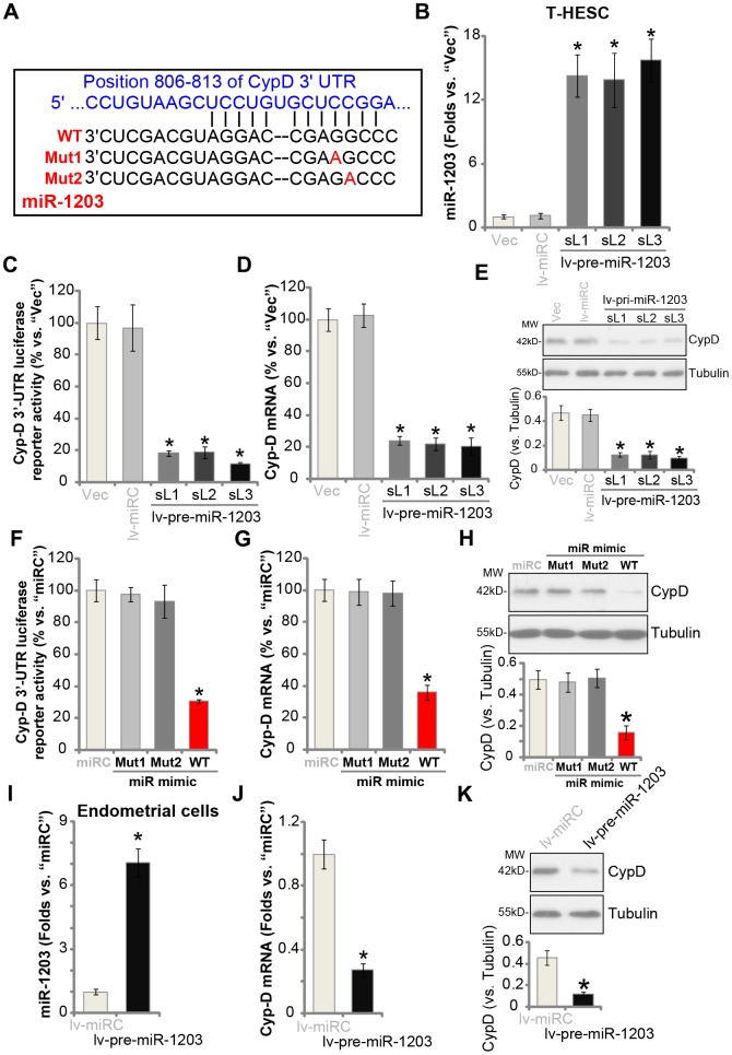
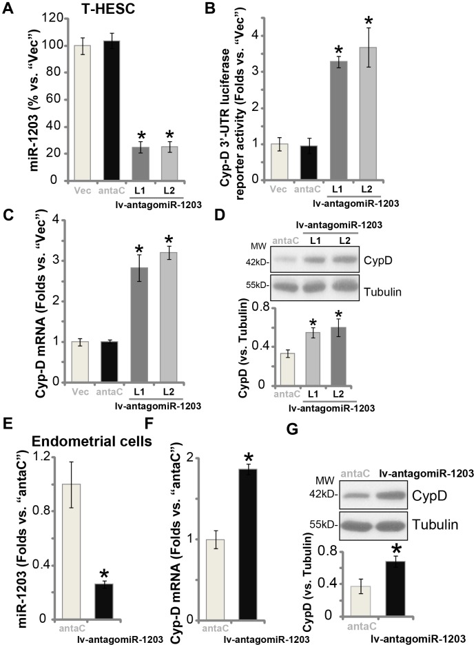
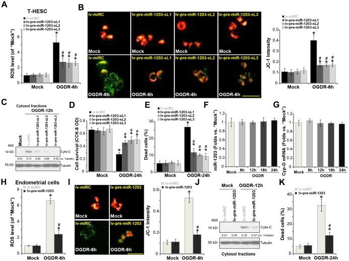
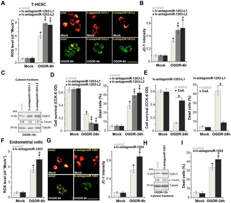
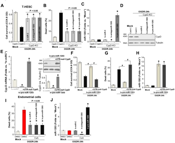
Oxygen and glucose deprivation (OGD)-re-oxygenation (OGDR) stimulation to the human endometrial cells mimics ischemia-reperfusion injury. Cyclophilin D (CypD)-dependent programmed necrosis pathway mediates OGDR-induced cytotoxicity to human endometrial cells. We here identified a novel CypD-targeting miRNA, microRNA-1203 (miR-1203). In T-HESC and primary human endometrial cells, ectopic overexpression of miR-1203, using a lentiviral construct, potently downregulated the CypD 3'-untranslated region (3'-UTR) activity and its expression. Both were however upregulated in endometrial cells with forced miR-1203 inhibition by its anti-sense sequence. Functional studies demonstrated that ectopic miR-1203 overexpression in endometrial cells alleviated OGDR-induced programmed necrosis, inhibiting mitochondrial CypD-p53-adenine nucleotide translocator 1 association, mitochondrial depolarization, reactive oxygen species production, and medium lactate dehydrogenase release. Contrarily OGDR-induced programmed necrosis and cytotoxicity were intensified with forced miR-1203 inhibition in endometrial cells. Significantly, ectopic miR-1203 overexpression or inhibition failed to change OGDR-induced cytotoxicity in CypD-knockout T-HESC cells. Furthermore, ectopic miR-1203 overexpression was unable to protect T-HESC endometrial cells from OGDR when CypD was restored by an UTR-depleted CypD construct. Collectively, these results show that miR-1203 targets and silences CypD to protect human endometrial cells from OGDR.

Keywords: cyclophilin D; human endometrial cells; miR-1203; oxygen and glucose deprivation-re-oxygenation (OGDR); programmed necrosis.


Conflict of interest statement

CONFLICTS OF INTEREST: The authors have no conflicts of interest.

Figures

Figure 1
Figure 1
miR-1203 targets and silences CypD in human endometrial cells. The wild-type (WT) microRNA-1203 (miR-1203) targets CypD 3’-UTR (3’-untranslated region) at position 806-813 (A). T-HESC endometrial cells were infected with pre-miR-1203-encoding lentivirus (“lv-pre-miR-1203”), following puromycin selection three stable cell lines were established: “sL1/sL2/sL3”. Control T-HESC cells were infected with microRNA control lentivirus (“lv-miRC”); Expression of mature miR-1203 and CypD mRNA was tested by qPCR assays (B and D); The relative CypD 3’-UTR luciferase reporter activity was examined (C), with CypD protein expression tested by Western blotting assays (E). T-HESC cells were transfected with 500 nM of control microRNA mimic (“miRC”), the wild-type (“WT-”) or the mutant (“Mut1/2”, see sequences in A) miR-1203 mimics for 48h, the relative CypD 3’-UTR luciferase reporter activity (F), CypD mRNA (G) and protein (H) levels were tested. The primary human endometrial cells (“Endometrial cells”, same for all Figures) were infected with lv-pre-miR-1203 or lv-miRC lentivirus for 48h, expression of mature miR-1203 (I), CypD mRNA (J) and protein (K) was shown. CypD protein expression was quantified and normalized to the loading control (E, H and K). “MW” stands for molecular weight (same for all Figures). “Vec” stands for the empty vector control (same for all Figures). Data were presented as mean ± SD (n=5). * P <0.05 vs. “Vec”/“miRC”/“lv-miRC” cells. Experiments in this figure were repeated three times with similar results obtained.
Figure 2
Figure 2
miR-1203 inhibition can elevate CypD expression in human endometrial cells. T-HESC endometrial cells were infected with pre-miR-1203 anti-sense lentivirus (“lv-antagomiR-1203”), following puromycin selection two stable cell lines were established: “L1/L2”. Control T-HESC cells were infected with microRNA anti-sense control lentivirus (“antaC”); Expression of mature miR-1203 and CypD mRNA was tested by qPCR assays (A and C); The relative CypD 3’-UTR luciferase reporter activity was examined (B), with CypD protein expression tested by Western blotting (D). The primary human endometrial cells were infected with lv-antagomiR-1203 or antaC for 48h, expression of mature miR-1203 (E), CypD mRNA (F) and protein (G) was shown. CypD protein expression was quantified and normalized to the loading control (D and G). Data were presented as mean ± SD (n=5), and results were normalized. * P <0.05 vs. “Vec”/“antaC” cells. Experiments in this figure were repeated five times with similar results obtained.
Figure 3
Figure 3
Forced miR-1203 overexpression protects human endometrial cells from OGDR-induced programmed necrosis. The stable T-HESC cells, with the pre-miR-1203-encoding lentivirus (“lv-pre-miR-1203-sL1/2/3”) or the control T-HESC cells with microRNA control lentivirus (“lv-miRC”), were subjected to OGD exposure for 4h, followed by re-oxygenation (“OGDR”) for applied time periods, ROS production (DCF-DA intensity, (A) mitochondrial depolarization (JC-1 green fluorescence accumulation, (B) cytochrome C release (C) testing cytosol proteins) were tested by the assays mentioned in the text; Cell survival and necrosis were tested by CCK-8 (D) and LDH release (E) assays, respectively. The parental control T-HESC cells were treated with the OGDR procedure for applied time periods, expression of mature miR-1203 (F) and CypD mRNA (G) was tested by qPCR assays. The primary human endometrial cells were infected with lv-pre-miR-1203 or lv-miRC lentivirus for 48h, followed by OGDR procedure for the applied time periods, ROS production (H), mitochondrial depolarization (I), cytochrome C release (J, testing cytosol proteins) and cell necrosis (K) were tested similarly. For the cytochrome C release assay, relative cytosol cytochrome C level (vs. Tubulin) was quantified (C and J). Data were presented as mean ± SD (n=5). “Mock” stands for non-OGDR treatment (same for all Figures). * P <0.05 vs. “Mock” treatment in “lv-miRC” cells. # P <0.05 vs. OGDR treatment in “lv-miRC” cells. Experiments in this figure were repeated three times with similar results obtained. Bar= 50 μm (B and I).
Figure 4
Figure 4
miR-1203 inhibition can exacerbate OGDR-induced cytotoxicity in human endometrial cells. Stable T-HESC cells with the pre-miR-1203 anti-sense lentivirus (“lv-antagomiR-1203-L1/L2”, two lines) or the microRNA anti-sense control lentivirus (“anta-C”) were subjected to OGDR for applied time periods, ROS production (DCF-DA intensity, (A) mitochondrial depolarization (JC-1 green fluorescence accumulation, (B) cytochrome C release (C) testing cytosol proteins) were tested by the assays mentioned in the text; Cell survival and necrosis were tested by CCK-8 and medium LDH release assays (D). Stable T-HESC cells with the pre-miR-1203 anti-sense lentivirus (“lv-antagomiR-1203-L1”) were pretreated with cyclosporin A (CsA, 10 μM) for 1h, followed by the OGDR stimulation for 24h, cell viability and necrosis were tested similarly (E). The primary human endometrial cells were infected with lv-antagomiR-1203 or anta-C lentivirus for 48h, followed by OGDR procedure for the applied time periods, ROS production (F), mitochondrial depolarization (G), cytochrome C release (H, testing cytosol proteins) and cell necrosis (I) were tested. For the cytochrome C release assay, relative cytosol cytochrome C level (vs. Tubulin) was quantified (C and H). Data were presented as mean ± SD (n=5). * P <0.05 vs. “Mock” treatment in “anta-C” cells. # P <0.05 vs. OGDR treatment in “anta-C” cells. $ P <0.05 (E). Experiments in this figure were repeated five times with similar results obtained. Bar= 50 μm (B and G).
Figure 5
Figure 5
Forced miR-1203 protects human endometrial cells from OGDR via silencing CypD. The stable T-HESC cells with the CRISPR/Cas9-CypD-KO construct (“CypD-KO” cells) were infected with microRNA control lentivirus (“lv-miRC”), pre-miR-1203-encoding lentivirus (“lv-pre-miR-1203”), or the pre-miR-1203 anti-sense lentivirus (“lv-antagomiR-1203”), with puromycin selection the stable cells established. These cells and the CRISPR/Cas9 vector control cells (“Cas9-C”) were subjected to OGDR for 24h, cell survival and necrosis were tested by CCK-8 assay (A) and LDH release assay (B), respectively, with miR-1203 (C) and CypD protein (D) expression respectively examined by qPCR and Western blotting assays. The lv-pre-miR-1203-expression stable T-HESC cells were further transfected with or without the UTR-depleted CypD construct (“+UTR-null CypD”), after 48h CypD mRNA and protein expression in these cells and also in lv-miRC-expressing control cells was shown (E); Cells were subjected to OGDR for 24h, cell survival and necrosis were respectively tested by CCK-8 (F) and LDH release (G) assays, with miR-1203 (H) expression examined by qPCR. The primary human endometrial cells, with/without cyclosporin A (CsA, 10 μM) pre-treatment, were infected with lv-miRC, lv-pre-miR-1203, or lv-antagomiR-1203. After 48h cells were treated with OGDR for indicated time periods, cell necrosis and miR-1203 expression were tested by LDH (I) and qPCR (J) assays, respectively. Data were presented as mean ± SD (n=5), and results were normalized. * P <0.05 vs. OGDR treatment in “Cas9-C” cells (AC). * P <0.05 vs. “lv-miRC” cells (E). # P <0.05 (F and G). * P <0.05 vs. “Mock” treatment (H). * P <0.05 vs. OGDR treatment with DMSO (vehicle control) pretreatment (I and J). Experiments in this figure were repeated four times with similar results obtained.

Similar articles

Cited by

References

    1. Mullins TL, Miller RJ, Mullins ES. Evaluation and Management of Adolescents with Abnormal Uterine Bleeding. Pediatr Ann. 2015; 44:e218–22. 10.3928/00904481-20150910-09 - DOI - PubMed
    1. Van de Velde M, Diez C, Varon AJ. Obstetric hemorrhage. Curr Opin Anaesthesiol. 2015; 28:186–90. 10.1097/ACO.0000000000000168 - DOI - PubMed
    1. Deering S, Rowland J. Obstetric emergency simulation. Semin Perinatol. 2013; 37:179–88. 10.1053/j.semperi.2013.02.010 - DOI - PubMed
    1. Ferrari RS, Andrade CF. Oxidative Stress and Lung Ischemia-Reperfusion Injury. Oxid Med Cell Longev. 2015; 2015:590987. 10.1155/2015/590987 - DOI - PMC - PubMed
    1. Kalogeris T, Bao Y, Korthuis RJ. Mitochondrial reactive oxygen species: a double edged sword in ischemia/reperfusion vs preconditioning. Redox Biol. 2014; 2:702–14. 10.1016/j.redox.2014.05.006 - DOI - PMC - PubMed

Publication types