. 2020 Feb 10;11(2):112.
doi: 10.1038/s41419-020-2273-y.

circRNA_0000140 suppresses oral squamous cell carcinoma growth and metastasis by targeting miR-31 to inhibit Hippo signaling pathway

Affiliations

circRNA_0000140 suppresses oral squamous cell carcinoma growth and metastasis by targeting miR-31 to inhibit Hippo signaling pathway

Qiu-Shi Peng et al. Cell Death Dis. .

Abstract

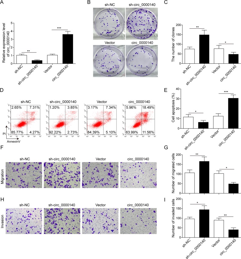
Oral squamous cell carcinoma (OSCC) is one of the most common malignancies and has a poor prognosis. Circular RNA (circRNA) has been increasingly recognized as a crucial contributor to carcinogenesis. circRNA_0000140 has been aberrantly expressed in OSCC, but its role in tumor growth and metastasis remains largely unclear. Sanger sequencing, actinomycin D, and RNase R treatments were used to confirm head-to-tail junction sequences and the stability of circ_0000140. In vitro cell activities, including proliferation, migration, invasion, and apoptosis, were determined by colony formation, transwell, and flow cytometry assays. The expression levels of circ_0000140, Hippo signaling pathway, and serial epithelial-mesenchymal transition (EMT) markers were measured by quantitative real-time PCR, western blotting, immunofluorescence, and immunohistochemistry. Dual luciferase reporter assays and Argonaute 2-RNA immunoprecipitation assays were performed to explore the interplay among circ_0000140, miR-31, and LATS2. Subcutaneous tumor growth was observed in nude mice, in which in vivo metastasis was observed following tail vein injection of OSCC cells. circ_0000140 is derived from exons 7 to 10 of the KIAA0907 gene. It was down-regulated in OSCC tissues and cell lines, and correlated negatively with poor prognostic outcomes in OSCC patients. Gain-of-function experiments demonstrated that circ_0000140 enhancement suppressed cell proliferation, migration, and invasion, and facilitated cell apoptosis in vitro. In xenograft mouse models, overexpression of circ_0000140 was able to repress tumor growth and lung metastasis. Furthermore, mechanistic studies showed that circ_0000140 could bind with miR-31 and up-regulate its target gene LATS2, thus affecting OSCC cellular EMT. Our findings demonstrated the roles of circ_0000140 in OSCC tumorigenesis as well as in metastasis, and circ_0000140 exerts its tumor-suppressing effect through miR-31/LATS2 axis of Hippo signaling pathway in OSCC.


Conflict of interest statement

The authors declare that they have no conflict of interest.

Figures

Fig. 1
Fig. 1. Expression and circRNA characterization of circ_0000140 in OSCC.
a The exonic information of circ_0000140 was illustrated as indicated. The specific primers of circ_0000140 were validated by Sanger sequencing. The length of circ_0000140 was 585 bp. The red arrow indicates the backsplice site. b The relative RNA levels were examined by qRT-PCR after treatment with actinomycin D at the indicated time points in HOK cells. c The relative RNA levels were examined by qRT-PCR after treatment with RNase R or mock in total RNAs derived from HOK cells. d The cellular distribution of circ_0000140 was analyzed by cellular RNA fractionation assays. GAPDH and U6 were used as cytoplasmic and nuclear positive controls, respectively. e The cellular distribution of circ_0000140 was analyzed by fluorescence in situ hybridization (FISH). Green indicates circ_0000140. Nuclei were stained with DAPI. Scale bar, 50 μm. The levels of KIAA0907 (f) and circ_0000140 (g) in 56 paired OSCC and matched adjacent normal tissues were examined by qRT-PCR. h Kaplan–Meier method with the log-rank test was used to analyze the overall survival of OSCC patients in high and low circ_0000140 expression groups. i The relative expression levels were examined by qRT-PCR after treatment with shKIAA0907. All the results were shown as mean ± SD. *P < 0.05, **p < 0.01, and ***p < 0.001.
Fig. 2
Fig. 2. circ_0000140 directly targeted miR-31 in OSCC cell lines.
a The expression level of circ_0000140 and miR-31 in Cal-27 and HSC-3 cell lines transfected with circ_0000140 overexpression vector. b The expression level of miR-31 in Cal-27 and HSC-3 cell lines transfected with miR-31 mimics, miR-31 inhibitors, and negative control. c The targeting sequence of circ_0000140 and miR-31. d Dual luciferase assay of miR-31 mimics, miR-31 inhibitors, and negative control on circ_0000140 wild type and mutated type. e Anti-AGO2 RIP assays were used in Cal-27 and HSC-3 cells to determine circ_0000140 and miR-31 RNA enrichment in IP complexes. Anti-IgG was used as a control. f, g Protein levels of the indicated Hippo pathway in circ_0000140-overexpressing OSCC cells, as detected by Western blotting. h Expression levels of YAP1 in circ_0000140-overexpressing OSCC cells as detected by immunofluorescence analyses. Scale bar, 50 μm. All the results were shown as mean ± SD (n = 3), which were three separate experiments performed in triplicate. *P < 0.05, **p < 0.01, and ***p < 0.001.
Fig. 3
Fig. 3. circ_0000140 regulated OSCC cancer proliferation, migration, invasion, and EMT process by directly targeting miR-31.
a, b Proliferation of Cal-27 and HSC-3 cells transfected with circ_0000140 overexpression vector, miR-31 mimics, or verteporfin treatment as detected by colony formation assay. c, d Apoptosis of Cal-27 and HSC-3 cells transfected with different treatments, as detected by Annexin V/PI assay. e–g Migration and invasion of Cal-27 and HSC-3 cells transfected with different treatments, as detected by transwell assay. h, i Protein levels of the indicated EMT markers in Cal-27 and HSC-3 cells transfected with different treatments, as detected by Western blotting. All the results were shown as mean ± SD (n = 3), which were three separate experiments performed in triplicate. *P < 0.05, **p < 0.01, and ***p < 0.001.
Fig. 4
Fig. 4. circ_0000140 inhibited proliferation, migration, invasion, and promoted apoptosis in HOK cells.
a The expresssion levels of circ_0000140 in HOK cells transfected with sh-circ_0000140 or circ_0000140 overexpression vector as detected by qRT-PCR. b, c Proliferation of HOK cells transfected with sh-circ_0000140 or circ_0000140 overexpression vector as detected by colony formation assay. d, e Apoptosis of HOK cells transfected with sh-circ_0000140 or circ_0000140 overexpression vector as detected by Annexin V/PI assay. f, g Migration of HOK cells transfected with sh-circ_0000140 or circ_0000140 overexpression vector as detected by transwell assay. h, i Invasion of HOK cells transfected with sh-circ_0000140 or circ_0000140 overexpression vector as detected by transwell assay. All the results were shown as mean ± SD (n = 3), which were three separate experiments performed in triplicate. *P < 0.05, **p < 0.01, and ***p < 0.001.
Fig. 5
Fig. 5. miR-31 activated Hippo pathway by directly targeting LATS2 in OSCC cell lines.
a The effect of miR-31 mimics and inhibitor on LATS2 mRNA level in Cal-27 and HSC-3 cells, as determined by qRT-PCR. b The targeting sequence of LATS2 mRNA and miR-31. c Dual luciferase assay of miR-31 mimics, miR-31 inhibitors, and negative control on LATS2 wild type and mutated type. d, e The effect of miR-31 on p53 and Hippo pathway-related proteins by Western blotting. f Expression levels of YAP1 in miR-31-overexpressing or miR-31-underexpressing OSCC cells as detected by immunofluorescence analysis. Scale bar, 50 μm. All the results were shown as mean ± SD (n = 3), which were three separate experiments performed in triplicate. *P < 0.05, **p < 0.01 and ***p < 0.001.
Fig. 6
Fig. 6. miR-31 accelerated proliferation and repressed apoptosis by directly targeting LATS2 in OSCC cells.
a, b Knockdown of LATS2 by shRNA in Cal-27 and HSC-3 cells as confirmed by qRT-PCR and Western blotting, respectively. c, d Proliferation of Cal-27 and HSC-3 cells transfected with miR-31 inhibitors or shLATS2, as detected by colony formation assay. e, f Apoptosis of Cal-27 and HSC-3 cells transfected with different treatments, as detected by Annexin V/PI assay. All the results were shown as mean ± SD (n = 3), which were three separate experiments performed in triplicate. *P < 0.05, **p < 0.01, and ***p < 0.001.
Fig. 7
Fig. 7. miR-31 promoted migration, invasion, and EMT by directly targeting LATS2 in OSCC cells.
a, b Migration of Cal-27 and HSC-3 cells transfected with different treatments, as detected by transwell assay. c, d Invasion of Cal-27 and HSC-3 cells transfected with different treatments, as detected by the transwell assay. e, f Protein levels of the indicated LATS2 and EMT markers in Cal-27 and HSC-3 cells transfected with different treatments, as detected by Western blotting. All the results were shown as mean ± SD (n = 3), which were three separate experiments performed in triplicate. *P < 0.05, **p < 0.01, and ***p < 0.001.
Fig. 8
Fig. 8. Overexpression of circ_0000140 inhibited the tumor growth and lung metastasis in nude mice.
a Typical OSCC tumors from tumor-bearing BALB/c mice after subcutaneous injection of Cal-27 and HSC-3 cells stably overexpression circ_0000140. b The volume of OSCC tumors from tumor-bearing BALB/c mice after treatment. c The weight of OSCC tumors from tumor-bearing BALB/c mice after treatment. d The expression level of circ_0000140, miR-31, and LATS2 in xenograft tumor tissues as determined by qRT-PCR. e The protein level of Ki-67, MMP-9, LATS2, and YAP1 in xenograft tumor tissues as determined by immunohistochemistry. Scale bar, 50 μm. f, g The protein level of Hippo pathway and EMT markers in xenograft tumor tissues as determined by western blotting. h Typical lung tissues with visualized metastatic nodules from tumor-bearing BALB/c mice after tail vein injection of Cal-27 and HSC-3 cells stably overexpressing circ_0000140. i The number of metastatic nodules of lungs from tumor-bearing BALB/c mice after treatment. j H&E for metastatic nodules of lungs in circ_0000140 overexpression group and vehicle group. Scale bar, 200 μm. All the results were shown as mean ± SD. *P < 0.05, **p < 0.01, and ***p < 0.001.

Similar articles

Cited by

References

    1. Choi S, Myers JN. Molecular pathogenesis of oral squamous cell carcinoma: implications for therapy. J. Dent. Res. 2008;87:14–32. doi: 10.1177/154405910808700104. - DOI - PubMed
    1. Sharma A, Boaz K, Natarajan S. Understanding patterns of invasion: a novel approach to assessment of podoplanin expression in the prediction of lymph node metastasis in oral squamous cell carcinoma. Histopathology. 2018;72:672–678. doi: 10.1111/his.13416. - DOI - PubMed
    1. Hannen EJ, Riediger D. The quantification of angiogenesis in relation to metastasis in oral cancer: a review. Int. J. Oral Maxillofac. Surg. 2004;33:2–7. doi: 10.1054/ijom.2003.0433. - DOI - PubMed
    1. Irani S. Distant metastasis from oral cancer: a review and molecular biologic aspects. J. Int. Soc. Prev. Community Dent. 2016;6:265–271. doi: 10.4103/2231-0762.186805. - DOI - PMC - PubMed
    1. Matsuura D, et al. Risk factors for salvage surgery failure in oral cavity squamous cell carcinoma. Laryngoscope. 2018;128:1113–1119. doi: 10.1002/lary.26935. - DOI - PubMed

Publication types

MeSH terms