. 2020 Feb 10;10(1):2277.
doi: 10.1038/s41598-020-59250-6.

Regulation of bone metabolism by megakaryocytes in a paracrine manner

Affiliations

Regulation of bone metabolism by megakaryocytes in a paracrine manner

Young-Sun Lee et al. Sci Rep. .

Abstract

Megakaryocytes (MKs) play key roles in regulating bone metabolism. To test the roles of MK-secreted factors, we investigated whether MK and promegakaryocyte (pro-MK) conditioned media (CM) may affect bone formation and resorption. K562 cell lines were differentiated into mature MKs. Mouse bone marrow macrophages were differentiated into mature osteoclasts, and MC3T3-E1 cells were used for osteoblastic experiments. Bone formation was determined by a calvaria bone formation assay in vivo. Micro-CT analyses were performed in the femurs of ovariectomized female C57B/L6 and Balb/c nude mice after intravenous injections of MK or pro-MK CM. MK CM significantly reduced in vitro bone resorption, largely due to suppressed osteoclastic resorption activity. Compared with pro-MK CM, MK CM suppressed osteoblastic differentiation, but stimulated its proliferation, resulting in stimulation of calvaria bone formation. In ovariectomized mice, treatment with MK CM for 4 weeks significantly increased trabecular bone mass parameters, such as bone volume fraction and trabecular thickness, in nude mice, but not in C57B/L6 mice. In conclusion, MKs may secrete anti-resorptive and anabolic factors that affect bone tissue, providing a novel insight linking MKs and bone cells in a paracrine manner. New therapeutic agents against metabolic bone diseases may be developed from MK-secreted factors.


Conflict of interest statement

The authors declare no competing interests.

Figures
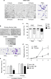
Figure 1
Figure 1
Differentiation of MK cells and suppression of bone resorption by MK CM. (a) K562 cells were incubated with phorbol 12-myristate 13-acetate (PMA) for the indicated times. (b) Cells from mouse fetal liver were differentiated with thrombopoietin (TPO) for the indicated days. Arrows indicate proplatelet-bearing megakaryocytes (MKs). Cell morphology was observed by microscope, and MK differentiation was detected by Wright-Giemsa staining at 3 and 4 days, respectively. Scale bars, 100 μm. (c) DNA polyploid content was analyzed by flow cytometry. K562 and primary cells were treated with PMA or TPO for 3 days or 4 days, respectively. The percentage of cells in each ploidy (2 N, 4 N, and ≥8 N) was shown. (d) Differentiation rates of K562 and primary cells. Mature MKs were scored by counting larger than 25 µm in diameter and extensive multinuclearity. (e) A resorption pit formation assay of mouse bone marrow macrophages (BMMϕ) cultured with M-CSF and RANKL to form osteoclasts in the presence or absence of 10% (v/v) conditioned media (CM) of MKs and pro-megakaryocytes (pro-MKs) on a dentine disc for the indicated times. MKs were derived from K562 cells. Resorbed areas were quantified as percentages of the total area. Data are presented as mean ± SEM. *P < 0.05 vs. non-conditioned media (non-CM, α-MEM media) or primary cells; #P < 0.05 vs. pro-MK CM. NS, not significant.
Figure 2
Figure 2
Suppression of osteoclastic resorption activity by MK CM. (a) Tartrate-resistant acid phosphatase (TRAP) staining of osteoclasts in the presence or absence of the indicated doses of conditioned medias (CMs) for 4 days. TRAP-positive cells with more than three nuclei were counted. (b) TRAP staining of osteoclasts in the presence of the indicated doses of phorbol 12-myristate 13-acetate (PMA) for 4 days. (c) TRAP staining of osteoclasts in the presence or absence of 30% (v/v) MK and pro-MK CM fractions derived from murine fetal livers for 4 days. (d) Viability of mouse BMMϕ was assessed using a CCK-8 assay in the presence or absence of the indicated doses of CM for 48 hours. (e) TRAP staining of osteoclasts in the presence of 10% (v/v) MK and pro-MK CMs for the indicated times. (f,g) Resorption activity of osteoclasts. After the full differentiation of BMMs into osteoclasts, cells were seeded on a dentine disc with M-CSF and RANKL, and then cultured in the presence of 10% (v/v) MK or pro-MK CM for the indicated times. MK and pro-MK cells were derived from K562 cells (f) or from murine fetal livers (g). Resorbed areas were quantified as percentages of the total area. MK and pro-MK were derived from K562 cells, unless otherwise specified. Data are presented as mean ± SEM. *P < 0.05 vs. non-CM (α-MEM media); #P < 0.05 vs. pro-MK CM. NS, not significant.
Figure 3
Figure 3
Stimulation of calvaria bone formation by MK CM. 30× Megakaryocyte (MK) and pro-megakaryocyte (pro-MK) conditioned media (CM), and non-CM were injected on the right side of the calvaria in 5-week-old female C57BL/6 mice for 4 weeks. PBS was injected on the left side of the calvaria as a negative control. Hematoxylin and eosin staining (a), and alkaline phosphatase immune histochemical (ALP, brown) and tartrate-resistant acid phosphatase (TRAP, purple) stainings (b) were performed with the sections of calvaria. (c) The osteoblastic surface percentage (OB.S/BS, %) and number per bone surface (OB.S/BS, N/mm), and the osteoclastic surface percentage (OC.S/BS, %) and number per bone surface (OC.S/BS, N/mm), were shown. (d) The width of the calvaria was quantitated by the average of 4 spots with the same interval (the indicated lines) of the midline between the sagittal suture and the site of muscle attachment. Arrows indicates osteoid lines as references of calvaria width measurements. Data are presented as mean ± SEM. *P < 0.05 vs. non-CM (α-MEM media); #P < 0.05 vs. pro-MK CM. Scale bar, 50 μm.
Figure 4
Figure 4
Effects of MK CM on osteoblastic proliferation and differentiation. (a) Viability of pre-osteoblast MC3T3-E1 cells was assessed using a CCK-8 assay in the presence or absence of 50% (v/v) conditioned media (CM) from megakaryocytes (MK) or pro-megakaryocytes (pro-MK) for 48 hours. MK and pro-MK cells were derived from K562 cells. (b,c) Viability of MC3T3-E1 cells was also determined with the indicated doses of phorbol 12-myristate 13-acetate (PMA) (b) or 50% (v/v) CMs of enriched MK and pro-MK derived from murine fetal livers (c) for 48 hours. (d) Proliferation of MC3T3-E1 cells was assessed using a BrdU incorporation assay in the presence or absence of 50% (v/v) MK and pro-MK CMs for 48 hours. (e,f) Alkaline phosphatase (ALP) activity (e) and osteocalcin secretion (f) of MC3T3-E1 cells in the presence or absence of 30% (v/v) MK and pro-MK CMs for 7 days. The ALP activity was normalized by total cellular protein amounts. (g) Bone nodule formation assay of MC3T3-E1 cells was assessed by Alizarin red S staining, and were quantified by extraction with cetylpyridinium chloride in the presence or absence of 30% (v/v) MK and pro-MK CMs for 14 days. MK and pro-MK were derived from K562 cells, unless otherwise specified. Data are presented as mean ± SEM. *P < 0.05 vs. non-CM (α-MEM media); #P < 0.05 vs. pro-MK CM. NS, not significant.
Figure 5
Figure 5
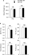
Micro-computed tomography analyses of the femurs in ovariectomized C57B/L6 mice. Female C57B/L6 mice were ovariectomized at 9 weeks of age (n = 8 in each group), and conditioned media (CM) of megakaryocytes (MK) or pro-megakaryocytes (pro-MK) were injected via the tail vein once a day starting at 13 weeks of age for 4 weeks. Mice were then sacrificed for analyses at 17 weeks of age. (a) Body weight before (Pre) and after (Post) the injections. (b) Trabecular bone parameters of the femurs were also measured. BV/TV, bone volume per tissue volume; Tb.Th, trabecular thickness; Tb.N, trabecular number; Tb.Sp, trabecular separation. Data are presented as mean ± SEM. NS, not significant.
Figure 6
Figure 6
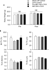
Micro-computed tomography analyses of the femurs in ovariectomized nude mice. Female nude mice were sham-operated or ovariectomized (OVX) at 9 weeks of age (n = 15 in each group), and conditioned media (CM) of megakaryocytes (MK) or pro-megakaryocytes (pro-MK) was injected via the tail vein once a day from 13 weeks of age for 4 weeks. Mice were then sacrificed for analyses at 17 weeks of age. The same volume of saline was injected in the control group. (a) Body weight before (Pre) and after (Post) the injections. (b) Trabecular bone parameters of the femurs were also measured. BV/TV, bone volume per tissue volume; Tb.Th, trabecular thickness; Tb.N, trabecular number; and Tb.Sp, trabecular separation. Data are presented as mean ± SEM. *P < 0.05 vs. untreated control; #P < 0.05 vs. pro-MK CM. NS, not significant.

Similar articles

Cited by

References

    1. Raisz LG. Pathogenesis of osteoporosis: concepts, conflicts, and prospects. J Clin Invest. 2005;115:3318–3325. doi: 10.1172/JCI27071. - DOI - PMC - PubMed
    1. Minaire P. Immobilization osteoporosis: a review. Clin Rheumatol. 1989;8:95–103. doi: 10.1007/BF02207242. - DOI - PubMed
    1. Amano H, Hackett NR, Rafii S, Crystal RG. Thrombopoietin gene transfer-mediated enhancement of angiogenic responses to acute ischemia. Circ Res. 2005;97:337–345. doi: 10.1161/01.RES.0000179534.17668.f8. - DOI - PubMed
    1. Geddis AE. Megakaryopoiesis. Semin Hematol. 2010;47:212–219. doi: 10.1053/j.seminhematol.2010.03.001. - DOI - PMC - PubMed
    1. Kacena MA, et al. Megakaryocytes regulate expression of Pyk2 isoforms and caspase-mediated cleavage of actin in osteoblasts. J Biol Chem. 2012;287:17257–17268. doi: 10.1074/jbc.M111.309880. - DOI - PMC - PubMed

Publication types

MeSH terms

Substances