Review
. 2020 Mar;21(3):153-168.
doi: 10.1038/s41583-019-0260-z. Epub 2020 Feb 10.

Dentate gyrus circuits for encoding, retrieval and discrimination of episodic memories

Affiliations
Review

Dentate gyrus circuits for encoding, retrieval and discrimination of episodic memories

Thomas Hainmueller et al. Nat Rev Neurosci. 2020 Mar.

Abstract

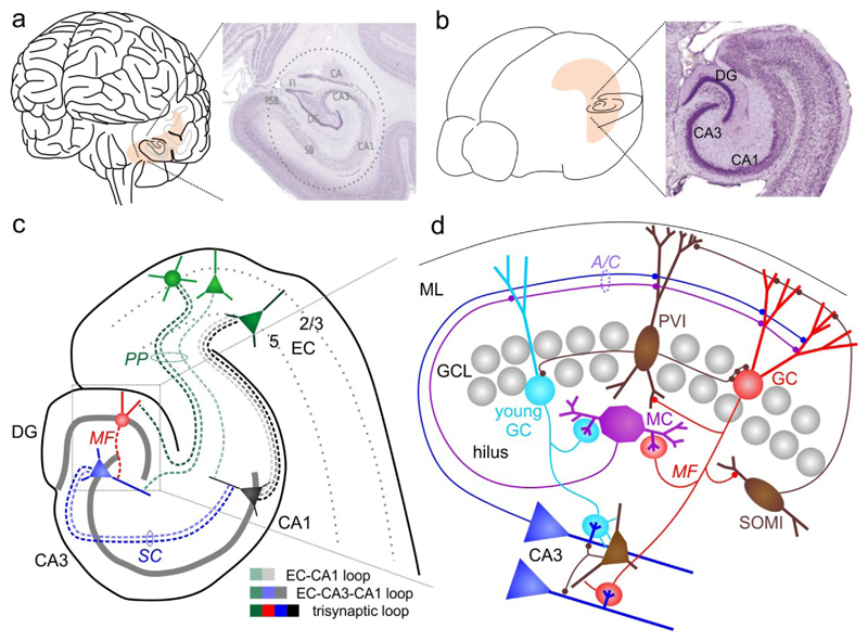
The dentate gyrus (DG) has a key role in hippocampal memory formation. Intriguingly, DG lesions impair many, but not all, hippocampus-dependent mnemonic functions, indicating that the rest of the hippocampus (CA1-CA3) can operate autonomously under certain conditions. An extensive body of theoretical work has proposed how the architectural elements and various cell types of the DG may underlie its function in cognition. Recent studies recorded and manipulated the activity of different neuron types in the DG during memory tasks and have provided exciting new insights into the mechanisms of DG computational processes, particularly for the encoding, retrieval and discrimination of similar memories. Here, we review these DG-dependent mnemonic functions in light of the new findings and explore mechanistic links between the cellular and network properties of, and the computations performed by, the DG.


Conflict of interest statement

Competing interests

The authors declare no competing interests.

Figures

Fig. 1
Fig. 1. Anatomical organization of the hippocampus.
a | Human brain and Nissl-stained section through the hippocampus. b | Same as in a for a mouse brain. c | Schematic of the mouse hippocampal formation and its main synaptic connections. d| Illustration of the DG microcircuitry, its intrinsic connections and outputs to CA3. A/C, associational-/commissural pathway; DG, dentate gyrus; EC, entorhinal cortex; GC, granule cell; GCL, granule cell layer; MC, mossy cell; MF, mossy fiber; ML, molecular layer; PP, perforant-path; PVI, parvalbumin-expressing interneuron; SC, schaffer-collateral; SOMI, somatostatin-positive interneuron. Photographs in a and b are derived from www.brainmaps.org and Franklin, K. B. J. & Paxinos, G., ‘The mouse brain in stereotaxic coordinates’, 3rd edition (Elsevier, New York, 2007), respectively.
Fig. 2
Fig. 2. Spatial, contextual and temporal firing characteristics of identified dentate gyrus neurons.
a | Schematic of juxtacellular recordings in freely moving rats and recorded signals (right). b | Left, Spikes of a granule cells (GC; red dots) plotted over the rat’s trajectory (grey line). Middle, heat-map plotting the firing frequency of this GC. Number indicates the maximum firing rate. Right, The GC was filled with neurobiotin after the recording to reconstruct the depicted morphology. c | As in b, but for a mossy cell. d | Schematic of a two-photon imaging setup for calcium imaging of DG neurons in head-fixed mice. The mice run on a treadmill to navigate along a virtual linear track displayed on monitors around them. Right, Fluorescence of a genetically encoded calcium indicator (GCaMP6f) expressed in GCs acquired in vivo. e | Mean activity (color-coded) of individual hippocampal place-cells (rows) plotted over distance on two visually different virtual linear tracks (context A and context B, respectively). The sorting of neurons is the same in both columns. A shift of place fields away from the central diagonal in the right plot signifies global remapping of the respective neuron. f | The same place cells were imaged over multiple days while the mouse ran on the same linear track on each day. Neurons were sorted by their place-fields on day 0 and this sorting is maintained for the other days shown. Note, GC place fields remain in the same location, whereas those of CA3 and CA1 cells change between days. GCL, Granule cell layer. Parts a-c are adapted from REF.41. Parts d-f are adapted from REF. 43.
Fig. 3
Fig. 3. DG functions in memory encoding and recall.
a | Schematic of memory encoding. Mossy-fiber (MF) input to a CA3 pyramidal cell dendrite can elicit dendritic spikes which may promote heterosynaptic potentiation of perforant-path (PP) and recurrent-collateral inputs. This process may potentially underlie the formation of new place-fields (lower row). b | Left, Rapid strengthening of PP-GC and MF-CA3 synapses may support the re-activation of CA3 pyramidal cells during memory recall minutes to hours after the original experience. Right, Once CA3 ensembles have been permanently established by durable plasticity of PP-CA3 and recurrent-synapses, the memory can be reliably recalled without MF input, which is reduced at this stage due to depotentiation of PP-GC synapses.
Fig. 4
Fig. 4. DG function in discriminatory contextual fear conditioning (CFC).
a | Schematic of discriminative CFC. b | Illustration of the physiological DG and CA3 activation patterns evoked by overlapping PP-inputs encoding similar contexts. Young and mature GC show similar activity patterns in both contexts. Young- and recently-active GCs potently recruit feed-forward inhibition, and may thus suppress firing of CA3 pyramidal cells with overlapping synaptic input-patterns between the two contexts. This could spare less overlapping CA3 ensembles forming context-unique representations. c | Activation pattern as in b in a mouse model where MF-plasticity onto CA3 feed-forward interneurons was disrupted genetically by knocking out the gene encoding adducin 2 (Add2). Releasing CA3 from MF-recruited feed-forward inhibition unmasks the activation of larger CA3 ensembles with more overlapping activity between the two contexts.

References

    1. Schacter DL, Tulving E. Memory systems 1994. MIT Press; 1994.
    1. Eichenbaum H. The cognitive neuroscience of memory: an introduction. Oxford University Press; 2012.
    1. Squire LR. Mechanisms of memory. Science. 1986;232:1612–1619. - PubMed
    1. Tonegawa S, Morrissey MD, Kitamura T. The role of engram cells in the systems consolidation of memory. Nature Reviews Neuroscience. 2018;19:485–498. - PubMed
    1. Corkin S. What’s new with the amnesic patient H.M.? Nat Rev Neurosci. 2002;3:153–160. - PubMed

Publication types