. 2020 Apr;38(4):420-425.
doi: 10.1038/s41587-019-0404-8. Epub 2020 Feb 10.

Antibody-mediated delivery of viral epitopes to tumors harnesses CMV-specific T cells for cancer therapy

Affiliations

Antibody-mediated delivery of viral epitopes to tumors harnesses CMV-specific T cells for cancer therapy

David G Millar et al. Nat Biotechnol. 2020 Apr.

Abstract

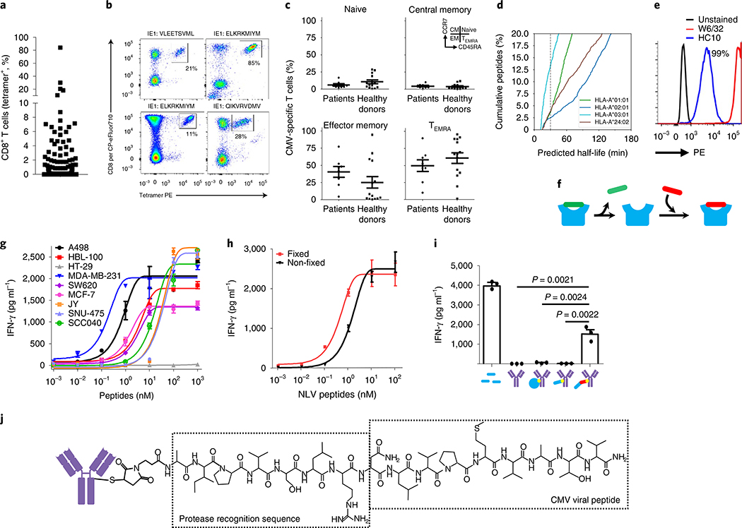
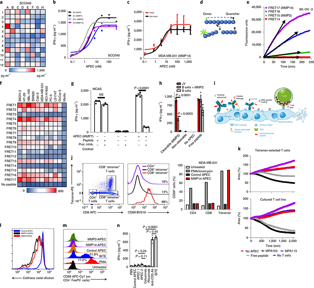
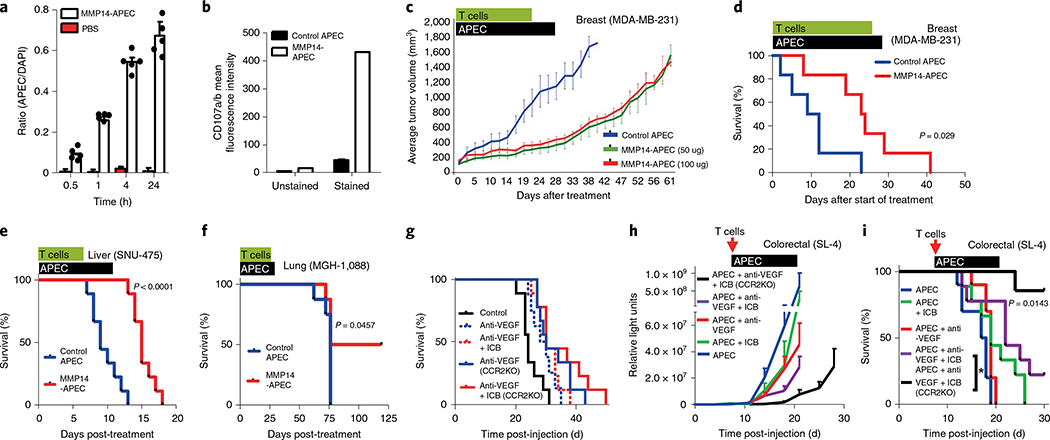
Several cancer immunotherapy approaches, such as immune checkpoint blockade and adoptive T-cell therapy, boost T-cell activity against the tumor, but these strategies are not effective in the absence of T cells specific for displayed tumor antigens. Here we outline an immunotherapy in which endogenous T cells specific for a noncancer antigen are retargeted to attack tumors. The approach relies on the use of antibody-peptide epitope conjugates (APECs) to deliver suitable antigens to the tumor surface for presention by HLA-I. To retarget cytomegalovirus (CMV)-specific CD8+ T cells against tumors, we used APECs containing CMV-derived epitopes conjugated to tumor-targeting antibodies via metalloprotease-sensitive linkers. These APECs redirect pre-existing CMV immunity against tumor cells in vitro and in mouse cancer models. In vitro, APECs activated specifically CMV-reactive effector T cells whereas a bispecific T-cell engager activated both effector and regulatory T cells. Our approach may provide an effective alternative in cancers that are not amenable to checkpoint inhibitors or other immunotherapies.


Figures

Fig. 1 |
Fig. 1 |. CMV-CTLs are found at high frequencies in patients with cancer and recognize antigen-modified cancer cells.
a, CMV-specific immunity in healthy donors (n = 30) and cancer patients (n = 104) is found at high frequencies. b, Example tetramer staining. Percentages denote tetramer+ cells of the total CD8 T-cell population. PE, phycoerythrin. c, Memory phenotype analysis of CMV-CTL populations between healthy donors (n = 14) and cancer patients (n = 8) using CCR7 and CD45RA expression. d, In silico prediction of dissociation rates for HLA-bound peptides from surface. e, Flow cytometric analysis of peptide-loaded (W6/32) and empty HLA molecules (HC10) at the cancer cell line surface. Percentages denote cells positive for HC10 staining. All cell lines tested were 100% positive for peptide-loaded HLA (W6/32). Staining was repeated (n = 3) in selected cell lines. f, Schematic of peptide dissociation and peptide loading into HLA at the cell surface. g, IFN-γ release depicting T-cell activation in response to eight tumor cell lines with exogenously added CMV peptide (n = 3 independent samples). h, T-cell cytokine release after recognition of peptide-loaded tumor cells that were loaded in either a fixed or unfixed state (n = 3 independent samples). i, T-cell recognition of tumor target cells pretreated with either exogenously added free CMV peptide (1 μM) or antibody conjugates (350 nM) where the conjugate was either no payload, whole-protein antigen, minimal CMV epitope peptide or protease-cleavable CMV peptide (n = 3 independent samples). g-i, Data shown as mean and error bars represent s.e.m. Significance was determined by unpaired two-tailed f-test. j, Chemistry schematic of APECs. In relevant panels, error bars represent s.e.m.
Fig. 2 |
Fig. 2 |. Engineering APECs to deliver peptides to the surface of tumor cells.
a, Tumor cell line treated with 96different cAPECs screened using a functional CMV-CTL cytokine release assay. b, APECs demonstrating positive responses in a were used in a dose-response assay to determine median effective concentration required to induce a 50% effect. CtsD, cathepsin D. c, Functional T-cell cytokine release assay demonstrating recognition of unfixed and lightly fixed cells treated with cAPEC (n = 3 independent samples). d, Schematic depicting FRET assay. e,f, Example data of peptide cleavage kinetics of FRET assay (maximum voltage depicted by arrows) (e) and with data from ten tumor cell lines shown (f). g, Antigenic reprogramming of tumor cells by APEC was inhibited by the co-incubation of protease inhibitors (n = 3 independent samples). Significance was determined by unpaired two-tailed f-test. h, MMP2 rAPEC-treated healthy B cells (350 nM) did not activate T cells whereas malignant B cells (JY) efficiently activated them. Healthy B cells become susceptible to APECs following the provision of exogenous MMP2 protease. Addition of exogenous free CMV peptide (1 μM) allowed recognition of both healthy and tumor B cells (n = 3 independent samples). Significance was determined by unpaired two-tailed f-test. i, Schematic of APEC showing the proposed mechanism of action at the tumor cell surface. j-l, APEC-treated tumor cells activated only the peptide antigen-specific T-cell population within PBMCs, as depicted by tetramer and CD69 flow cytometric analysis (j). Percentages denote activated T cells compared to untreated control (data from a single experiment). Comparison of cytotoxicity of CMV-CTL taken directly from PBMCs or in vitro expanded against APEC-treated MDA-MB-231 tumor cells (k); data from a single experiment. Proliferation of CellTrace violet-labeled CMV-CTLs isolated from PBMCs against APEC-treated MDA-MB-231 cells (l); data from a single experiment. m, Regulatory T cells were activated by BiTE but not by any of the APECs tested, as identified by flow cytometric analysis using the T-cell activation marker CD69. Percentages denote activated Treg compared to untreated control (data from a single experiment). n, Immobilized BiTE and the anti-CD3 antibody OKT3 activated T cells whereas no such activation was observed using immobilized APEC (n = 3 independent samples). Significance was determined by unpaired two-tailed f-test. c,g,h,n, Data shown as mean and error bars represent s.e.m.
Fig. 3 |
Fig. 3 |. APEC modulation of antigenicity in vivo.
a, In vivo tumor penetration of APEC-treated NOD/SCID mouse model of primary orthotopic human breast cancer (MDA-MB-231) using the ratio of APEC to DAPI (n = 4 independent samples). Data shown as mean and error bars represent s.d. b, In vivo T-cell cytotoxic capacity, as assayed by CD107 staining, demonstrated increased cytotoxic activity of freshly isolated CMV-CTL in MDA-MB-231 treated with MMP14-cAPEC compared with control cAPEC. c, NOD/SCID mouse model MDA-MB-231 (n = 8) demonstrated a delay in tumor growth following treatment with 50 μg or 100 μg MMP14 cAPEC compared with control cAPEC. d, MMP14-cAPEC improved the survival of mice (n = 8) in a breast cancer neoadjuvant model compared with control cAPEC (P = 0.029, Mantel-Cox two-sided test) after injection of ex vivo expanded CMV-CTL. e, Survival of mice orthotopically injected with hepatocellular carcinoma cell line SNU-475 (n = 8) treated with MMP14-cAPEC and ex vivo expanded CMV-CTL was improved compared to that of mice treated with control cAPEC (P< 0.0001, Mantel-Cox two-sided test). f, Improved survival of mice (n = 5) treated subcutaneously with MMP14 or control cAPEC and ex vivo expanded CMV-CTL in a PDX model of lung cancer (P = 0.0457, Mantel-Cox two-sided test). g, C57BL/6 or Ccr2−/− (C57 background) mouse model (n = 10) of metastatic colorectal carcinoma (SL4 clone) demonstrated only a marginal increase in survival when treated with either anti-VEGF or anti-VEGF in combination with ICB therapy (anti-PD-1+ anti-CTLA-4). All mice received ex vivo expanded OT-I T cells specific for SIINFEKL. h,i, Ccr2−/− mice (n = 8–10) treated with combination immune checkpoint blockade (therapy of anti-VEGF+ ICB+ murine APEC system demonstrated significant delay in tumor growth (h) and increased survival (i) compared with wild-type mice treated with the same reagents or with other APEC combination therapies (P = 0.0143, Mantel-Cox two-sided test). All mice received ex vivo expanded OT-I T cells specific for SIINFEKL. In relevant panels, error bars represent s.e.m.

Comment in

References

    1. Brahmer JR et al. Safety and activity of anti-PD-L1 antibody in patients with advanced cancer. N. Engl. J. Med 366, 2455–2465 (2012). - PMC - PubMed
    1. Yarchoan M, Hopkins A & Jaffee EM Tumor mutational burden and response rate to PD-1 inhibition. N. Engl. J. Med 377, 2500–2501 (2017). - PMC - PubMed
    1. Le DT et al. Mismatch-repair deficiency predicts response of solid tumors to PD-1 blockade. Science 357, 409–413 (2017). - PMC - PubMed
    1. Brown SD et al. Neo-antigens predicted by tumor genome meta-analysis correlate with increased patient survival. Genome Res. 24, 743–750 (2014). - PMC - PubMed
    1. Hu Z, Ott PA & Wu CJ Towards personalized, tumour-specific, therapeutic vaccines for cancer. Nat. Rev. Immunol 18, 168–182 (2018). - PMC - PubMed

Publication types

MeSH terms