. 2020 Mar;16(3):318-326.
doi: 10.1038/s41589-020-0467-3. Epub 2020 Feb 10.

Development of a covalent inhibitor of gut bacterial bile salt hydrolases

Affiliations

Development of a covalent inhibitor of gut bacterial bile salt hydrolases

Arijit A Adhikari et al. Nat Chem Biol. 2020 Mar.

Abstract

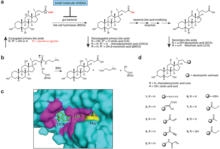
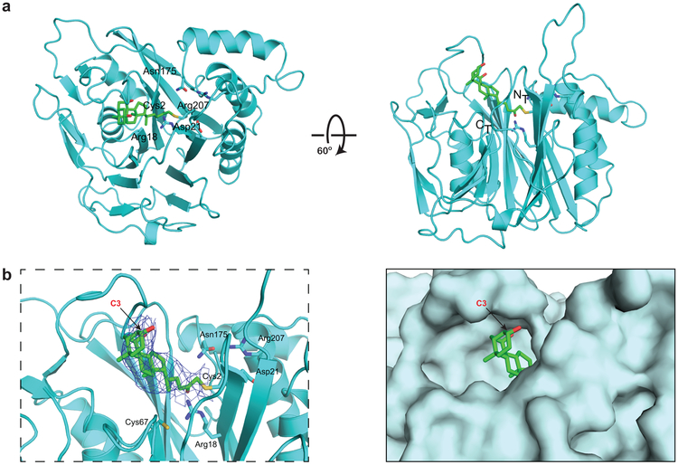
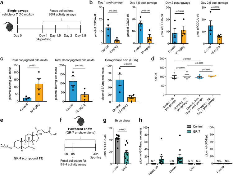
Bile salt hydrolase (BSH) enzymes are widely expressed by human gut bacteria and catalyze the gateway reaction leading to secondary bile acid formation. Bile acids regulate key metabolic and immune processes by binding to host receptors. There is an unmet need for a potent tool to inhibit BSHs across all gut bacteria to study the effects of bile acids on host physiology. Here, we report the development of a covalent pan-inhibitor of gut bacterial BSHs. From a rationally designed candidate library, we identified a lead compound bearing an alpha-fluoromethyl ketone warhead that modifies BSH at the catalytic cysteine residue. This inhibitor abolished BSH activity in conventional mouse feces. Mice gavaged with a single dose of this compound displayed decreased BSH activity and decreased deconjugated bile acid levels in feces. Our studies demonstrate the potential of a covalent BSH inhibitor to modulate bile acid composition in vivo.


Figures

Figure 1.
Figure 1.. Rational design of gut bacterial bile salt hydrolase (BSH) inhibitors.
(a) BSHs are the gateway enzymes in the conversion of primary (host-produced) to secondary (bacterially produced) bile acids. Inhibition of BSHs should result in a decrease in deconjugated primary and secondary bile acids. (b) Mechanism of enzymatic amide bond cleavage by BSHs. (c) A co-crystal structure of the BSH from the Gram positive gut bacterium Clostridium perfringens (strain 13 / type A) and deconjugated tauro-deoxycholic acid (PDB 2BJF) guided inhibitor design. While hydrophobic interactions orient the bile acid core in the active site, the D-ring side chain is exposed to solvent (magenta residues are within 4Å of bile acid, Cys2 is yellow). (d) Library of synthesized inhibitors. Electrophilic warheads were appended to the chenodeoxycholic acid bile core in order to create broad-spectrum BSH inhibitors.
Figure 2.
Figure 2.. Identification of 7 as a potent, non-toxic, pan-BSH inhibitor.
(a,b) Screen of inhibitors versus B. theta BSH (a) and B. longum BSH (b) showing % deconjugation of tauro bile acids at 2 and 21 hours (for concentrations of substrates and products, see Supplementary Fig. 4; for % deconjugation of each bile acid see Supplementary Table 2). Inhibitor (100 μM) was incubated with 200 nM rBSH for 30 mins followed by addition of taurine-conjugated bile acids (tauro-β-muricholic acid, TβMCA; tauro-cholic acid, TCA; tauro-ursodeoxycholic acid, TUDCA; and tauro-deoxycholic acid, TDCA, 25 μM each). Deconjugation of substrate was followed by UPLC-MS. (c) Compound 7 inhibited BSH activity in growing cultures of Gram negative (B. theta VPI 5482, Bacteroides fragilis ATCC 25285, and Bacteroides vulgatus ATCC 8482) and Gram positive (Lactobacillus plantarum WCFS1, Clostridium perfringens ATCC 13124, and Bifidobacterium adolescentis L2-32) bacteria. Inhibitor (100 μM of 7 or CAPE) and taurine-conjugated bile acids (TβMCA, TCA, TUDCA and TDCA, 25 μM each) were added to bacterial cultures at OD600 0.1. Cultures were allowed to grow into stationary phase and percent deconjugation of tauro bile acids at 21h was determined by UPLC-MS (for concentrations of substrates and products, see Supplementary Fig. 8; for % deconjugation of each bile acid see Supplementary Table 2). (d) Compound 7 is not bactericidal. Bacterial strains were incubated with conjugated bile acids (as described in panel c) and compound (100 μM) and plated at 21h to assess strain viability. CAPE decreased the cell viability of the Gram negative strains tested. Red downward arrows indicate fold decrease compared to DMSO control. For (c) and (d), one-way ANOVA followed by Dunnett’s multiple comparisons test. (e) Compound 7 inhibited BSH activity in a fecal slurry. All assays were performed in biological triplicate, and data are presented as mean ± SEM.
Figure 3.
Figure 3.. Compound 7 covalently modifies B. theta BSH at the active site cysteine residue.
(a) X-ray structure of 7 bound to B. theta BSH. The BSH (cyan) is shown in ribbon representation, with indicated side chains (cyan, with heteroatoms in CPK colors) rendered as sticks. Compound 7 (green, with heteroatoms in CPK colors) is rendered in stick form. There is electron density visible at the active site of one of the four subunits in the asymmetric unit, consistent with the conclusion that the inhibitor is covalently attached to Cys2. (b) Co-crystal structure of B. theta BSH and 7 shown in ribbon (left, with electron density of the compound shown as a blue net) and surface (right) representations. The A ring of 7, which includes the C3 hydroxyl group, is solvent-exposed. Panels a and b were prepared using PYMOL software (Schroedinger).
Figure 4.
Figure 4.. Compound 7 exhibits BSH engagement and minimal off-target effects.
(a) Structure of ‘clickable’ 7, 7-N3 (12), for on- and off-target studies. (b) 7-N3 displayed significant BSH inhibition in conventional mouse feces, showing that this probe retained its function as a BSH inhibitor. (c) Treatment of B. adolescentis L-32 culture with 7-N3 for 1 hour followed by cell lysis, click reaction with Fluor 488-alkyne, and visualization using in-gel fluorescence revealed labeling of only one protein ~35 kDa in size, the mass of the annotated B. adolescentis BSH. Proteins were loaded at a concentration of 1.5 mg/mL. B. theta recombinant BSH (BT BSH, 1 μM) was used as a positive control for the click reactions and as a standard to show 38 kDa. (d) Lysate from the treatment of B. adolescentis cultures with 7-N3 was reacted with desthiobiotin-alkyne, resolved by SDS-PAGE, and visualized by silver-staining. Arrow indicates a band in the probe-treated sample at the predicted molecular weight (~35 kDa) of BSH. (For full gels in triplicate, see Supplementary Fig. 15.) In-gel digestion followed by LC-MS/MS identified this band as BSH with high-confidence. Semi-quantitative analysis indicated a 4.5-fold enrichment in 7-N3-treated versus vehicle-treated bacterial cultures (see Supplementary Tables 4). (e) Treatment of B. adolescentis cultures with decreasing concentrations of 7 followed by treatment with 10 μM 7-N3 and click reaction with Fluor 488-alkyne resulted in a dose-dependent increase in fluorescence labeling of annotated B. adolescentis BSH. Experiment was repeated twice with similar results. (For full gels in triplicate, see Supplementary Fig. 15.) (f) One-hour treatment of NCI-H716 intestinal cells with 7-N3 followed by click reaction with Fluor 488-alkyne and visualization by in-gel fluorescence resulted in no significant labeling of proteins compared to control-treated cells (for triplicate gels, see Supplementary Fig. 16). For (b, c, d, and f), n=3 biological replicates per condition. For (b), data is presented as mean ± SEM.
Figure 5.
Figure 5.. Compound 7 inhibits BSH activity in vivo and can be gut-restricted.
(a-c) Treatment of conventional mice with a single dose of 7 resulted in recoverable inhibition of BSH activity and a shift toward conjugated bile acids. n=4 mice per group, Student’s t test. (a) Design of in vivo BSH inhibition experiment. Adult male C57BL/6 mice were gavaged with a single dose of 7 (10 mg/kg) or vehicle control. (b) BSH activity was measured in half-day increments starting 1 day post-gavage. Resuspended fresh feces from inhibitor- or vehicle-treated groups were incubated with substrate (GCDCA-d4, 100 μM) for 25 min and formation of product was quantified by UPLC-MS. n=4 mice per group, two-tailed Student’s t test. (c) Fecal bile acid composition 1 day post-gavage. Deconjugated bile acids, including the secondary bile acid deoxycholic acid (DCA), were decreased in the inhibitor-treated group. n=4 mice per group, two-tailed Student’s t test. (d) Bacterial OTUs (operational taxonomic units) did not differ between the inhibitor- and vehicle-treated groups 1 day post-gavage. n=4 mice per group, one-way ANOVA followed by Tukey’s multiple comparisons test. (e) Structure of gut-restricted compound 7 (GR-7, 13). (f) Design of proof-of-concept in vivo study with GR-7. Adult male C57BL/6 mice were fed powdered chow containing 0.09% (w/w) GR-7 or powdered chow alone for 30 hours. Fecal pellets were collected 8 hours post-diet change. n =10 mice per group. (g) Resuspended fresh feces (20 mg/mL) from inhibitor- or control-treated mice were incubated with substrate (GCDCA-d4, 100 μM) for 25 min and formation of product was quantified by UPLC-MS. Significant inhibition of BSH activity was observed in the feces of GR-7-treated compared to control-treated mice. Student’s t test. n=10 mice per group, two-tailed Student’s t test. (h) Quantification of GR-7 in tissues and plasma. Inhibitor was detected in feces 8 hours post-diet change and in cecal contents at sacrifice. No GR-7 was detected in the liver or plasma. N.D. = not detected. n=10 mice per group. All data are presented as mean ± SEM.

Similar articles

Cited by

References

    1. Ridlon JM, Kang D-J & Hylemon PB Bile salt biotransformations by human intestinal bacteria. J. Lipid Res 47, 241–259 (2006). - PubMed
    1. Fiorucci S & Distrutti E Bile Acid-Activated Receptors, Intestinal Microbiota, and the Treatment of Metabolic Disorders. Trends Mol Med 21, 702–714 (2015). - PubMed
    1. Setchell KD, Lawson AM, Tanida N & Sjövall J General methods for the analysis of metabolic profiles of bile acids and related compounds in feces. J. Lipid Res 24, 1085–1100 (1983). - PubMed
    1. Hamilton JP et al. Human cecal bile acids: concentration and spectrum. Am. J. Physiol. Gastrointest. Liver Physiol 293, G256–G263 (2007). - PubMed
    1. Modica S, Gadaleta RM & Moschetta A Deciphering the nuclear bile acid receptor FXR paradigm. Nucl Recept Signal 8, e005 (2010). - PMC - PubMed

Publication types

LinkOut - more resources