

Signature mRNA markers in extracellular vesicles for the accurate diagnosis of colorectal cancer
- PMID: 32042310
- PMCID: PMC7001337
- DOI: 10.1186/s13036-020-0225-9
Signature mRNA markers in extracellular vesicles for the accurate diagnosis of colorectal cancer
Abstract
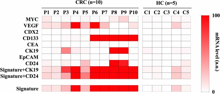
Background: With the increasing incidence of colorectal cancer (CRC), its accurate diagnosis is critical and in high demand. However, conventional methods are not ideal due to invasiveness and low accuracy. Herein, we aimed to identify efficient CRC mRNA markers in a non-invasive manner using CRC-derived extracellular vesicles (EVs). The expression levels of EV mRNAs from cancer cell lines were compared with those of a normal cell line using quantitative polymerase chain reaction. Eight markers were evaluated in plasma EVs from CRC patients and healthy controls. The diagnostic value of each marker, individually or in combination, was then determined using recessive operating characteristics analyses and the Mann-Whitney U test.
Results: Eight mRNA markers (MYC, VEGF, CDX2, CD133, CEA, CK19, EpCAM, and CD24) were found to be more abundant in EVs derived from cancer cell lines compared to control cell lines. A combination of VEGF and CD133 showed the highest sensitivity (100%), specificity (80%), and accuracy (93%) and an area under the curve of 0.96; hence, these markers were deemed to be the CRC signature. Moreover, this signature was found to be highly expressed in CRC-derived EVs compared to healthy controls.
Conclusions: VEGF and CD133 mRNAs comprise a unique CRC signature in EVs that has the potential to act as a novel, non-invasive, and accurate biomarker that would improve the current diagnostic platform for CRC, while also serving to strengthen the value of EV mRNA as diagnostic markers for myriad of diseases.
Keywords: CD133; Colorectal cancer; Extracellular vesicle; Non-invasive biomarker; VEGF; mRNA.
© The Author(s). 2020.
Conflict of interest statement
Competing interestsThe authors declare that they have no competing interests.
Figures


Similar articles
-
Cancer-Type OATP1B3 mRNA in Extracellular Vesicles as a Promising Candidate for a Serum-Based Colorectal Cancer Biomarker.Biol Pharm Bull. 2018;41(3):445-449. doi: 10.1248/bpb.b17-00743. Biol Pharm Bull. 2018. PMID: 29491222
-
Colorectal cancer cell-derived extracellular vesicles transfer miR-221-3p to promote endothelial cell angiogenesis via targeting suppressor of cytokine signaling 3.Life Sci. 2021 Nov 15;285:119937. doi: 10.1016/j.lfs.2021.119937. Epub 2021 Sep 8. Life Sci. 2021. PMID: 34508764
-
A circulating extracellular vesicles-based novel screening tool for colorectal cancer revealed by shotgun and data-independent acquisition mass spectrometry.J Extracell Vesicles. 2020 Apr 14;9(1):1750202. doi: 10.1080/20013078.2020.1750202. eCollection 2020. J Extracell Vesicles. 2020. PMID: 32363013 Free PMC article.
-
Diagnostic Role of Extracellular Vesicles in Cancer: A Comprehensive Systematic Review and Meta-Analysis.Front Cell Dev Biol. 2021 Oct 15;9:705791. doi: 10.3389/fcell.2021.705791. eCollection 2021. Front Cell Dev Biol. 2021. PMID: 34722499 Free PMC article.
-
Exosomal Components and Modulators in Colorectal Cancer: Novel Diagnosis and Prognosis Biomarkers.Biomedicines. 2021 Jul 31;9(8):931. doi: 10.3390/biomedicines9080931. Biomedicines. 2021. PMID: 34440135 Free PMC article. Review.
Cited by
-
Extracellular vesicle biomarkers in circulation for colorectal cancer detection: a systematic review and meta-analysis.BMC Cancer. 2024 May 22;24(1):623. doi: 10.1186/s12885-024-12312-8. BMC Cancer. 2024. PMID: 38778252 Free PMC article.
-
Extracellular vesicle biomarkers in circulation for the diagnosis of gastric cancer: A systematic review and meta‑analysis.Oncol Lett. 2023 Aug 11;26(4):423. doi: 10.3892/ol.2023.14009. eCollection 2023 Oct. Oncol Lett. 2023. PMID: 37664665 Free PMC article.
-
Regulatory role of exosomes in colorectal cancer progression and potential as biomarkers.Cancer Biol Med. 2023 Aug 8;20(8):575-98. doi: 10.20892/j.issn.2095-3941.2023.0119. Cancer Biol Med. 2023. PMID: 37553810 Free PMC article. Review.
-
Extracellular vesicles as carriers of mRNA: Opportunities and challenges in diagnosis and treatment.Theranostics. 2024 Mar 11;14(5):2265-2289. doi: 10.7150/thno.93115. eCollection 2024. Theranostics. 2024. PMID: 38505610 Free PMC article. Review.
-
Small extracellular vesicles in cancer.Bioact Mater. 2021 Apr 7;6(11):3705-3743. doi: 10.1016/j.bioactmat.2021.03.015. eCollection 2021 Nov. Bioact Mater. 2021. PMID: 33898874 Free PMC article. Review.
References
-
- American Cancer Society . Cancer Facts & Figures 2017. Atlanta: American Cancer society; 2017.
-
- American Cancer Society . Colorectal Cancer Facts & Figures 2017–2019. Atlanta: American Cancer society; 2017.
-
- Amin MF, Hassanin AM. Diagnostic performance of CT colonography with limited cathartic preparation in colorectal cancer screening; comparison with conventional colonoscopy. Egypt J Radiol Nucl Med. 2015;46(3):591–598. doi: 10.1016/j.ejrnm.2015.05.012. - DOI
LinkOut - more resources
Full Text Sources
Research Materials
Miscellaneous
