

SREBP1 siRNA enhance the docetaxel effect based on a bone-cancer dual-targeting biomimetic nanosystem against bone metastatic castration-resistant prostate cancer
- PMID: 32042326
- PMCID: PMC6993241
- DOI: 10.7150/thno.40489
SREBP1 siRNA enhance the docetaxel effect based on a bone-cancer dual-targeting biomimetic nanosystem against bone metastatic castration-resistant prostate cancer
Abstract
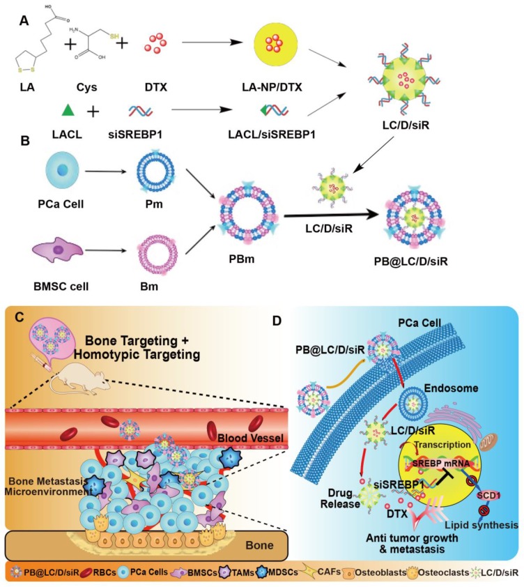
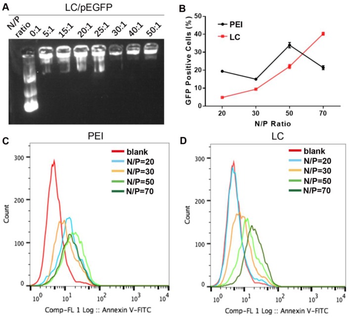
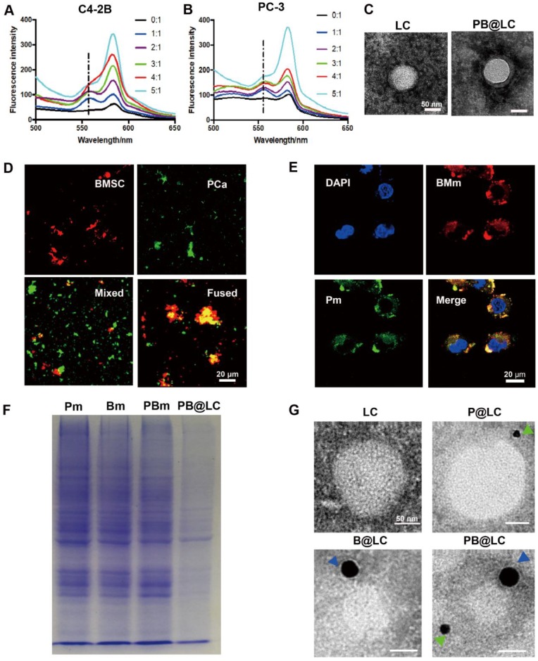
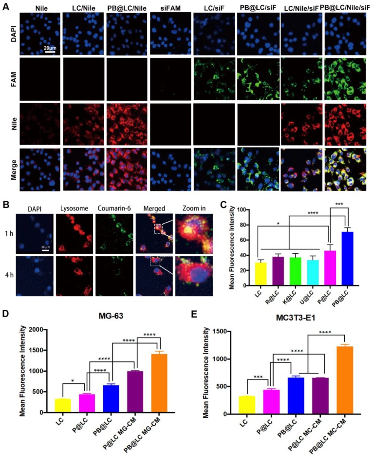
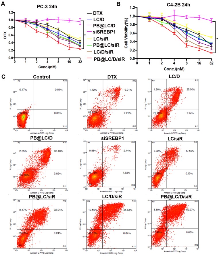
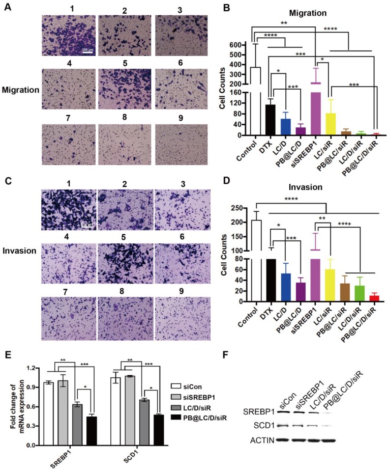
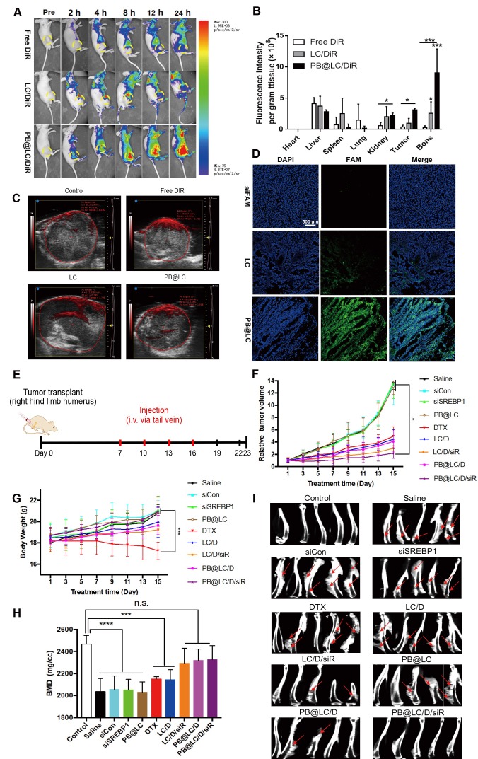
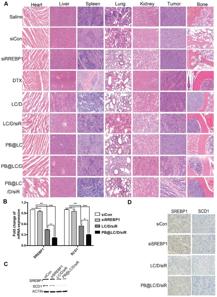
Until recently, there have been limited options for patients with bone metastatic castration-resistant prostate cancer (BmCRPC) following the failure of or development of resistance to docetaxel (DTX), which is one of the frontline treatments. Sterol regulatory element-binding protein 1 (SREBP1) is reported to regulate abnormal lipid metabolism and to promote the progression and metastasis of prostate cancer (PCa). The siRNA interferes SREBP1 may provide an efficient treatment when combined with DTX. Methods: In this study, lipoic acid (LA) and cross-linked peptide-lipoic acid micelles were cross-linked (LC) for DTX and siSREBP1 delivery (LC/D/siR). Then, cell membrane of PCa cells (Pm) and bone marrow mesenchymal stem cells (Bm) were fused for cloaking LC/D/siR (PB@LC/D/siR). Finally, the synthesized PB@LC/D/siR was evaluated in vitro and in vivo. Results: PB@LC/D/siR is internalized in PCa cells by a mechanism of lysosome escape. Tumor targeting and bone homing studies are evaluated using bone metastatic CRPC (BmCRPC) models, both in vitro and in vivo. Moreover, the enhanced anti-proliferation, anti-migration and anti-invasion capacities of DTX- and siSREBP1- loaded PB@LC (PB@LC/D/siR) were observed in vitro. Furthermore, PB@LC/D/siR was able to suppress the growth of the tumor effectively with deep tumor penetration, high safety and good protection of the bone at the tumor site. Additionally, the mRNA levels and protein levels of SREBP1 and SCD1 were able to be significantly downregulated by PB@LC/D/siR. Conclusion: This study presented a bone-cancer dual-targeting biomimetic nanodelivery system for bone metastatic CRPC.
Keywords: SREBP1 siRNA; bone marrow mesenchymal stem cells; bone metastatic prostate cancer; docetaxel; fused cell membrane.
© The author(s).
Conflict of interest statement
Competing Interests: The authors have declared that no competing interest exists.
Figures
Similar articles
-
Codelivery of miR-4638-5p and Docetaxel Based on Redox-Sensitive Polypeptide Micelles as an Improved Strategy for the Treatment of Castration-Resistant Prostate Cancer.Mol Pharm. 2019 Jan 7;16(1):437-447. doi: 10.1021/acs.molpharmaceut.8b01074. Epub 2018 Dec 4. Mol Pharm. 2019. PMID: 30452268
-
Dual CXCR4 and E-Selectin Inhibitor, GMI-1359, Shows Anti-Bone Metastatic Effects and Synergizes with Docetaxel in Prostate Cancer Cell Intraosseous Growth.Cells. 2019 Dec 20;9(1):32. doi: 10.3390/cells9010032. Cells. 2019. PMID: 31877673 Free PMC article.
-
Codelivery of GRP78 siRNA and docetaxel via RGD-PEG-DSPE/DOPA/CaP nanoparticles for the treatment of castration-resistant prostate cancer.Drug Des Devel Ther. 2019 Apr 29;13:1357-1372. doi: 10.2147/DDDT.S198400. eCollection 2019. Drug Des Devel Ther. 2019. PMID: 31118572 Free PMC article.
-
[Recent advances in treatment of patients with castration-resistant prostate cancer after docetaxel failure].Zhejiang Da Xue Xue Bao Yi Xue Ban. 2014 Jan;43(1):115-8. doi: 10.3785/j.issn.1008-9292.2014.01.002. Zhejiang Da Xue Xue Bao Yi Xue Ban. 2014. PMID: 24616470 Review. Chinese.
-
Bone-targeting radiopharmaceuticals for the treatment of bone-metastatic castration-resistant prostate cancer: exploring the implications of new data.Oncologist. 2014 Oct;19(10):1012-8. doi: 10.1634/theoncologist.2013-0472. Epub 2014 Sep 17. Oncologist. 2014. PMID: 25232039 Free PMC article. Review.
Cited by
-
Targeting the SREBP-1/Hsa-Mir-497/SCAP/FASN Oncometabolic Axis Inhibits the Cancer Stem-like and Chemoresistant Phenotype of Non-Small Cell Lung Carcinoma Cells.Int J Mol Sci. 2022 Jun 30;23(13):7283. doi: 10.3390/ijms23137283. Int J Mol Sci. 2022. PMID: 35806291 Free PMC article.
-
RNA Drug Delivery Using Biogenic Nanovehicles for Cancer Therapy.Front Pharmacol. 2021 Dec 24;12:734443. doi: 10.3389/fphar.2021.734443. eCollection 2021. Front Pharmacol. 2021. PMID: 35002692 Free PMC article. Review.
-
Intravenous delivery of enzalutamide based on high drug loading multifunctional graphene oxide nanoparticles for castration-resistant prostate cancer therapy.J Nanobiotechnology. 2020 Mar 18;18(1):50. doi: 10.1186/s12951-020-00607-4. J Nanobiotechnology. 2020. PMID: 32188463 Free PMC article.
-
Nrf2 Signaling Pathway in Chemoprotection and Doxorubicin Resistance: Potential Application in Drug Discovery.Antioxidants (Basel). 2021 Feb 26;10(3):349. doi: 10.3390/antiox10030349. Antioxidants (Basel). 2021. PMID: 33652780 Free PMC article. Review.
-
PSMA-Targeting Reduction-Cleavable Hyperbranched Polyamide-Amine Gene Delivery System to Treat the Bone Metastases of Prostate Cancer.Int J Nanomedicine. 2020 Sep 28;15:7173-7184. doi: 10.2147/IJN.S268398. eCollection 2020. Int J Nanomedicine. 2020. PMID: 33061374 Free PMC article.
References
-
- Body JJ, Casimiro S, Costa L. Targeting bone metastases in prostate cancer: improving clinical outcome. Nat Rev Urol. 2015;12:340–356. - PubMed
-
- Bubendorf L, Schöpfer A, Wagner U, Sauter G, Moch H, Willi N. et al. Metastatic patterns of prostate cancer: an autopsy study of 1,589 patients. Hum Pathol. 2000;31:578–83. - PubMed
-
- Alibhai SMH, Zukotynski K, Walker-Dilks C, Emmenegger U, Finelli A, Morgan SC. et al. Bone health and bone-targeted therapies for prostate cancer: a programme in evidence-based care cancer care ontario clinical practice guideline. Clin Oncol. 2017;29:348–55. - PubMed
-
- Fitzpatrick JM, Wit R. Taxane mechanisms of action: potential implications for treatment sequencing in metastatic castration-resistant prostate cancer. Eur Urol. 2014;65:1198–204. - PubMed
Publication types
MeSH terms
Substances
LinkOut - more resources
Full Text Sources
Medical
Research Materials
