. 2020 Jan 1;10(4):1694-1707.
doi: 10.7150/thno.41309. eCollection 2020.

Sex Differences Revealed in a Mouse CFA Inflammation Model with Macrophage Targeted Nanotheranostics

Affiliations

Sex Differences Revealed in a Mouse CFA Inflammation Model with Macrophage Targeted Nanotheranostics

Lu Liu et al. Theranostics. .

Abstract

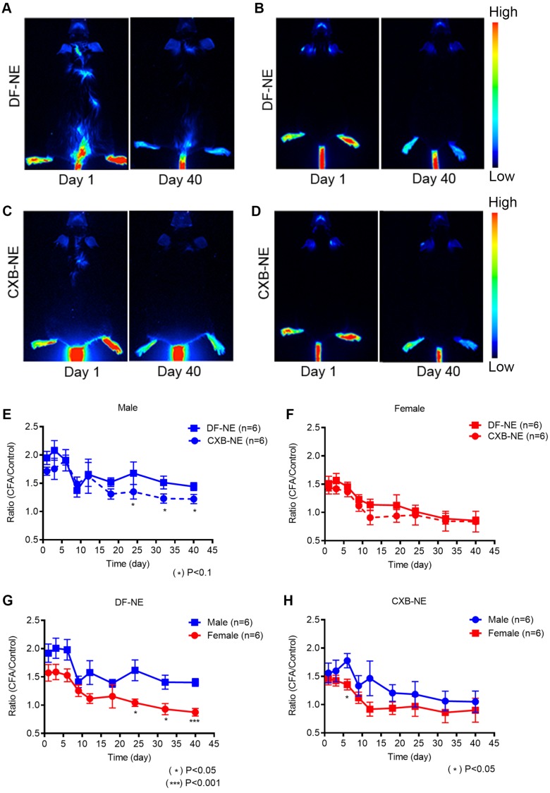
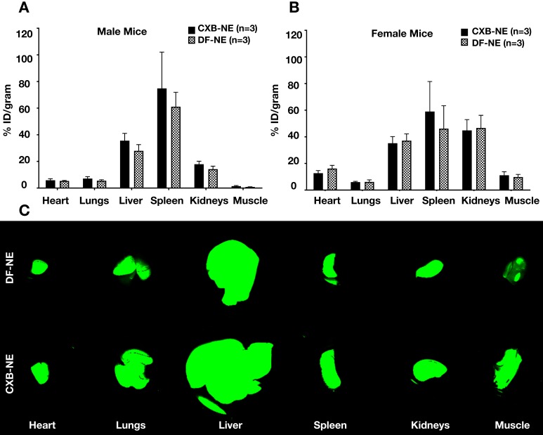
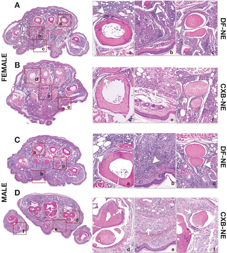
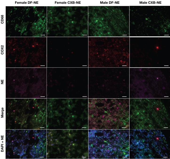
Monocyte derived macrophages (MDMs) infiltrate sites of infection or injury and upregulate cyclooxygenase-2 (COX-2), an enzyme that stimulates prostaglandin-E2 (PgE2). Nanotheranostics combine therapeutic and diagnostic agents into a single nanosystem. In previous studies, we demonstrated that a nanotheranostic strategy, based on theranostic nanoemulsions (NE) loaded with a COX-2 inhibitor (celecoxib, CXB) and equipped with near-infrared fluorescent (NIRF) reporters, can specifically target circulating monocytes and MDMs. The anti-inflammatory and anti-nociceptive effects of such cell-specific COX-2 inhibition lasted several days following Complete Freund's Adjuvant (CFA) or nerve injury in male mice. The overall goal of this study was to investigate the extended (up to 40 days) impact of MDM-targeted COX-2 inhibition and any sex-based differences in treatment response; both of which remain unknown. Our study also evaluates the feasibility and efficacy of a preclinical nanotheranostic strategy for mechanistic investigation of the impact of such sex differences on clinical outcomes. Methods: CFA was administered into the right hind paws of male and female mice. All mice received a single intravenous dose of NIRF labeled CXB loaded NE twelve hours prior to CFA injection. In vivo whole body NIRF imaging and mechanical hypersensitivity assays were performed sequentially and ex vivo NIRF imaging and immunohistopathology of foot pad tissues were performed at the end point of 40 days. Results: Targeted COX-2 inhibition of MDMs in male and female mice successfully improved mechanical hypersensitivity after CFA injury. However, we observed distinct sex-specific differences in the intensity or longevity of the nociceptive responses. In males, a single dose of CXB-NE administered via tail vein injection produced significant improved mechanical hypersensitivity for 32 days as compared to the drug free NE (DF-NE) (untreated) control group. In females, CXB-NE produced similar, though less prominent and shorter-lived effects, lasting up to 11 days. NIRF imaging confirmed that CXB-NE can be detected up to day 40 in the CFA injected foot pad tissues of both sexes. There were distinct signal distribution trends between males and females, suggesting differences in macrophage infiltration dynamics between the sexes. This may also relate to differences in macrophage turnover rate between the sexes, a possibility that requires further investigation in this model. Conclusions: For the first time, this study provides unique insight into MDM dynamics and the early as well as longer-term targeted effects and efficacy of a clinically translatable nanotheranostic agent on MDM mediated inflammation. Our data supports the potential of nanotheranostics as presented in elucidating the kinetics, dynamics and sex-based differences in the adaptive or innate immune responses to inflammatory triggers. Taken together, our study findings lead us closer to true personalized, sex-specific pain nanomedicine for a wide range of inflammatory diseases.

Keywords: Pain nanomedicine; inflammatory pain; macrophages; nanotheranostic.; sex differences.


Conflict of interest statement

Competing Interests: The authors have declared that no competing interest exists.

Figures

Figure 1
Figure 1
Mechanical hypersensitivity monitoring for 40 days with von Frey in male and female mice following tail vein injection with 200 µL of CXB-NE or DF-NE 12h prior to i.pl injection with CFA. A) Mechanical hypersensitivity responses in male mice, show significant difference between mice administered CXB-NE versus DF-NE (control, drug free NE) for 18 days and trend continued up to 32 days post injection; B) Mechanical hypersensitivity responses in female mice show significant difference between mice administered CXB-NE over DF-NE for 6 days; C) CXB-NE show marked improvement in mechanical hypersensitivity in males at days 1 and 32 as compared to free drug (CXB) solution control at matched low dose injected i.v. There is no significant difference between DF-NE and free drug control as both do not produce any pain relief. D) CXB-NE results in statistically significant improvement in pain behavior in females at days 1 and 11 over free drug (CXB) control. E) Male and female mice administered DF-NE reveal no statistical significance in mechanical hypersensitivity during the course of the experiment except at day 12 post CFA injection (*p<0.05). F) Male and female mice reveal significant differences between footpad withdrawal responses. There is a marked difference during the course of the experiment for 32 days showing that CXB-NE results in greater improvement in mechanical hypersensitivity in males than females. G) Mechanical hypersensitivity responses show significant difference for male mice administered CFA i.p. injection vs. saline i.p. injection in right hind footpads. H) Mechanical hypersensitivity responses show significant difference for female mice administered CFA i.p. injection vs. saline i.p. injection in right hind footpads. GraphPad Prism 8 was used for statistical analyses and graphical representation. Data represents average ± SEM, n = 5 across all treatment groups.
Figure 2
Figure 2
NIRF whole body imaging of CXB-NE and DF-NE accumulation at the site of inflammation following CFA injection in males and females. The NEs were injected 12h prior to the CFA induction at the right hind paw in male and female mice. Whole body NIRF images were collected post-CFA induction at distinct time points: days 1, 3, 6, 9, 12, 15, 18, 24, 32, and 40. A-B) Representative NIRF images showing the accumulation of DF-NE (drug free NE) at the site of inflammation (hind right paw) at days 1 and 40 post-CFA induction in males (A) and females (B). C-D) Accumulation of CXB-NE at the site of inflammation (hind right paw) at days 1 and 40 post-CFA injection in males (C) and females (D); E-H) The ratio of quantified fluorescence of inflamed hind right paw footpad is calculated to that of the control (contralateral, left paw) footpad in both between male and females at days 1, 3, 6, 9, 12, 15, 18, 24, 32, and 40 post-CFA. E) DF-NE and CXB-NE associated signal distribution in males over 40 days follow up. CXB-NE treated animals show decreasing signal over time, though not statistically different from DF-NE treatments. This indicate the CXB-NE may impact macrophage infiltration dynamics in males. F) Comparison of signal distribution in DF-NE and CXB-NE treated female mice shows no statistical significance between the treatment groups. G) DF-NE accumulation at the site of CFA insult differs between males and females with a lower overall trend of accumulation in females. There is a lower (statistically significant) signal in females versus males at later time points (Days 24, 32 (p<0.05) and day 40 (p<0.001). H) CXB-NE shows no significant difference in accumulation patterns between males and females over 40 days of follow up. GraphPad Prism 8 was used for statistical analyses and graphical representation. Data represents average ± SEM, n = 5 across all treatment g roups. Two-way ANOVA was used to establish statistical significance between all groups.
Figure 3
Figure 3
Whole Body NIRF of CXB-NE and DF-NE at the ratio of CFA footpad/ itself pre-injury in males and females. A) In DF-NE treated female group, the ratio of CFA/pre-injury showed statistically significant difference at day 1, 3, and 6 post CFA injection compare to the ratio of control/pre-injury; B) In CXB-NE treated female group, the ratio of CFA/pre-injury showed statistically significant difference at day 1, 3, and 6 post CFA injection compare to the ratio of control/pre-injury; C) In DF-NE treated male group, the ratio of CFA/pre-injury showed statistically significant difference at day 1, 3, and 6 post CFA injection compare to the ratio of control/pre-injury; D) In CXB-NE treated male group, the ratio of CFA/pre-injury only showed statistically significant difference at day 6 post CFA injection compare to the ratio of control/pre-injury (Figure 3D).
Figure 4
Figure 4
Biodistribution of theranostic NEs. A. Representative fluorescence image quantification of indicated organs showing NE biodistribution at 40 days post CFA-induced inflammation in male animals ex vivo as evaluated by Odyssey CLx imaging system. B. Representative fluorescence image quantification of indicated organs showing NE biodistribution at 40 days post CFA-induced inflammation in female animals ex vivo as evaluated by Pearl Trilogy Small Animal Imaging System. C. Representative fluorescent images of biodistribution in the organs.
Figure 5
Figure 5
H&E staining of inflamed paw sections collected at end-point from male and female mice treated with CXB-NE or DF-NE. A) Coronal Section of Female Mouse Hind Paw at 40 days following CFA Injection: Panel A shows a coronal section of the mid foot in a female mouse receiving DF-NE injection. Significant inflammatory infiltration (white arrowheads) is seen in the periosteal zone around the metatarsals (4th metatarsal is shown in [a]), soft tissue (including epidermo-dermal structures, interfaces and adnexa in [b]) and the synovial sheaths of the flexor tendons (the flexor digitorum brevis tendons and sheath are shown for the 2nd metatarsal in [c]) Panel B shows a coronal section of the mid foot in a female mouse receiving CXB-NE injection. There is only partial improvement or resolution of inflammation (white arrows) as manifested by infiltration in the periosteal zone around the metatarsals (2nd metatarsal is shown in [d]), soft tissue (including epidermo-dermal structures, interfaces and adnexa in [e]) and the synovial sheaths of the flexor tendons (the flexor digitorum brevis tendons and sheath are shown for the 3rd metatarsal in [f]); B) Coronal Section of Male Mouse Hind Paw at 40 days following CFA Injection: Panel C shows a coronal section of the mid foot in a male mouse receiving DF-NE injection. Significant inflammatory infiltration (white arrowheads) is seen in the periosteal zone around the metatarsals (2nd metatarsal is shown in [a]), soft tissue (including epidermo-dermal structures, interfaces and adnexa in [b]) and the synovial sheaths of the flexor tendons (the flexor digitorum brevis tendons and sheath are shown for the 4th metatarsal in [c]) Panel D shows a coronal section of the mid foot in a male mouse receiving CXB-NE injection. Compared to that in the female mouse hind paw, there is marked improvement or resolution of inflammation (white arrows) as shown by discernible reduction in infiltration in the periosteal zone around the metatarsals (4th metatarsal is shown in [d]), soft tissue (including epidermo-dermal structures, interfaces and adnexa in [e]) and the synovial sheaths of the flexor tendons (the flexor digitorum brevis tendons and sheath are shown for the 1st metatarsal in [f]).
Figure 6
Figure 6
Representative images of immunofluorescence staining of excised tissues from CFA-treated footpad in males and females on day 40 post CFA induction. Sections were stained with Rat anti mouse-CD68 (selective macrophage marker, green) and Goat anti mouse-COX-2 (COX-2, red). DF-NE and CXB-NE shown as purple. The merged panel shows the co-localization of CXB-NE or DF-NE with COX-2 expressing macrophages, confirming the persistence of both NEs in affected tissues at day 40 post-CFA injection. The lowermost panel shows DAPI nuclear counterstaining co-localized with NE in both DF-NE and CXB-NE groups, confirming the intracellular (peri-nuclear) localization of NE inside macrophages (CD68 positive or negative).
Figure 7
Figure 7
Coronal section of the mouse hind paw showing contextual anatomical localization of CFA-induced inflammation. A coronal section of the mid foot is shown with the arrow demonstrating the location of CFA injection on the plantar aspect of the sole of the hind paw. The metatarsals of the big toe (m1), and other toes are labeled (m2, m3, m4, m5). The flexor digitorum brevis (FDB) tendons are shown for each toe (t1 for great toe, and t2, t3, t4, t5 for other toes). The FDB arises from the tendon of plantaris as three slender muscles which pass over into long tendons. Each tendon, at the base of the first phalanx, divides into two and insert on the proximal end of the second phalanx of the second, third and fourth toes. The plantar interossei for the 2nd, 3rd and 4th toe are shown (i2, i3 [2nd digit]; i4, i5 [3rd digit]; i6, i7 [4th digit]). These are small slender muscles on the volar surface of the metatarsals. There are two interossei for 2nd, 3rd and 4th metatarsals, and one each for the great toe and little toe.

Similar articles

Cited by

References

    1. Mantyh PW, Clohisy DR, Koltzenburg M, Hunt SP. Molecular mechanisms of cancer pain. Nature reviews Cancer. 2002;2:201–9. - PubMed
    1. Davignon JL, Hayder M, Baron M, Boyer JF, Constantin A, Apparailly F. et al. Targeting monocytes/macrophages in the treatment of rheumatoid arthritis. Rheumatology (Oxford) 2013;52:590–8. - PubMed
    1. Siouti E, Andreakos E. The many facets of macrophages in rheumatoid arthritis. Biochem Pharmacol; 2019. - PubMed
    1. Griffin TM, Scanzello CR. Innate inflammation and synovial macrophages in osteoarthritis pathophysiology. Clin Exp Rheumatol. 2019;37(Suppl 120):57–63. - PMC - PubMed
    1. Meroni E, Stakenborg N, Viola MF, Boeckxstaens GE. Intestinal macrophages and their interaction with the enteric nervous system in health and inflammatory bowel disease. Acta Physiol (Oxf); 2018. e13163. - PMC - PubMed

Publication types

MeSH terms