

The Influence of Glycans-Specific Bioconjugation on the FcγRI Binding and In vivo Performance of 89Zr-DFO-Pertuzumab
- PMID: 32042334
- PMCID: PMC6993239
- DOI: 10.7150/thno.39089
The Influence of Glycans-Specific Bioconjugation on the FcγRI Binding and In vivo Performance of 89Zr-DFO-Pertuzumab
Abstract
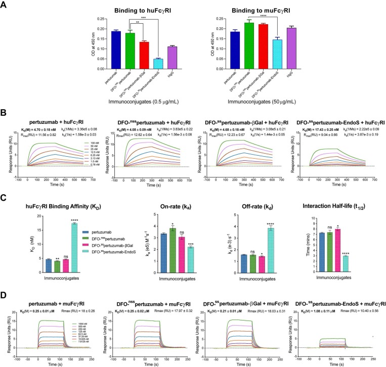
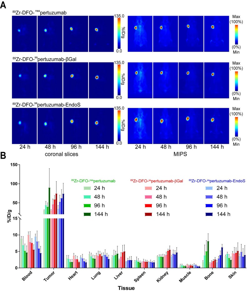
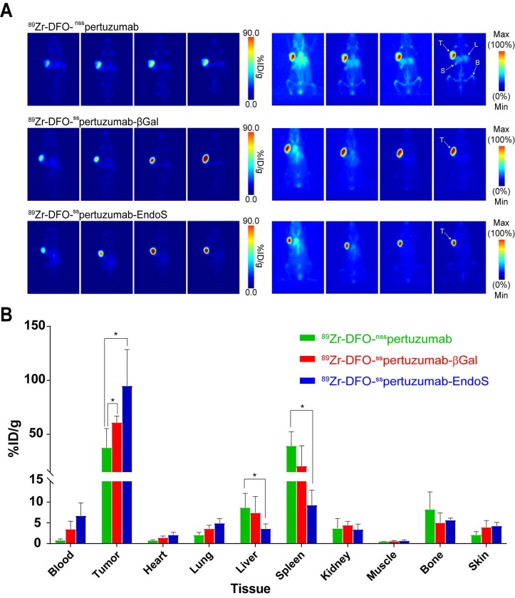
Rationale: The overwhelming majority of radioimmunoconjugates are produced via random conjugation methods predicated on attaching bifunctional chelators to the lysines of antibodies. However, this approach inevitably produces poorly defined and heterogeneous immunoconjugates because antibodies have several lysines distributed throughout their structure. To circumvent this issue, we have previously developed a chemoenzymatic bioconjugation strategy that site-specifically appends cargoes to the biantennary heavy chain glycans attached to CH2 domains of the immunoglobulin's Fc region. In the study at hand, we explore the effects of this approach to site-specific bioconjugation on the Fc receptor binding and in vivo behavior of radioimmunoconjugates. Methods: We synthesized three desferrioxamine (DFO)-labeled immunoconjugates based on the HER2-targeting antibody pertuzumab: one using random bioconjugation methods (DFO-nsspertuzumab) and two using variants of our chemoenzymatic protocol (DFO-sspertuzumab-EndoS and DFO-sspertuzumab-βGal). Subsequently, we characterized these constructs and evaluated their ability to bind HER2, human FcγRI (huFcγRI), and mouse FcγRI (muFcγRI). After radiolabeling the immunoconjugates with zirconium-89, we conducted PET imaging and biodistribution studies in two different mouse models of HER2-expressing breast cancer. Results: MALDI-ToF and SDS-PAGE analysis confirmed the site-specific nature of the bioconjugation, and flow cytometry and surface plasmon resonance (SPR) revealed that all three immunoconjugates bind HER2 as effectively as native pertuzumab. Critically, however, SPR experiments also illuminated that DFO-sspertuzumab-EndoS possesses an attenuated binding affinity for huFcγRI (17.4 ± 0.3 nM) compared to native pertuzumab (4.7 ± 0.2 nM), DFO-nsspertuzumab (4.1 ± 0.1 nM), and DFO-sspertuzumab-βGal (4.7 ± 0.2 nM). ImmunoPET and biodistribution experiments in athymic nude mice bearing HER2-expressing BT474 human breast cancer xenografts yielded no significant differences in the in vivo behavior of the radioimmunoconjugates. Yet experiments in tumor-bearing humanized NSG mice revealed that 89Zr-DFO-sspertuzumab-EndoS produces higher activity concentrations in the tumor (111.8 ± 39.9 %ID/g) and lower activity concentrations in the liver and spleen (4.7 ± 0.8 %ID/g and 13.1 ± 4.0 %ID/g, respectively) than its non-site-specifically labeled cousin, a phenomenon we believe stems from the altered binding of the former to huFcγRI. Conclusion: These data underscore that this approach to site-specific bioconjugation not only produces more homogeneous and well-defined radioimmunoconjugates than traditional methods but may also improve their in vivo performance in mouse models by reducing binding to FcγRI.
Keywords: 89Zr; HER2; glycans; immunoPET; pertuzumab; radioimmunoconjugate; site-specific.
© The author(s).
Conflict of interest statement
Competing Interests: The authors have declared that no competing interest exists.
Figures

Similar articles
-
Lysine-Directed Site-Selective Bioconjugation for the Creation of Radioimmunoconjugates.Bioconjug Chem. 2022 Sep 21;33(9):1750-1760. doi: 10.1021/acs.bioconjchem.2c00354. Epub 2022 Aug 10. Bioconjug Chem. 2022. PMID: 35946495 Free PMC article.
-
Site-Specific Photoaffinity Bioconjugation for the Creation of 89Zr-Labeled Radioimmunoconjugates.Mol Imaging Biol. 2023 Dec;25(6):1104-1114. doi: 10.1007/s11307-023-01818-5. Epub 2023 Apr 13. Mol Imaging Biol. 2023. PMID: 37052759 Free PMC article.
-
The Impact of FcγRI Binding on Immuno-PET.J Nucl Med. 2019 Aug;60(8):1174-1182. doi: 10.2967/jnumed.118.223636. Epub 2019 Feb 7. J Nucl Med. 2019. PMID: 30733320 Free PMC article.
-
89Zr-Immuno-Positron Emission Tomography in Oncology: State-of-the-Art 89Zr Radiochemistry.Bioconjug Chem. 2017 Sep 20;28(9):2211-2223. doi: 10.1021/acs.bioconjchem.7b00325. Epub 2017 Aug 24. Bioconjug Chem. 2017. PMID: 28767228 Free PMC article. Review.
-
Site-Specifically Labeled Immunoconjugates for Molecular Imaging--Part 2: Peptide Tags and Unnatural Amino Acids.Mol Imaging Biol. 2016 Apr;18(2):153-65. doi: 10.1007/s11307-015-0920-y. Mol Imaging Biol. 2016. PMID: 26754791 Free PMC article. Review.
Cited by
-
Lysine-Directed Site-Selective Bioconjugation for the Creation of Radioimmunoconjugates.Bioconjug Chem. 2022 Sep 21;33(9):1750-1760. doi: 10.1021/acs.bioconjchem.2c00354. Epub 2022 Aug 10. Bioconjug Chem. 2022. PMID: 35946495 Free PMC article.
-
Synthesis and In Vivo Evaluation of a Site-specifically Labeled Radioimmunoconjugate for Dual-Modal (PET/NIRF) Imaging of MT1-MMP in Sarcomas.Bioconjug Chem. 2022 Aug 17;33(8):1564-1573. doi: 10.1021/acs.bioconjchem.2c00306. Epub 2022 Jul 22. Bioconjug Chem. 2022. PMID: 35867034 Free PMC article.
-
Removal of Fc Glycans from [89Zr]Zr-DFO-Anti-CD8 Prevents Peripheral Depletion of CD8+ T Cells.Mol Pharm. 2020 Jun 1;17(6):2099-2108. doi: 10.1021/acs.molpharmaceut.0c00270. Epub 2020 May 12. Mol Pharm. 2020. PMID: 32330387 Free PMC article.
-
Exploring the Interplay Between Radioimmunoconjugates and Fcγ Receptors in Genetically Engineered Mouse Models of Cancer.ACS Pharmacol Transl Sci. 2024 Oct 17;7(11):3452-3461. doi: 10.1021/acsptsci.4c00275. eCollection 2024 Nov 8. ACS Pharmacol Transl Sci. 2024. PMID: 39539260
-
Detection of HER2-Low Lesions Using HER2-Targeted PET Imaging in Patients with Metastatic Breast Cancer: A Paired HER2 PET and Tumor Biopsy Analysis.J Nucl Med. 2025 Jun 2;66(6):873-879. doi: 10.2967/jnumed.124.269227. J Nucl Med. 2025. PMID: 40341092
References
-
- Dijkers EC, Oude Munnink TH, Kosterink JG, Brouwers AH, Jager PL, Jong JR. et al. Biodistribution of 89Zr-trastuzumab and PET imaging of HER2-positive lesions in patients with metastatic breast cancer. Clin. Pharmacol. Ther. 2010;87:586–592. - PubMed
-
- Gaykema SBM, Brouwers AH, Lub-de Hooge MN, Pleijhuis RG, Timmer-Bosscha H, Pot L. et al. 89Zr-Bevacizumab PET imaging in primary breast cancer. J. Nucl. Med. 2013;54:1014–1018. - PubMed
Publication types
MeSH terms
Substances
Grants and funding
LinkOut - more resources
Full Text Sources
Medical
Research Materials
Miscellaneous
