. 2020 Jan 1;10(4):1758-1776.
doi: 10.7150/thno.39013. eCollection 2020.

CBP mediated DOT1L acetylation confers DOT1L stability and promotes cancer metastasis

Affiliations

CBP mediated DOT1L acetylation confers DOT1L stability and promotes cancer metastasis

Chaohua Liu et al. Theranostics. .

Abstract

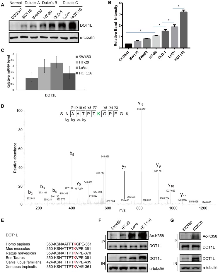
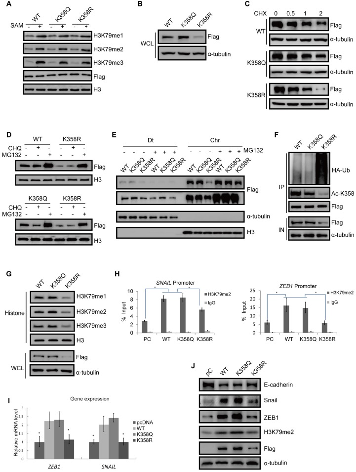
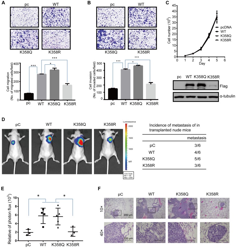
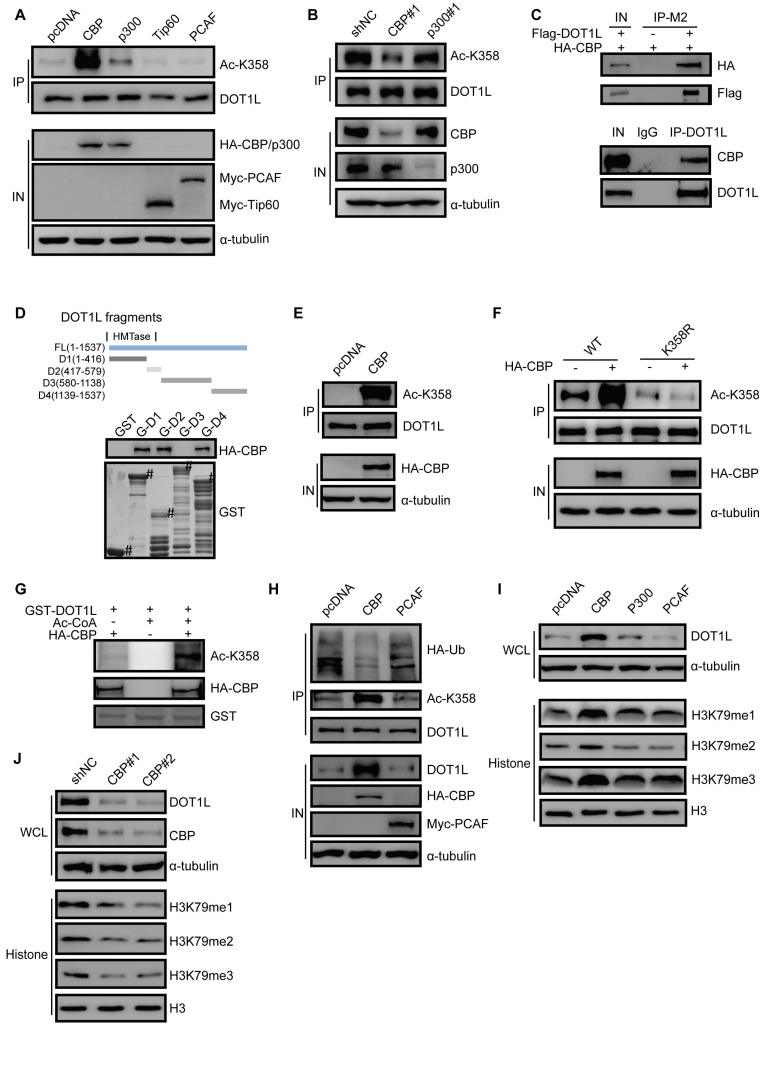
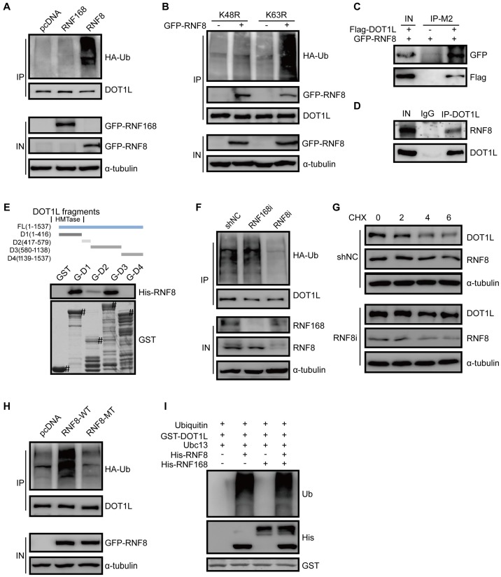
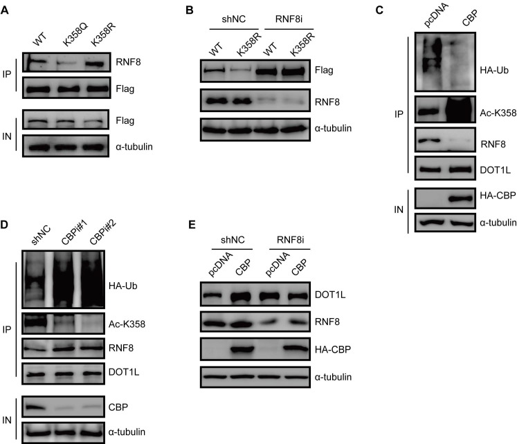
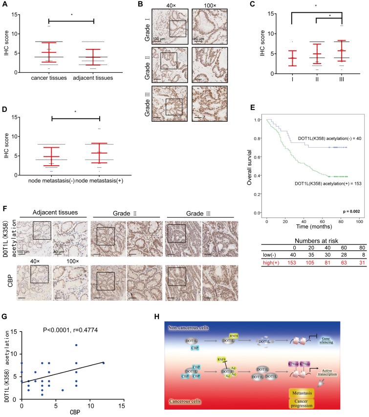
Background and Aim: DOT1L regulates various genes involved in cancer onset and progression by catalyzing H3K79 methylation, but how DOT1L activity itself is regulated is unclear. Here, we aimed to identify specific DOT1L post-translational modifications that might regulate DOT1L activity and thus impact on colorectal cancer (CRC) progression. Methods: We conducted affinity purification and mass spectrometry to explore DOT1L post-translational modifications. We then established transwell migration and invasion assays to specifically investigate the role of DOT1L(K358) acetylation on CRC cellular behavior in vitro and a bioluminescence imaging approach to determine the role of DOT1L(K358) acetylation in CRC metastasis in vivo. We performed chromatin immunoprecipitation to identify DOT1L acetylation-controlled target genes. Finally, we used immunohistochemical staining of human tissue arrays to examine the relevance of DOT1L(K358) acetylation in CRC progression and metastasis and the correlation between DOT1L acetylation and CBP. Results: We found that CBP mediates DOT1L K358 acetylation in human colon cancer cells and positively correlates with CRC stages. Mechanistically, DOT1L acetylation confers DOT1L stability by preventing the binding of RNF8 to DOT1L and subsequent proteasomal degradation, but does not affect its enzyme activity. Once stabilized, DOT1L can catalyze the H3K79 methylation of genes involved in epithelial-mesenchymal transition, including SNAIL and ZEB1. An acetylation mimic DOT1L mutant (Q358) could induce a cancer-like phenotype in vitro, characterized by metastasis and invasion. Finally, DOT1L(K358) acetylation correlated with CRC progression and a poor survival rate as well as with high CBP expression. Conclusions: DOT1L acetylation by CBP drives CRC progression and metastasis. Targeting DOT1L deacetylation signaling is a potential therapeutic strategy for DOT1L-driven cancers.

Keywords: Acetylation; CBP; DOT1L; Degradation; Metastasis.


Conflict of interest statement

Competing Interests: The authors have declared that no competing interest exists.

Figures

Figure 1
Figure 1
DOT1L acetylation at K358 positively correlates with CRC stage. (A) DOT1L protein levels in normal and colon cancer cell lines at different Duke's stages were examined by western blotting. (B) The quantification of the results of (A) is shown, and the relative DOT1L protein levels are presented as means ± SD (n = 3). *p < 0.05. (C) DOT1L mRNA levels in different colon cancer cell lines were extracted with Trizol and were analyzed by real-time PCR. The data are presented as the means ± SD (n = 3). (D) HCT116 cells were transfected with Flag-DOT1L and Flag-immunoprecipitates were separated by SDS-PAGE and stained with CBB. The Flag-DOT1L band was analyzed by mass spectrometry (MS). (E) Alignment of MS-characterized putative DOT1L K358 acetylation residues among different species. (F) DOT1L(K358) acetylation levels in different colon cancer cell lines were analyzed by western blotting. (G) DOT1L(K358) acetylation levels in human primary (SW480) and metastatic (SW620) colon cancer cell lines were analyzed by western blotting.
Figure 2
Figure 2
DOT1L acetylation confers DOT1L stability to regulate EMT transcription factor expression. (A) HCT116 cells were transfected with Flag-DOT1L(WT), Flag-DOT1L(K358Q) or Flag-DOT1L(K358R), and the Flag immunoprecipitates were incubated with histones extracted from HCT116 cells separately. Western blotting was performed to detect H3K79me1/2/3 levels. (B) Whole cell lysate (WCL) were extracted from HCT116 cells transfected with Flag-DOT1L(WT), Flag-DOT1L(K358Q) or Flag-DOT1L(K358R), and then analyzed by western blotting with the indicated antibodies. (C) HCT116 cells were transfected with Flag-DOT1L(WT), Flag-DOT1L(K358Q) or Flag-DOT1L(K358R) for 36 h, and then incubated with 20 μg/ml cycloheximide (CHX) for the indicated times and analyzed by western blotting. (D) HCT116 cells were transfected with Flag-DOT1L(WT), Flag-DOT1L(K358Q), or Flag-DOT1L(K358R) for 24 h, and subsequently treated with PBS, MG132 (10 μM) or chloroquine (CHQ) (50 μM) for 24 h. WCL were extracted and analyzed by western blotting with the indicated antibodies. (E) HCT116 cells were transfected with Flag-DOT1L(WT), Flag-DOT1L(K358Q) or Flag-DOT1L(K358R) for 24 h, and subsequently treated with PBS or MG132 (10 μM) for 24 h, soluble nucleoplasm proteins (Dt), and chromatin proteins (Chr) were extracted and analyzed by western blotting with the indicated antibodies. (F) DOT1L(K358Q), DOT1L(K358R) or DOT1L(WT) were transfected in HCT116 cells. The lysates were extracted, and DOT1L ubiquitination was detected by co-IP and western blotting with the indicated antibodies. (G) WCL and histones were extracted from HCT116 cells transfected with Flag-DOT1L(WT), Flag-DOT1L(K358Q) or Flag-DOT1L(K358R), and H3K79 methylation was detected by western blotting with the indicated antibodies. (H) ChIP-qPCR showing the level of the indicated proteins recruited to the SNAIL (left) and ZEB1 (right) promoter regions. The data represent the means ± SD (n=3). *p < 0.05. (I) SNAIL and ZEB1 mRNA levels in pcDNA-, DOT1L(WT)-, DOT1L(K358Q)- or DOT1L(K358R)-transfected HCT116 cells were analyzed by RT-qPCR. The data represent the means ± SD (n = 3). *p < 0.05. (J) EMT marker expression was measured by western blotting in HCT116 cells transfected with pcDNA, DOT1L(WT), DOT1L(K358Q) or DOT1L(K358R).
Figure 3
Figure 3
DOT1L acetylation regulates CRC migration, invasion and metastasis in vivo. (A, B) Transwell cell migration (A) and matrigel cell invasion (B) assays in HCT116 cells transfected with either pcDNA, DOT1L(WT), DOT1L(K358Q) or DOT1L(K358R) plasmids (upper). The data represent the means ± SEM (n = 3) (lower). *p < 0.05, ***p < 0.001. (C) Comparative growth assay for pcDNA, DOT1L(WT), DOT1L(K358Q) and DOT1L(K358R) overexpressing HCT116 cells. The cell number of each sample was counted at the indicated times. (D, E) HCT116 cells stably expressing pHBLV-luci control (pC), pHBLV-luci-DOT1L(WT) pHBLV-luci-DOT1L(K358Q) or pHBLV-luci-DOT1L(K358R) plasmids were injected intravenously via the tail vein into 6-week-old male nude mice (n = 6 mice per group). Lung metastasis was monitored by bioluminescent imaging after 7 weeks of injection. Representative in vivo bioluminescent images and the incidence of lung metastasis from the different groups are shown (D); the bioluminescent quantitation of lung metastases is given (E). *p < 0.05 compared with the DOT1L(WT) and DOT1L(K358Q). The data represent the means ± SD. (F) Representative lung metastasis specimens were sectioned and stained with H&E. Scale bars: 200 μm.
Figure 4
Figure 4
CBP mediates DOT1L acetylation in vivo and in vitro and confers DOT1L stability. (A) DOT1L(K358) acetyltransferase screen by over-expressing a series of acetyltransferases in HCT116 cells followed by western blotting. (B) HCT116 cells were transfected with CBP or p300 siRNAs prior to detecting DOT1L acetylation levels by immunoprecipitation (IP). (C) Endogenous and an exogenous co-IP to detect the interaction between DOT1L and CBP in HCT116 cells. (D) Purified GST or GST-DOT1L fragments were incubated with HA-CBP prior to western blotting to detect the HA-CBP and GST-DOT1L fragment interactions. (E) In vivo acetylation assay. HCT116 cells were transfected with pcDNA or HA-CBP, and the DOT1L immunoprecipitates were analyzed by western blotting with the indicated antibodies. (F) HCT116 cells were co-transfected with pcDNA or HA-CBP and DOT1L(WT) or DOT1L(K358R), then DOT1L acetylation levels were detected by western blotting. (G) HCT116 cells were transfected with control pcDNA or HA-CBP, and the HA-CBP immunoprecipitates were incubated with GST-DOT1L. DOT1L acetylation levels were analyzed by western blotting with the indicated antibodies. (H) HCT116 cells co-transfected with control pcDNA, HA-CBP or Myc-PCAF, and DOT1L immunoprecipitates were analyzed by western blotting with the indicated antibodies. (I) Whole cell lysate (WCL) and histones were extracted from HCT116 cells transfected with pcDNA, HA-CBP, HA-p300 or Myc-PCAF, and then analyzed by western blotting with the indicated antibodies. (J) Whole cell lysate (WCL) and histones were extracted from HCT116 cells transfected with non-specific siRNA negative control shRNA (shNC), CBP#1 or CBP#2 siRNAs and then analyzed by western blotting with the indicated antibodies.
Figure 5
Figure 5
E3 ligase RNF8 ubiquitinates DOT1L prior to degradation. (A) HCT116 cells were transfected with HA-tagged ubiquitin (Ub) and control pcDNA, GFP-RNF8 or GFP-RNF168 separately, and the cell lysates were subjected to western blotting with the indicated antibodies to detect DOT1L ubiquitination. (B) HCT116 cells were transfected with control pcDNA or GFP-RNF8 together with HA-Ub(K48R) or HA-Ub(K63R), and western blotting was performed with the indicated antibodies to detect DOT1L ubiquitination. (C, D) Nuclear proteins were extracted from HCT116 cells, and endogenous (C) and exogenous (D) co-IPs were performed to detect the interaction between DOT1L and RNF8. (E) Purified GST or GST-DOT1L fragments were incubated with His-RNF8. Western blotting was performed to detect His-RNF8 protein levels, and CBB staining was performed to detect GST or GST-tagged proteins (#). (F) HCT116 cells were transfected with a non-specific shRNA negative control (shNC), RNF8 siRNA or RNF168 siRNA for 72 h. DOT1L immunoprecipitates were subjected to western blotting with the indicated antibodies to detect DOT1L ubiquitination. (G) HCT116 cells were transfected with shNC or RNF8 siRNA. The protein lysates were extracted from 0-6 h following CHX treatment and the DOT1L, RNF8 and α-tubulin protein levels were examined by western blotting. (H) HCT116 cells were co-transfected with HA-Ub, pcDNA, GFP-RNF8(WT) or enzymatically defective GFP-RNF8 (RNF8-MT), and the DOT1L immunoprecipitates were subjected to western blotting with the indicated antibodies. (I) Combinations of GST-DOT1L, UBE1 (E1), Ubc13 (E2), His-RNF8 (E3), and His-RNF168 (E3) were incubated at 37°C for 1 h, and detected by western blotting.
Figure 6
Figure 6
DOT1L acetylation protects DOT1L from degradation via preventing the interaction between RNF8 and DOT1L. (A) Co-IPs were performed to detect the interaction between RNF8 and DOT1L(WT), DOT1L(K358Q) or DOT1L(K358R) in HCT116 cells. (B) HCT116 cells were transfected with a non-specific shRNA negative control (shNC) or an RNF8 siRNA for 24 h followed by transfection with Flag-DOT1L(WT) and Flag-DOT1L(K358R) for 48 h. The whole cell lysates were analyzed by western blotting with the indicated antibodies. (C) HCT116 cells were transfected with pcDNA or HA-CBP, and then subjected to immunoprecipitation (IP) and western blotting with the indicated antibodies. (D) HCT116 cells were transfected with non-specific shNC or CBP siRNAs, and the proteins were analyzed by western blotting with the indicated antibodies. (E) HCT116 cells were transfected with shNC or RNF8 siRNA for 24 h, and then transfected with HA-CBP for 48 h. The whole cell lysates were extracted and analyzed by western blotting with the indicated antibodies.
Figure 7
Figure 7
DOT1L acetylation levels positively correlates with CBP expression and is associated with CRC metastasis and progression. (A) DOT1L(K358) acetylation staining on tissue microarrays (TMAs) containing 155 normal and CRC tissues. The relative DOT1L(K358) acetylation levels were compared between normal and CRC tissues (Student's t-test, n =155; the data represent the means ± SD, error bars are shown in red; *P<0.05). (B) IHC staining of TMAs containing 193 colon carcinoma samples (grades I, II, III) for DOT1L(K358) acetylation levels. Representative images of anti-DOT1L(K358) acetylation staining. Scale bars: 100 μm (left); 40 μm (right). (C) DOT1L(K358) acetylation staining scores were determined by evaluating the extent and intensity of immune-positivity and were analyzed by two-tailed unpaired student's t-test; the data represent the means ± SD, error bars are shown in red; *p < 0.05. (D) The relative DOT1L(K358) acetylation levels were compared between colon adenocarcinoma tissues with and without lymph node metastasis. The data were analyzed by Student's t-test, n =193; the data represent the means ± SD, error bars are shown in red; *P<0.05. (E) Kaplan-Meier curve showing the percentage overall survival of patients with colon cancer, stratified by DOT1L(K358) acetylation expression. (F) Samples from adjacent normal tissues and CRC cancers were immuno-stained with antibodies against DOT1L(K358) acetylation or CBP. Representative images are shown. Scale bars: 100 μm (left); 40 μm (right). (G) The CBP scores were plotted against the DOT1L(K358) acetylation scores, and the correlation coefficients were calculated by Pearson correlation analysis. (H) A proposed model of the DOT1L(K358) regulatory pathway in CRC carcinogenesis. Excessive CBP-mediated DOT1L acetylation in cancerous cells protects DOT1L from degradation via RNF8 and induces DOT1L enrichment. This effect enables H3K79 methylation of target genes and transcriptional activation of EMT transcription factors. The overall effect is promotion of tumorigenesis and metastasis.

Similar articles

Cited by

References

    1. Torre LA, Bray F, Siegel RL, Ferlay J, Lortet-Tieulent J, Jemal A. Global cancer statistics, 2012. CA Cancer J Clin. 2015;65:87–108. - PubMed
    1. O'Connell JB, Maggard MA, Ko CY. Colon cancer survival rates with the new american joint committee on cancer sixth edition staging. J Natl Cancer Inst. 2004;96:1420–5. - PubMed
    1. Liefers GJ, Cleton-Jansen AM, van de Velde CJ, Hermans J, van Krieken JH, Cornelisse CJ. et al. Micrometastases and survival in stage ii colorectal cancer. N Engl J Med. 1998;339:223–8. - PubMed
    1. Jorissen RN, Gibbs P, Christie M, Prakash S, Lipton L, Desai J. et al. Metastasis-associated gene expression changes predict poor outcomes in patients with dukes stage b and c colorectal cancer. Clin Cancer Res. 2009;15:7642–51. - PMC - PubMed
    1. Kuo CC, Ling HH, Chiang MC, Chung CH, Lee WY, Chu CY. et al. Metastatic colorectal cancer rewrites metabolic program through a glut3-yap-dependent signaling circuit. Theranostics. 2019;9:2526–40. - PMC - PubMed

Publication types

MeSH terms