. 2020 Jan 17:18:271-286.
doi: 10.1016/j.csbj.2020.01.002. eCollection 2020.

Computational saturation mutagenesis to predict structural consequences of systematic mutations in the beta subunit of RNA polymerase in Mycobacterium leprae

Affiliations

Computational saturation mutagenesis to predict structural consequences of systematic mutations in the beta subunit of RNA polymerase in Mycobacterium leprae

Sundeep Chaitanya Vedithi et al. Comput Struct Biotechnol J. .

Abstract

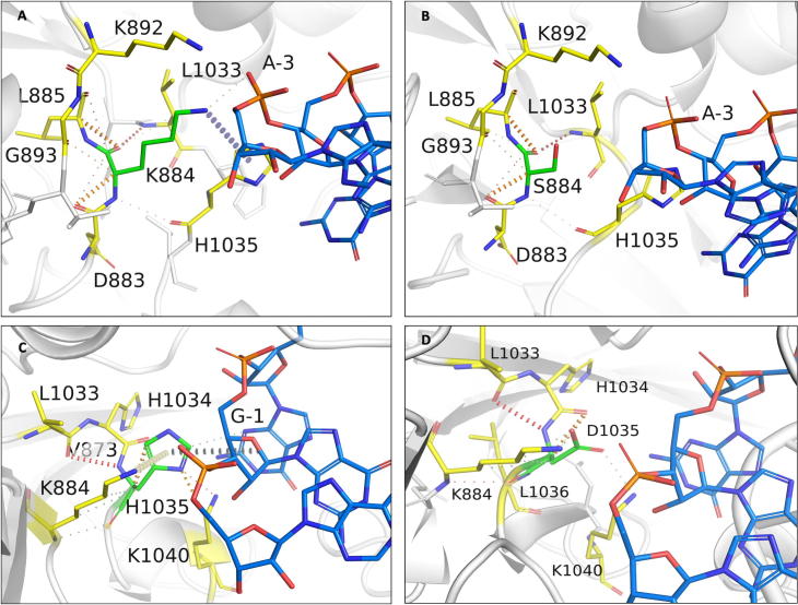
Rifampin resistance in leprosy may remain undetected due to the lack of rapid and effective diagnostic tools. A quick and reliable method is essential to determine the impacts of emerging detrimental mutations in the drug targets. The functional consequences of missense mutations in the β-subunit of RNA polymerase (RNAP) in Mycobacterium leprae (M. leprae) contribute to phenotypic resistance to rifampin in leprosy. Here, we report in-silico saturation mutagenesis of all residues in the β-subunit of RNAP to all other 19 amino acid types (generating 21,394 mutations for 1126 residues) and predict their impacts on overall thermodynamic stability, on interactions at subunit interfaces, and on β-subunit-RNA and rifampin affinities (only for the rifampin binding site) using state-of-the-art structure, sequence and normal mode analysis-based methods. Mutations in the conserved residues that line the active-site cleft show largely destabilizing effects, resulting in increased relative solvent accessibility and a concomitant decrease in residue-depth (the extent to which a residue is buried in the protein structure space) of the mutant residues. The mutations at residue positions S437, G459, H451, P489, K884 and H1035 are identified as extremely detrimental as they induce highly destabilizing effects on the overall protein stability, and nucleic acid and rifampin affinities. Destabilizing effects were predicted for all the clinically/experimentally identified rifampin-resistant mutations in M. leprae indicating that this model can be used as a surveillance tool to monitor emerging detrimental mutations that destabilise RNAP-rifampin interactions and confer rifampin resistance in leprosy.

Author summary: The emergence of primary and secondary drug resistance to rifampin in leprosy is a growing concern and poses a threat to the leprosy control and elimination measures globally. In the absence of an effective in-vitro system to detect and monitor phenotypic resistance to rifampin in leprosy, diagnosis mainly relies on the presence of mutations in drug resistance determining regions of the rpoB gene that encodes the β-subunit of RNAP in M. leprae. Few labs in the world perform mouse food pad propagation of M. leprae in the presence of drugs (rifampin) to determine growth patterns and confirm resistance, however the duration of these methods lasts from 8 to 12 months making them impractical for diagnosis. Understanding molecular mechanisms of drug resistance is vital to associating mutations to clinically detected drug resistance in leprosy. Here we propose an in-silico saturation mutagenesis approach to comprehensively elucidate the structural implications of any mutations that exist or that can arise in the β-subunit of RNAP in M. leprae. Most of the predicted mutations may not occur in M. leprae due to fitness costs but the information thus generated by this approach help decipher the impacts of mutations across the structure and conversely enable identification of stable regions in the protein that are least impacted by mutations (mutation coolspots) which can be a potential choice for small molecule binding and structure guided drug discovery.

Keywords: In-silico Saturation Mutagenesis; Mutation Coolspots; Mycobacterium leprae; RNA Polymerase; Rifampin; Thermodynamic stability.


Conflict of interest statement

The authors declare that they have no known competing financial interests or personal relationships that could have appeared to influence the work reported in this paper.

Figures

None
Graphical abstract
Fig. 1
Fig. 1
[A] Methodology and study design. [B] A lollipop plot with stability predictions for mutations reported in the literature and are known to confer rifampin resistance in Leprosy.
Fig. 2
Fig. 2
[A] The β-subunit of RNAP with residues where mutations were reported experimentally from patient samples in various studies (Supplementary Table 2) (highlighted in red). [B] Each residue in the β-subunit of RNAP that is colored by the conservations scores determined by CONSURF. The residues in green are variable (conservations scores greater than 1) and are usually surface exposed. The residues in red are conserved with conservation scores less than 1 and usually form the core of the protein. The rifampin binding site is highly conserved in M. leprae. [C] The maximum destabilizing effect (predicted by mCSM) on the protein stability for any mutation at each residue position, is mapped on the structure. Red are the regions that are largely destabilized by mutations while the white regions are relatively stable with mutations. [D] The converse of B where the regions, whose stability is least impacted by mutations, are coloured in blue and we called them “Mutation CoolSpots”. (For interpretation of the references to colour in this figure legend, the reader is referred to the web version of this article.)
Fig. 3
Fig. 3
[A] The interfacial region of the β-subunit of RNAP highlighted in Maroon. [B]. The maximum destabilizing effect a mutation can induce on the interface stability, is predicted by mCSM-PPI and mapped on the structure. Red indicates regions that are highly destabilized by mutations (-5.108 Kcal/mol) while the blue indicates stable regions. (For interpretation of the references to colour in this figure legend, the reader is referred to the web version of this article.)
Fig. 4
Fig. 4
[A] Change in relative solvent accessibility for maximum destabilizing mutants in the rifampin binding pocket (mCSM). [B]. Change in depth of the highly destabilizing mutant residue in the rifampin binding pocket (mCSM).
Fig. 5
Fig. 5
[A] The change in relative side chain solvent accessibility with mutations was mapped on to the structure. Blue indicates a decrease in RSA while red indicates an increase. [B] The changes in depth with highly destabilizing mutations at each residue position was mapped on the structure. (For interpretation of the references to colour in this figure legend, the reader is referred to the web version of this article.)
Fig. 6
Fig. 6
[A] Stability changes in β-subunit -RNA and β-subunit- rifampin [B] interactions due to mutations in the binding sites as predicted by mCSM-NA2 and mCSM-lig. The maximum destabilizing effect a mutation can cause at each residue position in the binding site is depicted on the structure.
Fig. 7
Fig. 7
[A] Interactions of S437 with the surrounding residue environment in the wildtype and of H437 in the S437H mutant [B]. [C] Interactions of G459 with the surrounding residue environment and [D] W459 in the mutant G459W. The red dotted lines represent hydrogen bonds. Orange dotted lines represent weak hydrogen bond interactions. Ring-Ring and intergroup interactions are depicted in cyan. Aromatic interactions are represented in sky-blue and carbonyl interactions in pink dotted lines. Green dotted lines represent hydrophobic interactions. (For interpretation of the references to colour in this figure legend, the reader is referred to the web version of this article.)
Fig. 8
Fig. 8
[A] Interactions of P489 with the surrounding residue environment in the wildtype and of G489 in the P489G mutant [B]. [C] Interactions of H451 with the surrounding residue environment and [D] S451 in the mutant H451S.
Fig. 9
Fig. 9
[A] Interactions of K884 with the surrounding residue environment in the wildtype and of S884 in the K884S mutant [B]. [C] Interactions of H1035 with the surrounding residue environment and [D] D1035 in the mutant H1035D. The blue dotted lines represent cation-π interaction. (For interpretation of the references to colour in this figure legend, the reader is referred to the web version of this article.)
Fig. 10
Fig. 10
[A] The maximum destabilizing effects on the protein stability, a mutation can induce at each residue position in the flexible conformations (as predicted by ENCoM [A] and DynaMut [B]), are mapped on the structure. Regions in red represent highly destabilizing while the blue regions are relatively stable with mutations. (For interpretation of the references to colour in this figure legend, the reader is referred to the web version of this article.)
Fig. 11
Fig. 11
Fragment hotspots were mapped on the structure which was coloured with maximum destabilizing effects of systematic mutations at each residue positions. Blue represents regions which are least impacted by any mutations. Stable and potential small molecule binding sites “A” and “B” are depicted on the structure.
Supplementary figure 1
Pairplot depicting correlations between mCSM, SDM and other structural predictors of protein stability changes upon mutations in the b-subunit of RNAP. Each datapoint corresponds to maximum destabilizing effect noted at each residue position in the b subunit when systematically mutated to other 19 residues. The data points in orange correspond to predictions at rifampin interacting residues.
Supplementary figure 2
Pairplot depicting correlations between mCSM, SDM and other sequence-based predictors of protein stability changes upon mutations in the b-subunit of RNAP. Each data point corresponds to maximum destabilizing effect noted at each residue position in the b subunit when systematically mutated to other 19 residues. The data points in orange correspond to predictions at rifampin interacting residues.
Supplementary figure 3
Pairplot depicting correlations between mCSM, SDM and other NMA-based predictors of protein stability changes upon mutations in the beta-subunit of RNAP. Each data point corresponds to average destabilizing effect noted at each residue position in the beta-subunit when systematically mutated to other 19 residues.
Supplementary figure 4
Heatmap showing variation in the RMSD among 100 models generated to validate the computational modelling of the beta-subunit of RNAP.
Supplementary figure 5
Heatmap showing variation in the TM-Align score among 100 models generated to validate the computational modelling of the beta-subunit of RNAP.
Supplementary figure 6
Heatmaps showing RMSD and TM-Align scores between M. leprae b-subunit of RNAP models and <span data-format=M. tb beta-subunit of RNAP structure (PDB Id: 5UHC).

Similar articles

Cited by

References

    1. World Health Organization. Global tuberculosis report 2018 [Internet]. World Health Organization; 2018 [cited 2018 Dec 19]. 231 p. Available from: http://apps.who.int/iris/handle/10665/274453
    1. Han X.Y., Sizer K.C., Thompson E.J., Kabanja J., Li J., Hu P. Comparative sequence analysis of Mycobacterium leprae and the new leprosy-causing Mycobacterium lepromatosis. J Bacteriol. 2009;191(19):6067–6074. - PMC - PubMed
    1. WHO | Weekly Epidemiological Record, 31 August 2018, vol. 93, 35 (pp. 444–456) [Internet]. WHO. [cited 2018 Dec 19]. Available from: http://www.who.int/wer/2018/wer9335/en/.
    1. Vedithi S.C., Malhotra S., Das M., Daniel S., Kishore N., George A. Structural implications of mutations conferring rifampin resistance in Mycobacterium leprae. Sci Rep. 2018;8(1):5016. - PMC - PubMed
    1. Nakata N., Kai M., Makino M. Mutation analysis of mycobacterial rpoB genes and rifampin resistance using recombinant Mycobacterium smegmatis. Antimicrob Agents Chemother. 2012;56(4):2008–2013. - PMC - PubMed

LinkOut - more resources