. 2020 Mar;12(3):99-108.
doi: 10.3892/br.2019.1269. Epub 2019 Dec 30.

Sulfur-containing amino acids in aged garlic extract inhibit inflammation in human gingival epithelial cells by suppressing intercellular adhesion molecule-1 expression and IL-6 secretion

Affiliations

Sulfur-containing amino acids in aged garlic extract inhibit inflammation in human gingival epithelial cells by suppressing intercellular adhesion molecule-1 expression and IL-6 secretion

Masahiro Ohtani et al. Biomed Rep. 2020 Mar.

Abstract

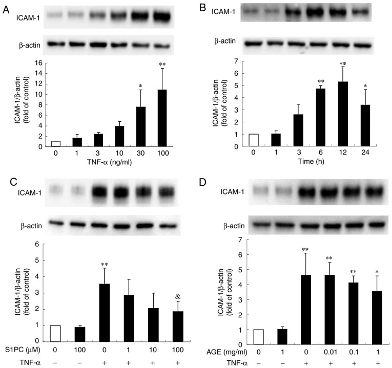
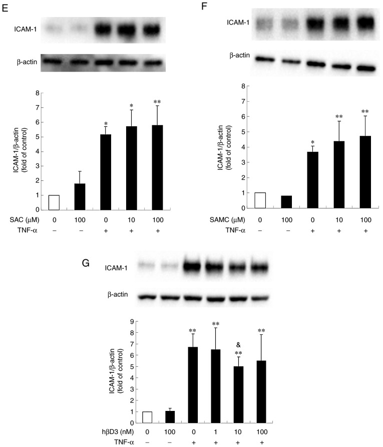
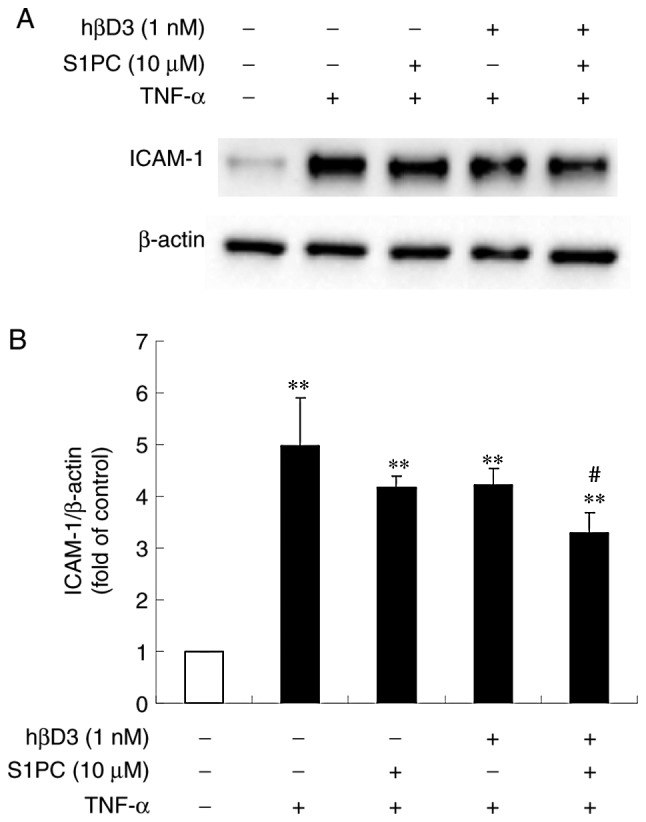
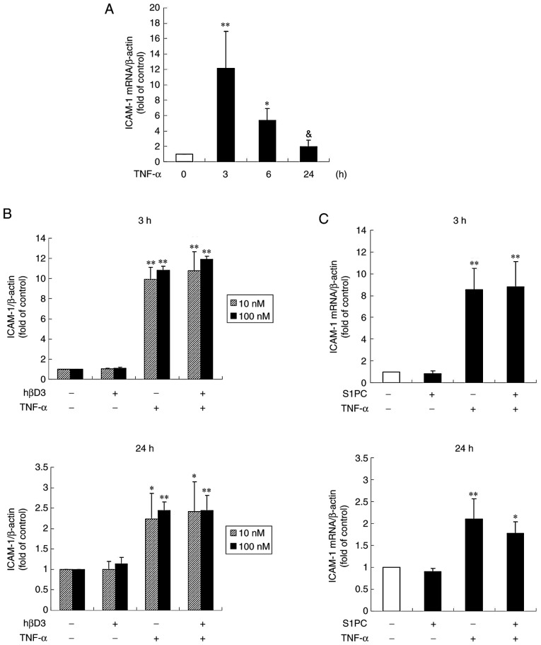
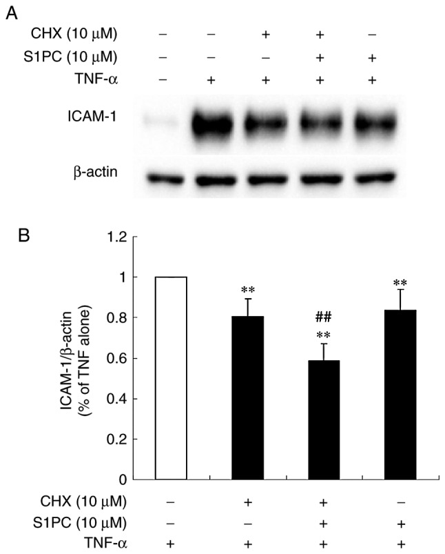
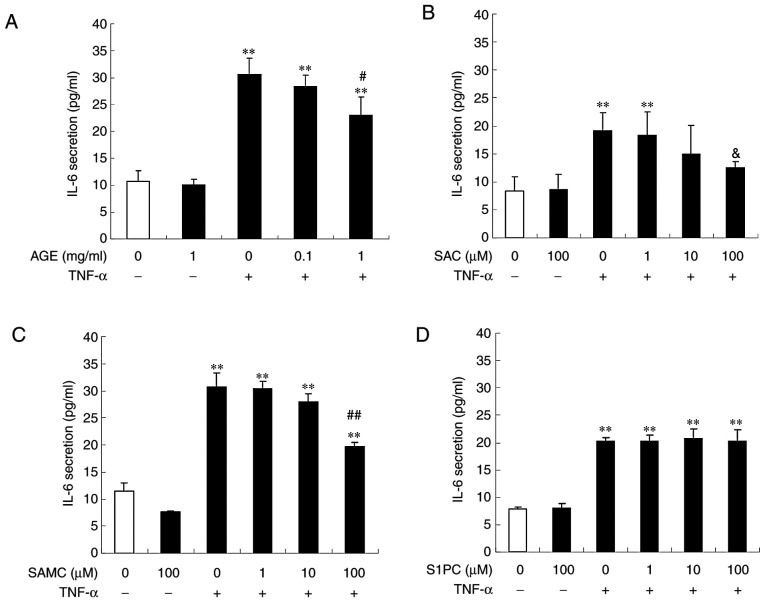
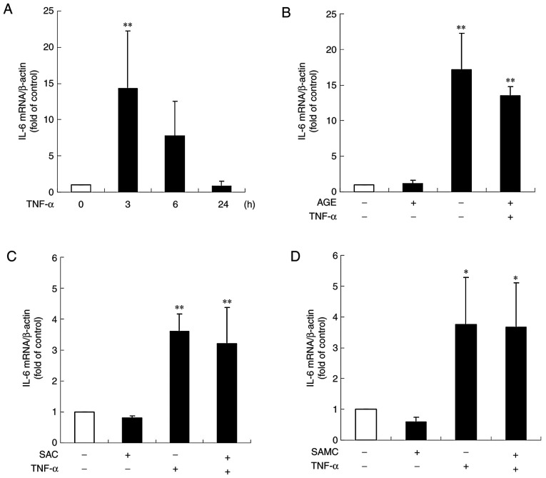
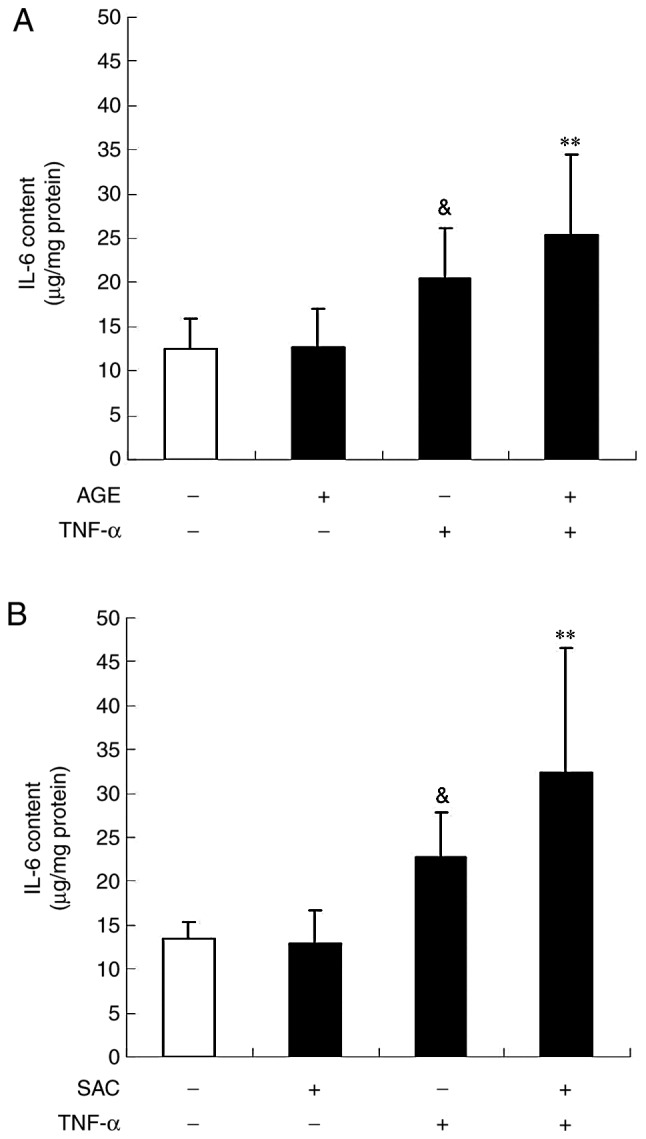
Aged garlic extract (AGE) contains various biologically active sulfur-containing amino acids, such as S-allylcysteine (SAC), S-1-propenylcysteine (S1PC) and S-allylmercaptocysteine (SAMC). These amino acids have been demonstrated to lower hypertension, improve atherosclerosis and enhance immunity through their anti-inflammatory and antioxidant activities. It was recently reported that the administration of AGE alleviated gingivitis in a clinical trial. In this study, to gain insight into this effect of AGE, the authors examined whether AGE and the three above-mentioned sulfur compounds influence the effects of tumor necrosis factor-α (TNF-α) in inducing intercellular adhesion molecule-1 (ICAM-1) expression and interleukin-6 (IL-6) secretion in Ca9-22 human gingival epithelial cells. It was found that S1PC reduced the level of ICAM-1 protein induced by TNF-α possibly through post-translational levels without affecting the TNF-α-induced mRNA expression. However, SAC and SAMC had no effect. It was also confirmed the inhibitory effect of an antimicrobial peptide [human-β defensin-3 (hβD3)] and found that the inhibitory effects of hbD3 and S1PC were synergistic. On the other hand, the TNF-α-induced IL-6 secretion was attenuated by SAC and SAMC in a dose-dependent manner, whereas S1PC was ineffective. In addition, SAC and SAMC, but not S1PC inhibited the phosphorylation of the transcription factor nuclear factor κ-light-chain-enhancer of activated B cells (NF-κB), which is involved in the expression of inflammatory molecules, suggesting that the anti-inflammatory effects of SAC and SAMC are mediated, at least partly, by NF-κB. On the whole, the findings of this study suggest that the three sulfur amino acids in AGE function synergistically in alleviating inflammation in human gingival epithelial cells.

Keywords: ICAM-1; IL-6; aged garlic extract; gingival epithelial cells; inflammation; sulfur-containing amino acid.


Figures

Figure 1.
Figure 1.
Effect of S1PC and hβD3 on the level of ICAM-1 protein induced by TNF-α. (A) The concentration (1 to 100 ng/ml, 12 h)- and (B) time (1 to 24 h, 100 ng/ml)-dependent induction of ICAM-1 protein stimulated by TNF-α. The cells were left untreated (control) or treated with TNF-α in the absence or presence of (C) S1PC (1 to 100 µM), (D) AGE (0.01 to 1 mg/ml), (E) SAC (10, 100 µM), (F) SAMC (10, 100 µM) or (G) hβD3 (1 to 100 nM) for 24 h and total protein was extracted. ICAM-1 proteins in the extract were detected by western blot analysis. Each bar in the graphs represents the mean ± SD of the band intensity relative to β-actin (n=3). *P<0.05, **P<0.01 in comparison to the control (Tukey's multiple comparison test). &P<0.05 in comparison to TNF-α alone (Student's t-test). S1PC, S-1-propenylcysteine; hβD3, human β-defensin-3; TNF-α, tumor necrosis factor-α; ICAM-1, intercellular adhesion molecule-1; AGE, aged garlic extract; SAC, S-allylcysteine; SAMC, S-allylmercaptocysteine.
Figure 2.
Figure 2.
Effect of S1PC on the level of ICAM-1 protein induced by TNF-α in the presence of hβD3. (A) The cells were left untreated (control) or treated with TNF-α (100 ng/ml) in the absence or presence of S1PC (10 µM) and/or hβD3 (1 nM) for 24 h and total protein was extracted. ICAM-1 proteins in the extract were detected by western blot analysis. (B) Quantitative analysis of the band intensity relative to β-actin. Each bar in the graph represents the mean ± SD (n=3). **P<0.01 in comparison to the control and #P<0.05 in comparison to TNF-α alone (Tukey's multiple comparisons test). S1PC, S-1-propenylcysteine; hβD3, human β-defensin-3; TNF-α, tumor necrosis factor-α; ICAM-1, intercellular adhesion molecule-1.
Figure 3.
Figure 3.
Effect of hβD3 and S1PC on the ICAM-1 gene expression induced by TNF-α. The cells were untreated (control) or treated with (A) TNF-α (100 ng/ml) alone for 3, 6 or 24 h or in the presence of (B) hβD3 (10, 100 nM) or (C) S1PC (100 µM) for 3 h or 24 h, and total RNA was extracted. The ICAM-1 mRNA expression level in total RNA was evaluated by RT-qPCR. Each bar in graphs represents the mean ± SD (n=3-5). *P<0.05, **P<0.01 (Tukey's multiple comparison test) or &P<0.05 (Student's t-test) in comparison to the control. S1PC, S-1-propenylcysteine; hβD3, human β-defensin-3; TNF-α, tumor necrosis factor-α; ICAM-1, intercellular adhesion molecule-1.
Figure 4.
Figure 4.
Effect of S1PC on the level of ICAM-1 protein induced by TNF-α in the presence of cycloheximide (CHX). (A) The cells were left untreated (control) or pretreated with TNF-α (100 ng/ml) for 12 h in the absence or presence of CHX (10 µM) and/or S1PC (10 µM) for 6 h and total protein was extracted. ICAM-1 proteins in the extract were detected by western blot analysis. (B) The quantitative analysis of the band intensity relative to β-actin. Each bar in the graph represents the mean ± SD (n=3). **P<0.01 in comparison to control and ##P<0.01 in comparison to TNF-α alone (Tukey's multiple comparisons test). S1PC, S-1-propenylcysteine; hβD3, human β-defensin-3; TNF-α, tumor necrosis factor-α; ICAM-1, intercellular adhesion molecule-1.
Figure 5.
Figure 5.
Effect of AGE, SAC and SAMC on IL-6 secretion induced by TNF-α. The cells were left untreated (control) or treated with TNF-α (100 ng/ml) in the absence or presence of (A) AGE, (B) SAC, (C) SAMC or (D) S1PC (each at 1 to 100 µM) for 6 h and culture medium was collected. The IL-6 concentration in the medium was determined by ELISA. Each bar in graphs represents the mean ± SD (n=3-4). **P<0.01 in comparison to the control and #P<0.05, ##P<0.01 (Tukey's multiple comparison test), &P<0.05 in comparison to TNF-α alone (Student's t-test). S1PC, S-1-propenylcysteine; hβD3, human β-defensin-3; TNF-α, tumor necrosis factor-α; IL-6, interleukin-6; AGE, aged garlic extract; SAC, S-allylcysteine; SAMC, S-allylmercaptocysteine.
Figure 6.
Figure 6.
Effect of AGE, SAC and SAMC on the IL-6 gene expression induced by TNF-α. The cells were untreated (control) or treated with (A) TNF-α (100 ng/ml) alone for 3, 6 or 24 h or in the presence of (B) AGE (1 mg/ml) or (B) SAC, (C) SAC, (D) SAMC (each at 100 µM) for 3 h and total RNA was extracted. The IL-6 mRNA expression level in total RNA was evaluated by RT-qPCR. Each bar in graphs represents the mean ± SD (n=3-6). *P<0.05, **P<0.01 (Tukey's multiple comparison test) in comparison to the control. S1PC, S-1-propenylcysteine; hβD3, human β-defensin-3; TNF-α, tumor necrosis factor-α; IL-6, interleukin-6; AGE, aged garlic extract; SAC, S-allylcysteine; SAMC, S-allylmercaptocysteine.
Figure 7.
Figure 7.
Effect of AGE and SAC on the intracellular IL-6 content induced by TNF-α. The cells were left untreated (control) or treated with TNF-α (100 ng/ml) in the absence or presence of (A) AGE or (B) SAC (100 µM) for 3 h and total protein was extracted. The IL-6 content in the protein extract was determined by ELISA. Each bar in the graphs represents the mean ± SD (n=6). **P<0.01 (Tukey's multiple comparison test), &P<0.05 (Student's t-test) in comparison to the control. TNF-α, tumor necrosis factor-α; IL-6, interleukin-6; AGE, aged garlic extract; SAC, S-allylcysteine.
Figure 8.
Figure 8.
Effect of hβD3, S1PC, AGE, SAC and SAMC on the level of phosphorylated NF-κB p65 protein induced by TNF-α. The cells were left untreated (control) or treated with TNF-α (100 ng/ml) in the absence or presence of (A) hβD3 (10, 100 nM) or (B) S1PC (100 µM) for 24 h, (C) AGE (1 mg/ml), (D) SAC or (E) SAMC (each at 100 µM) for 6 h and total protein was extracted. Total NF-κB (NF-κB) and phosphorylated NF-κB (pNF-κB) p65 proteins in the extract were detected by western blot analysis. Each bar in graphs represents the mean ± SD of the band intensity relative to β-actin (n=3-4). *P<0.05, **P<0.01 in comparison to the control and #P<0.05, ##P<0.01 in comparison to TNF-α alone (Tukey's multiple comparisons test). S1PC, S-1-propenylcysteine; hβD3, human β-defensin-3; TNF-α, tumor necrosis factor-α; IL-6, interleukin-6; AGE, aged garlic extract; SAC, S-allylcysteine; SAMC, S-allylmercaptocysteine.

Similar articles

Cited by

References

    1. Zini A, Mann J, Mazor S, Vered Y. The efficacy of aged garlic extract on gingivitis-A randomized clinical trial. J Clin Dentist. 2018;29:52–56. - PubMed
    1. Sojod B, Chateau D, Mueller CG, Babajko S, Berdal A, Lézot F, Castaneda B. RANK/RANKL/OPG signalization implication in periodontitis: New evidence from a RANK transgenic mouse model. Front Physiol. 2017;8(338) doi: 10.3389/fphys.2017.00338. - DOI - PMC - PubMed
    1. Tzach-Nahman R, Nashef R, Fleisigg O, Palmon A, Shapira L, Wilensky A, Nussbaum G. Oral fibroblasts modulate the macrophage response to bacterial challenge. Sci Rep. 2017;7(11516) doi: 10.1038/s41598-017-11771-3. - DOI - PMC - PubMed
    1. Wu YH, Kuraji R, Taya Y, Ito H, Numabe Y. Effects of theaflavins on tissue inflammation and bone resorption on experimental periodontitis in rats. J Periodontal Res. 2018;53:1009–1019. doi: 10.1111/jre.12600. - DOI - PMC - PubMed
    1. Morihara N, Hino A, Yamaguchi T, Suzuki JI. Aged garlic extract suppresses the development of atherosclerosis in apolipoprotein E-knockout mice. J Nutr. 2016;146:460S–463S. doi: 10.3945/jn.114.206953. - DOI - PubMed