

Primary Umbilical Endometriosis in an Adolescent Girl: Unsuspected Pathology
- PMID: 32042547
- PMCID: PMC7007811
- DOI: 10.1055/s-0039-1700987
Primary Umbilical Endometriosis in an Adolescent Girl: Unsuspected Pathology
Abstract
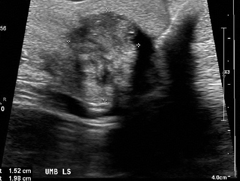
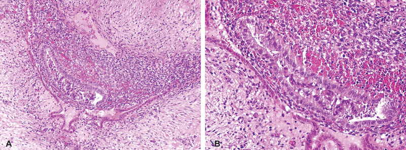
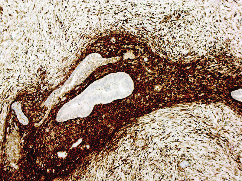
Endometriosis affects 7 to 10% of women of reproductive age. Primary umbilical endometriosis (PUE) is even rarer with unclear pathogenesis. We report a case of PUE possibly the youngest patient reported in the literature. A 16-year-old girl of African origin presented with painful umbilical lump for 2 to 3 months duration with background history of precocious puberty, cyclical vomiting, and menorrhagia. Clinical examination showed dark-colored, tender, irreducible umbilical lump. A provisional diagnosis of incarcerated umbilical hernia was made. Abdominal X-ray showed no features of intestinal obstruction. Ultrasound scan of the abdomen showed lump containing heterogeneous echogenic material measuring 2.0 × 1.5cm within the umbilicus with no visible bowel loops or peristalsis. This was reported as consistent with an umbilical hernia with narrow neck possibly containing mesentery or intra-abdominal fat. The patient underwent urgent exploration of umbilicus under general anesthetic. At operation, a dark-colored, firm mass was excised and sent for histology. The underlying fascia and peritoneum were repaired. Histological examination confirmed the excised tissue was endometriosis. Follow-up continues in the endometriosis clinic. Umbilical endometriosis should be considered in differential diagnoses of painful umbilical lesion in adolescent girls and women of reproductive age. Complete excision and histology are highly recommended for obtaining a definitive diagnosis, to exclude malignancy and to prevent recurrence.
Keywords: adolescent; endometriosis; primary; umbilical.
Conflict of interest statement
Conflict of Interest None.
Figures
Similar articles
-
Spontaneous umbilical endometriosis: Rare occurrence in nulliparous women - Case report & literature review.Int J Surg Case Rep. 2024 Sep;122:110155. doi: 10.1016/j.ijscr.2024.110155. Epub 2024 Aug 13. Int J Surg Case Rep. 2024. PMID: 39142186 Free PMC article.
-
Umbilical Hernia as Forerunner of Primary Umbilical Endometriosis: A Case Report.Medeni Med J. 2021 Dec 19;36(4):348-351. doi: 10.4274/MMJ.galenos.2021.66990. Medeni Med J. 2021. PMID: 34939402 Free PMC article.
-
Primary umbilical endometriosis: A painful swelling in the umbilicus concomitantly with menstruation.Int J Surg Case Rep. 2016;28:78-80. doi: 10.1016/j.ijscr.2016.09.029. Epub 2016 Sep 23. Int J Surg Case Rep. 2016. PMID: 27689524 Free PMC article.
-
Primary umbilical endometrioma: Analyzing the pathogenesis of endometriosis from an unusual localization.Taiwan J Obstet Gynecol. 2015 Jun;54(3):306-12. doi: 10.1016/j.tjog.2014.03.011. Taiwan J Obstet Gynecol. 2015. PMID: 26166347 Review.
-
Endometriosis of umbilical cicatrix: case report and review of the literature.Acta Dermatovenerol Croat. 2008;16(4):218-21. Acta Dermatovenerol Croat. 2008. PMID: 19111147 Review.
Cited by
-
Clinical Features and Management of Umbilical Endometriosis: A 30 Years' Monocentric Retrospective Study.Int J Environ Res Public Health. 2022 Dec 14;19(24):16754. doi: 10.3390/ijerph192416754. Int J Environ Res Public Health. 2022. PMID: 36554635 Free PMC article.
-
Writing a Case Report in Pediatric Surgery: A Comprehensive Guideline.European J Pediatr Surg Rep. 2022 Feb 10;10(1):e13-e19. doi: 10.1055/s-0041-1740935. eCollection 2022 Jan. European J Pediatr Surg Rep. 2022. PMID: 35155079 Free PMC article.
References
-
- Papavramidis T S, Sapalidis K, Michalopoulos N et al.Spontaneous abdominal wall endometriosis: a case report. Acta Chir Belg. 2009;109(06):778–781. - PubMed
-
- Chatzikokkinou P, Thorfinn J, Angelidis I K, Papa G, Trevisan G. Spontaneous endometriosis in an umbilical skin lesion. Acta Dermatovenerol Alp Panonica Adriat. 2009;18(03):126–130. - PubMed
-
- Bektaş H, Bilsel Y, Sari Y S et al.Abdominal wall endometrioma; a 10-year experience and brief review of the literature. J Surg Res. 2010;164(01):e77–e81. - PubMed
Publication types
LinkOut - more resources
Full Text Sources