. 2019 Sep 26;8(10):e1247-e1253.
doi: 10.1016/j.eats.2019.06.014. eCollection 2019 Oct.

Anterior Capsule Reconstruction of the Native Hip: A Technique Guide

Affiliations

Anterior Capsule Reconstruction of the Native Hip: A Technique Guide

Adrian Z Kurz et al. Arthrosc Tech. .

Abstract

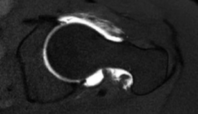
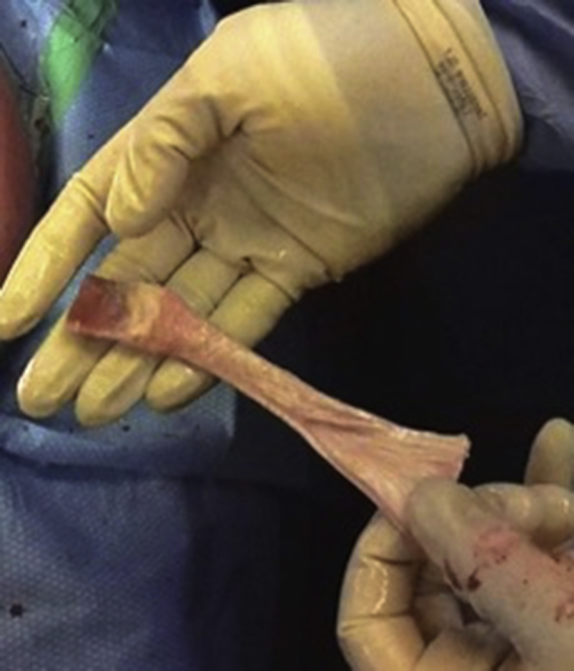
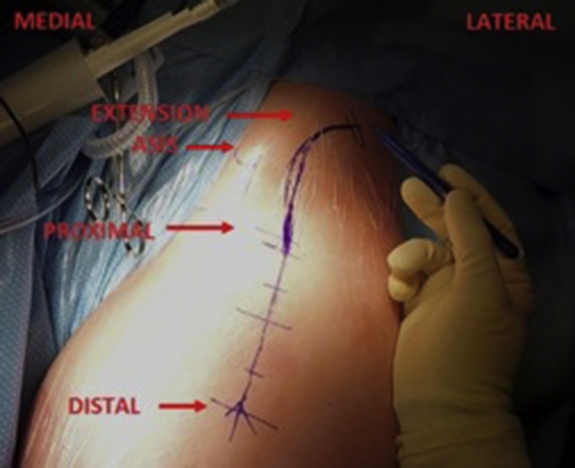
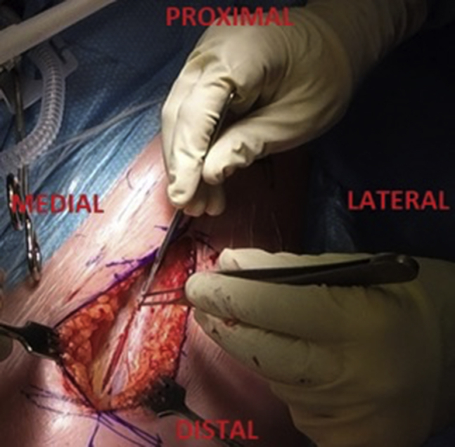
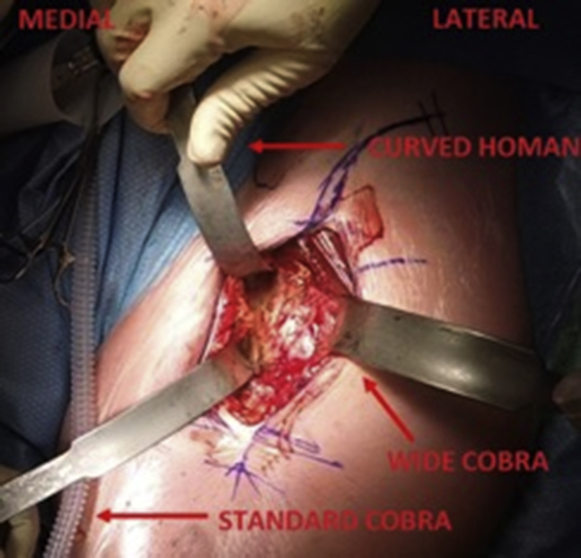
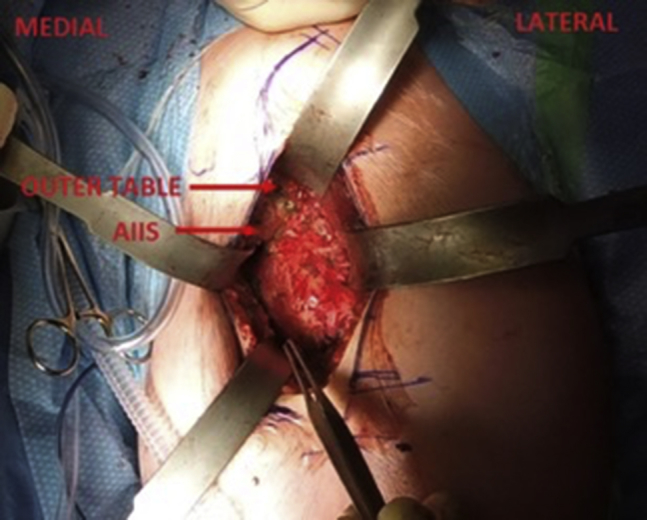
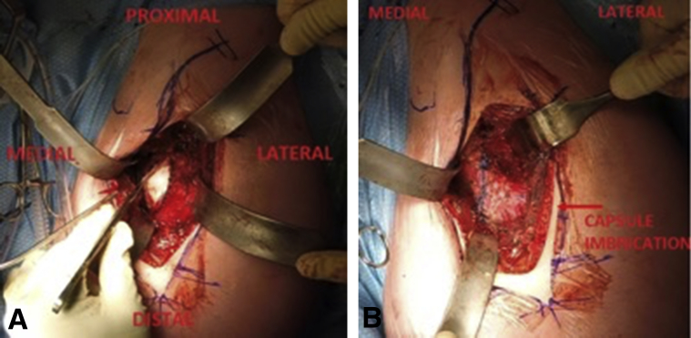
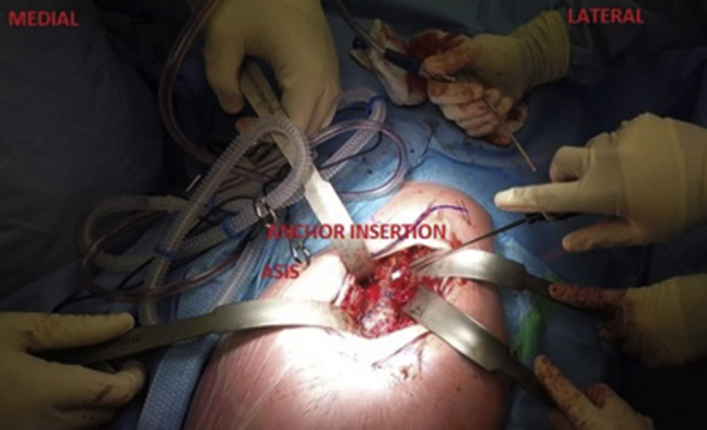
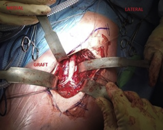
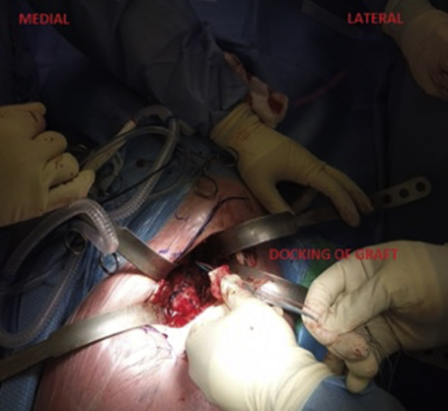
This is a technique guide for open anterior capsule reconstruction in unstable native hips due to Ehlers-Danlos syndrome. Ehlers-Danlos syndrome is a connective tissue disease that predisposes an individual to hypermobility of their soft tissues and recurrent subluxations and dislocations of their joints. This may lead to constant instability, guarding of the hip, and subsequent arthritic changes and poor quality of life. We demonstrate anterior capsule reconstruction of the hip using an Achilles tendon allograft performed through a direct anterior approach to the hip (Heuter's approach). This technique guide outlines in detail the surgical steps required to accomplish this reconstructive procedure. Furthermore, a narrated surgical video is provided that clearly depicts the anticipated result of the increased constrained provided to the hip from the anterior capsule reconstruction using an Achilles tendon allograft.


Figures

Fig 1
Fig 1
Axial-cut magnetic resonance (MR) arthrogram of the left hip demonstrating a patulous capsule and deficient iliofemoral ligament anteriorly. The anterior capsule is heterogeneous in nature and not a homogenous black band as seen on a normal MR imaging study.
Fig 2
Fig 2
Stryker direct anterior approach retractor set. From left to right: Standard Cobra, Wide Cobra, Standard Homan, and Curved Homan.
Fig 3
Fig 3
Stryker retractors. Cobras are blunt (left), and Homans are sharp (right).
Fig 4
Fig 4
Type of graft used. Achilles tendon allograft with the calcaneal tuberosity still attached. It originally comes with a large bone block and a single tendinous structure. The bone block is fashioned to a width and length of 2 cm each and a depth of 0.8 cm. The single tendinous structure is split longitudinally 50% of its length into 2 strands starting at the free end (non-bony end) to replicate the iliofemoral ligament (allowing for a Y-configuration). The figures depicting the surgical procedure are in a supine position, on a left hip. Proximal, distal, medial, and lateral are the top, bottom, left, and right of the image, respectively.
Fig 5
Fig 5
Surgical step 1. Location of the skin incision is 2 finger breadths lateral and 2 finger breadths distal to the anterior superior iliac spine. The tensor fascia lata muscle belly is accentuated with internal rotation of the leg, and the skin incision is centrally based on the muscle belly. Proximal extension above the flexor crease is marked out, which can help with exposure of the anterior inferior iliac spine (AIIS) and the gluteal pillar on the outer table of the pelvis where the graft will be docked. The figures depicting the surgical procedure are in a supine position, on a left hip. Proximal, distal, medial, and lateral are the top, bottom, left, and right of the image, respectively.
Fig 6
Fig 6
Surgical step 2. Incision of the tensor fascia lata (TFL) superficial fascia is in line with the skin incision. The Hueter's approach through the TFL fascia helps to protect the lateral femoral cutaneous nerve as it lies medial to the superficial fascial incision. The figures depicting the surgical procedure are in a supine position, on a left hip. Proximal, distal, medial, and lateral are the top, bottom, left, and right of the image, respectively.
Fig 7
Fig 7
Surgical step 3. Stryker direct anterior approach retractor placement to exposure the anterior hip capsule includes the wide Cobra at the lateral femoral neck, the standard Cobra at the medial femoral neck, and the curved Homan over the pelvic brim. The curved Homan needs to stay perpendicular to the inguinal ligament to prevent a femoral nerve palsy. The figures depicting the surgical procedure are in a supine position, on a left hip. Proximal, distal, medial, and lateral are the top, bottom, left, and right of the image, respectively.
Fig 8
Fig 8
Surgical step 4. Exposure of the anterior inferior iliac spine (AIIS) and outer table of the pelvis. Just adjacent to the AIIS is where the docking site of the graft is prepared. This 2-cm × 2-cm area is decorticated with an oscillating saw until bleeding bone is visualized. The figures depicting the surgical procedure are in a supine position, on a left hip. Proximal, distal, medial, and lateral are the top, bottom, left, and right of the image, respectively.
Fig 9
Fig 9
Surgical step 5. The capsule imbrication is an augmentation to the procedure. A superolateral to inferomedial cut in the capsule is made with a new No. 15 scalpel and the femoral head is exposed (left). When performing the capsulotomy, heavy pickups are used to lift the capsule off the surface of the femoral head to avoid iatrogenic damage to the underlying cartilage. The capsule is imbricated with a pants over vest technique using #1 Vicryl suture (right). The figures depicting the surgical procedure are in a supine position, on a left hip. Proximal, distal, medial, and lateral are the top, bottom, left, and right of the image, respectively.
Fig 10
Fig 10
Surgical step 6. Proximal anchor insertion is done at a 45° distal angle at the docking site, just adjacent and lateral to the anterior inferior iliac spine (AIIS). The 45° distal angle of anchor insertion helps with pullout strength. A triple-loaded anchor is used. The figures depicting the surgical procedure are in a supine position, on a left hip. Proximal, distal, medial, and lateral are the top, bottom, left, and right of the image, respectively.
Fig 11
Fig 11
Surgical step 8. The Achilles tendon allograft shown fixed in place by use of proximal, medial, and lateral triple-loaded suture anchors. The Y-limbs are spun 180° to allow for the corkscrew effect and reciprocal tightening of the iliofemoral ligament. The medial suture anchor is positioned just anterior to the lesser trochanter, and the lateral suture anchor is positioned on the anterior prominence of the greater trochanter crest. The limbs are tensioned in flexion and internal rotation of the hip. The wound is then irrigated, and closed in layers starting with the tensor fascia lata fascia, subcutaneous layer, and finally skin. The figures depicting the surgical procedure are in a supine position, on a left hip. Proximal, distal, medial, and lateral are the top, bottom, left, and right of the image, respectively.
Fig 12
Fig 12
Surgical step 7. The bone block attachment of the Achilles tendon allograft is docked by passing all 6 strands of the triple loaded anchor to allow for horizontal mattress suture fixation over a bone bridge. The figures depicting the surgical procedure are in a supine position, on a left hip. Proximal, distal, medial, and lateral are the top, bottom, left, and right of the image, respectively.

Similar articles

Cited by

References

    1. Castori M. Ehlers-Danlos syndrome, hypermobility type: An underdiagnosed hereditary connective tissue disorder with mucocutaneous, articular, and systemic manifestations. ISRN Dermatol. 2012;2012:751768. - PMC - PubMed
    1. Reinstein E., Pariani M., Lachman R.S., Nemec S., Rimoin D.L. Early-onset osteoarthritis in Ehlers-Danlos syndrome type VIII. Am J Med Genet A. 2012;158A:938–941. - PMC - PubMed
    1. Yeung M., Khan M., Williams D., Ayeni O.R. Anterior hip capsuloligamentous reconstruction with Achilles allograft following gross hip instability post-arthroscopy. Knee Surg Sports Traumatol Arthrosc. 2017;25:3–8. - PubMed
    1. Telleria J.J.M., Lindsey D.P., Giori N.J., Safran M.R. A quantitative assessment of the insertional footprints of the hip joint capsular ligaments and their spanning fibers for reconstruction. Clin Anat. 2014;27:489–497. - PubMed
    1. Larson C.M., Stone R.M., Grossi E.F., Giveans M.R., Cornelsen G.D. Ehlers-Danlos syndrome: Arthroscopic management for extreme soft-tissue hip instability. Arthroscopy. 2015;31:2287–2294. - PubMed

LinkOut - more resources