. 2019 Dec;7(24):799.
doi: 10.21037/atm.2019.12.71.

In vivo gait kinematics of the knee after anatomical and non-anatomical single-bundle anterior cruciate ligament reconstruction-a prospective study

Affiliations

In vivo gait kinematics of the knee after anatomical and non-anatomical single-bundle anterior cruciate ligament reconstruction-a prospective study

Wenqiang Yan et al. Ann Transl Med. 2019 Dec.

Abstract

Background: The factors that influence functions of knees after anterior cruciate ligament reconstruction (ACLR) still remains uncertain. The functional restoration of knees after ACLR can be reflected on gait kinematics restoration. The purpose of this study was to evaluate the gait kinematics and clinical outcomes of knees after anatomical and non-anatomical single-bundle ACLR during level walking.

Methods: Thirty-four patients with unilateral primary single-bundle ACLR and 18 healthy people were recruited. Patients were divided into anatomical reconstruction group (AR group; n=13) and non-anatomical reconstruction group (Non-AR group; n=21) according to Bernard Quadrant method. The ACL graft orientations on coronal and sagittal planes were measured on 3D models from medical images. The 6 degrees of freedom (DOF) kinematics of knees and range of motion (ROM) of 6 DOF kinematics were measured with a portable optical tracking system. The comparison of 6 DOF kinematics and ROM of 6 DOF kinematics were performed between the ACLR knees and contralateral knees. The following assessments were also performed including clinical examination, KT-2000 arthrometer measurement, International Knee Documentation Committee (IKDC) and Lysholm scores.

Results: All patients reached a minimum follow-up of 6 months (10±4 months). For AR group and Non-AR group, no statistically significant differences were observed in gait kinematics between the ACLR knees and contralateral knees. No statistically significant differences between the ACLR knees and contralateral knees were observed in terms of ROM of 6 DOF kinematics in AR group. However, in Non-AR group, the ACLR knees exhibited significant ROM of anterior-posterior translation by approximately 0.5 cm than contralateral knees (P=0.0080). No statistically significant differences between the two groups were observed regarding IKDC subjective score, Lysholm score and KT-2000 arthrometer test.

Conclusions: The anatomical ACLR can restore close to normal gait kinematics and ROM of 6 DOF kinematics compared with non-anatomical ACLR. The ACL graft after anatomical ACLR simulated native ACL fibers to function in terms of graft orientation.

Keywords: Anatomical anterior cruciate ligament reconstruction (ACLR); anterior cruciate ligament graft orientation; knee kinematics; non-anatomical reconstruction (non-AR); single-bundle.


Conflict of interest statement

Conflicts of Interest: The authors have no conflicts of interest to declare.

Figures
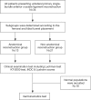
Figure 1
Figure 1
Study design flow diagram.
Figure 2
Figure 2
Femoral and tibial tunnel position determination. (A) The center (red point) of femoral tunnel aperture was measured by Bernard quadrant method (4×4 grid). The tunnel position was calculated as (x/t%; y/h%); (B) tibial tunnel position was measured by the method described by Amis et al. (25). the anatomical tibial footprint (A’P’) was within 25–62% (anterior to posterior); (C) the distribution of femoral tunnel center of all included patients. The green circle represented the standard area of anatomical femoral footprint center described by Xu et al. (26). The red points within the green circle represented the anatomical femoral reconstruction, the yellow points outside the green circle represented the non-anatomical femoral reconstruction; (D) the corresponding tibial tunnel distribution. The color of the points was in accordance with the femoral tunnel. t, total sagittal distance of lateral condyle along Blumensaat’s line; h, maximum intercondylar notch height perpendicular to Blumensaat’s line; x, distance from the center to proximal border line; y, distance from the center to Blumensaat’s line; A, anterior; P, posterior.
Figure 3
Figure 3
The measurement of graft orientation between the simulated ACL graft and tibial plateau on the coronal and sagittal plane. (A) 3D model of the knee with a cylinder simulating the ACL graft from the back view; (B,C) the graft orientation was defined as the angle between the long axis of ACL graft and tibial plateau projected in coronal (α) and sagittal plane (β). ACL, anterior cruciate ligament; a, the center of femoral tunnel aperture; b, the center of tibial tunnel aperture.
Figure 4
Figure 4
Gait kinematics acquisition procedure. (A) The 3D knee kinematics analysis instrumentation; (B) identification of the femoral and tibial anatomic bone markers with an infrared light reflecting probe to setup knee local coordinate systems before kinematics data collection.
Figure 5
Figure 5
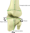
Definition of femoral and tibial coordinate system. The midpoint of transepicondylar axis was defined as the origin of femoral coordinate system. A line crossing the transepicondylar axis was defined as the medial-lateral axis. The anterior-posterior axis was perpendicular to the plane composed of the transepicondylar axis and greater trochanter. The proximal-distal axis was perpendicular to the other two axes. The origin of tibial coordinate system was defined as the center of the line combining the most medial and lateral points of tibial plateau. A line crossing the medial-lateral tibial plateau line was defined as the medial-lateral axis. The anterior-posterior axis was perpendicular to the plane composed of the medial-lateral axis and lateral malleolus. The proximal-distal axis was perpendicular to the other two axes.
Figure 6
Figure 6
The outcomes of IKDC, Lysholm Score and manual maximum laxity test. (A) The post-operative IKDC and Lysholm score of two groups at follow up time; (B) the post-operative manual maximum laxity test by KT-2000 of two groups at follow up time. The upper and lower bound of the box represented 95% CI. ns, non-statistical significance; IKDC, International Knee Documentation Committee.
Figure 7
Figure 7
The ensemble average curve of DOF and SPM1D analysis results of left (dashed black circle lines) and right (dashed grey triangle lines) knees of healthy people. The grey shadow represented standard deviation. The SPM1D analysis results indicated no statistically significant differences between left and right knees, where the SnPM values below the dashed red lines (alpha level threshold of 0.05). DOF, degrees of freedom.
Figure 8
Figure 8
The ensemble average curve of DOF and SPM1D analysis results of contralateral normal knees (dashed black circle lines) and ACLR knees (dashed grey triangle lines) after anatomical ACL reconstruction. The grey shadow represented standard deviation. The SPM1D analysis results indicated no statistically significant differences between contralateral normal knees and ACLR knees, where the SnPM{t} values below the dashed red lines (alpha level threshold of 0.05). DOF, degrees of freedom.
Figure 9
Figure 9
The ensemble average curve of DOF and SPM1D analysis results of contralateral normal knees (dashed black circle lines) and ACLR knees (dashed grey triangle lines) after Non-anatomical ACL reconstruction. The grey shadow represented standard deviation. The SPM1D analysis results indicated no statistically significant differences between contralateral normal knees and ACLR knees, where the SnPM{t} values below the dashed red lines (alpha level threshold of 0.05). DOF, degrees of freedom; ACLR, anterior cruciate ligament reconstruction.

Similar articles

Cited by

References

    1. Andersen HN, Dyhre-Poulsen P. The anterior cruciate ligament does play a role in controlling axial rotation in the knee. Knee Surg Sports Traumatol Arthrosc 1997;5:145-9. 10.1007/s001670050042 - DOI - PubMed
    1. Siegel L, Vandenakker-Albanese C, Siegel D. Anterior cruciate ligament injuries: anatomy, physiology, biomechanics, and management. Clin J Sport Med 2012;22:349-55. 10.1097/JSM.0b013e3182580cd0 - DOI - PubMed
    1. Zhang Y, Huang W, Yao Z, et al. Anterior Cruciate Ligament Injuries Alter the Kinematics of Knees With or Without Meniscal Deficiency. Am J Sports Med 2016;44:3132-9. 10.1177/0363546516658026 - DOI - PubMed
    1. Levy BA. Is early reconstruction necessary for all anterior cruciate ligament tears? N Engl J Med 2010;363:386-8. 10.1056/NEJMe1004397 - DOI - PubMed
    1. Fithian DC, Paxton EW, Stone ML, et al. Prospective trial of a treatment algorithm for the management of the anterior cruciate ligament-injured knee. Am J Sports Med 2005;33:335-46. 10.1177/0363546504269590 - DOI - PubMed