. 2018 Nov 28;6(3):296-303.
doi: 10.1016/j.gendis.2018.11.003. eCollection 2019 Sep.

Down-regulation of argininosuccinate lyase induces hepatoma cell apoptosis through activating Bax signaling pathway

Affiliations

Down-regulation of argininosuccinate lyase induces hepatoma cell apoptosis through activating Bax signaling pathway

Rui Gong et al. Genes Dis. .

Abstract

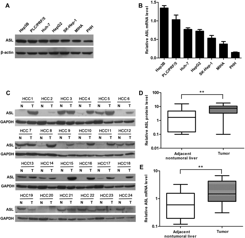
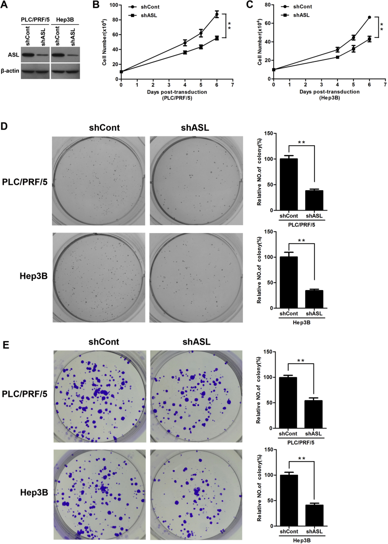
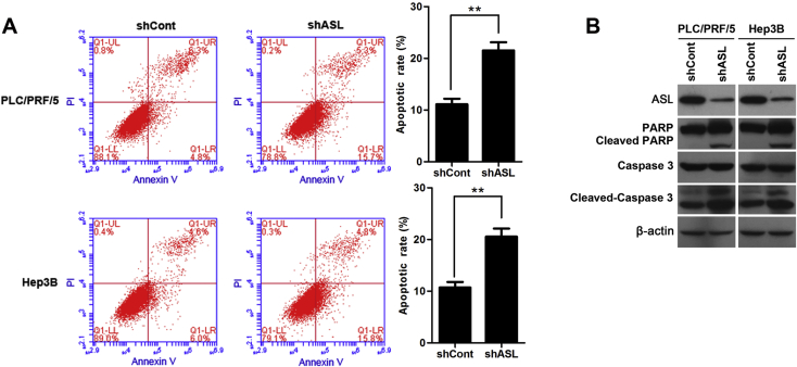
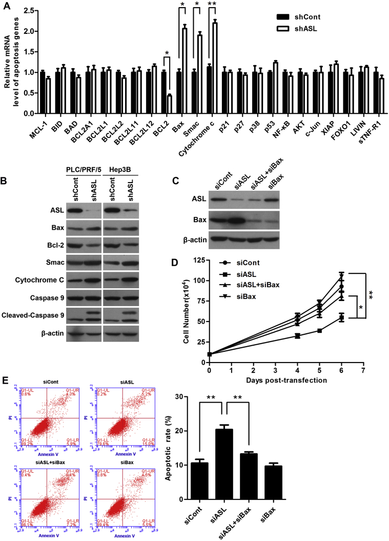
Argininosuccinate lyase (ASL) plays an important role in the hepatic urea cycle, and can catalyze the reversible reaction of argininosuccinate to arginine and fumarate. However, the function of ASL in hepatocellular carcinoma (HCC) is not fully understood. In this study, we found that ASL expression was frequently upregulated in HCC tissues and HCC cell lines. Knock down of ASL inhibited cell proliferation and induced apoptosis in HCC cells. Mechanistic studies revealed the BCL2-associated X protein (Bax) signaling pathway which determines cancer cell apoptosis was regulated by ASL. Moreover, the depletion of Bax restored the inhibition of cell growth and reduced apoptosis initiated by ASL silencing. Together, the study demonstrated that ASL regulated HCC cell growth and apoptosis by modulating Bax signaling. Thus, the therapeutic targeting of ASL may offer options for HCC treatment.

Keywords: ASL; Apoptosis; Bax; HCC; Proliferation.


Figures

Figure 1
Figure 1
ASL expression in HCC. (A and B) ASL protein and mRNA expression in five liver cancer cell lines, the immortalized liver cell line (MIHA), and PHH were measured by western blot and qRT-PCR, respectively. β-actin was used as a loading control for Western blot and as a reference gene for qRT-PCR. (C) Western blot analysis of ASL protein levels in 24 paired frozen HCC tissues (T) and adjacent nontumoral liver tissues (N). GAPDH was used as a loading control. (D) Quantitative analysis of ASL protein levels in 24 paired HCC tissues. **P < 0.01. (E) qRT-PCR analysis of ASL mRNA levels in 24 paired HCC tissues and adjacent nontumoral tissues. GAPDH was used as a reference gene. **P < 0.01.
Figure 2
Figure 2
ASL knockdown inhibited HCC cell proliferation. (A) PLC/PRF/5 and Hep3B cells were infected with lentivirus expressing ASL-targeting shRNA (shASL) or control shRNA (shCont) for 3 days. ASL protein level was detected by western blot. β-actin was used as a loading control. (B and C) ASL knockdown significantly inhibited the proliferation of PLC/PRF/5 and Hep3B cells at indicated time points. Cell numbers were counted using a trypan blue exclusion assay. **P < 0.01. (D) Soft agar assay. PLC/PRF/5 and Hep3B cells were infected with lentiviruses as indicated and were grown for 3 weeks under antibiotic selection. The quantification of colonies was performed by counting visible colonies in each well and expressing then as a percentage relative to control cells. Columns show the mean values of triplicate experiments. **P < 0.01. (E) Colony-formation assay. PLC/PRF/5 and Hep3B cells infected with lentiviruses as indicated were cultured for 2 weeks and stained with crystal violet. All histograms show mean values from three independent experiments. **P < 0.01.
Figure 3
Figure 3
ASL knockdown induced apoptosis in HCC cells. (A) ASL depletion induced apoptosis in PLC/PRF/5 and Hep3B cells was analyzed by flow cytometry with Annexin V/PI. Columns show the mean values of triplicate experiments. **P < 0.01. (B) Apoptosis in PLC/PRF/5 and Hep3B cells infected with shASL or shCont was analyzed by PARP cleavage and caspase 3 cleavage analysis. β-actin was used as a loading control.
Figure 4
Figure 4
ASL regulated Bax signaling in HCC cells. (A) PLC/PRF/5 cells were infected with lentiviruses as indicated for 3 days. mRNA microarray analysis indicated alternations in several key regulators in the Bax-mediated apoptotic pathway. β-actin mRNA expression was used as an internal control. *P < 0.05. **P < 0.01. (B) Western blot analysis of several components of the Bax-mediated apoptotic pathway in PLC/PRF/5 and Hep3B cells transduced with shASL or shCont. β-actin was used as a loading control. (C) PLC/PRF/5 cells were transfected with indicated siRNA for 3 days. The ASL and Bax protein levels were determined by western blot. β-actin was used as a loading control. (D) Gene silencing of Bax rescued the growth inhibition induced by ASL knockdown in PLC/PRF/5 cells. Cell numbers were counted using a trypan blue exclusion assay at indicated time points. *P < 0.05. **P < 0.01. (E) Bax knockdown abolished the induction of apoptosis in ASL-silenced PLC/PRF/5 cells. Apoptosis rate was analyzed by flow cytometry with Annexin V/PI. Columns show the mean values of triplicate experiments. **P < 0.01.

Similar articles

Cited by

References

    1. Ploeger C., Waldburger N., Fraas A. Chromosome 8p tumor suppressor genes SH2D4A and SORBS3 cooperate to inhibit interleukin-6 signaling in hepatocellular carcinoma. Hepatology (Baltimore Md.) 2016;64(3):828–842. - PMC - PubMed
    1. Peng L., Yuan X.Q., Zhang C.Y. The emergence of long non-coding RNAs in hepatocellular carcinoma: an update. J Cancer. 2018;9(14):2549–2558. - PMC - PubMed
    1. Petrizzo A., Mauriello A., Tornesello M.L., Buonaguro F.M., Tagliamonte M., Buonaguro L. Cellular prognostic markers in hepatitis-related hepatocellular carcinoma. Infect Agent Cancer. 2018;13:10. - PMC - PubMed
    1. Petruzziello A. Epidemiology of hepatitis B Virus (HBV) and hepatitis C Virus (HCV) related hepatocellular carcinoma. Open Virol J. 2018;12:26–32. - PMC - PubMed
    1. Budny A., Kozlowski P., Kaminska M. Epidemiology and risk factors of hepatocellular carcinoma. Polski merkuriusz lekarski – Organ Polskiego Towarzystwa Lekarskiego. 2017;43(255):133–139. - PubMed