. 2019 Oct 30;5(6):e369.
doi: 10.1212/NXG.0000000000000369. eCollection 2019 Dec.

Clinical spectrum of POLR3-related leukodystrophy caused by biallelic POLR1C pathogenic variants

Affiliations

Clinical spectrum of POLR3-related leukodystrophy caused by biallelic POLR1C pathogenic variants

Laurence Gauquelin et al. Neurol Genet. .

Abstract

Objective: To determine the clinical, radiologic, and molecular characteristics of RNA polymerase III-related leukodystrophy (POLR3-HLD) caused by biallelic POLR1C pathogenic variants.

Methods: A cross-sectional observational study involving 25 centers worldwide was conducted. Clinical and molecular information was collected on 23 unreported and previously reported patients with POLR3-HLD and biallelic pathogenic variants in POLR1C. Brain MRI studies were reviewed.

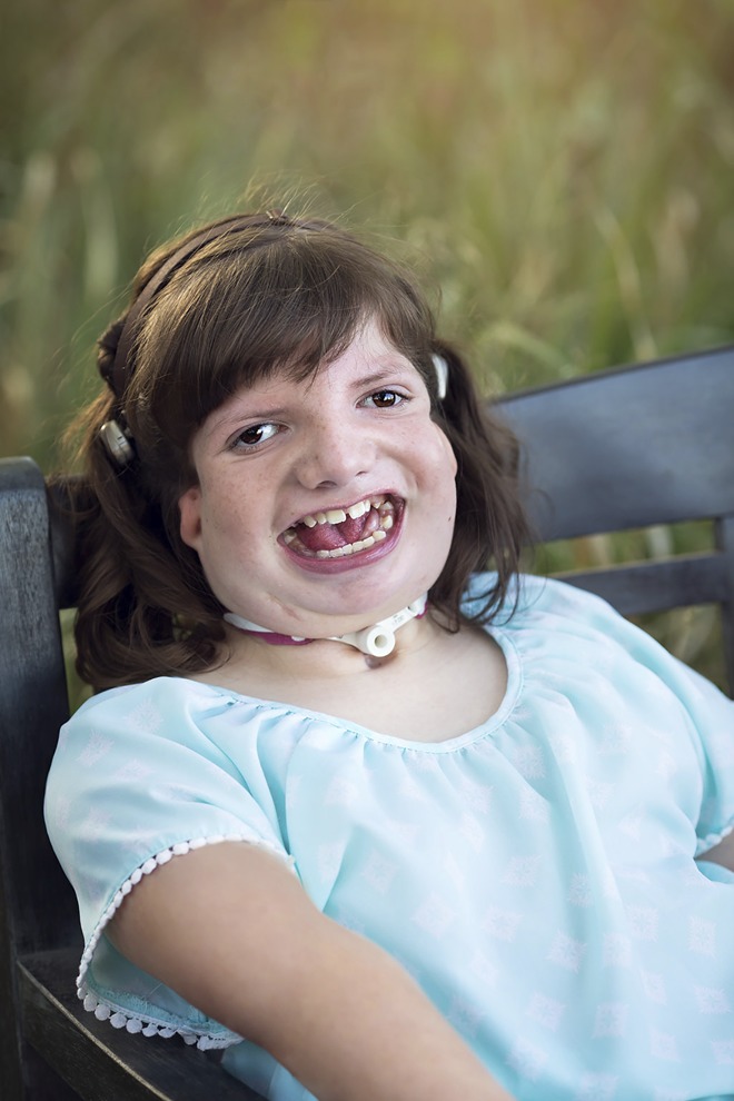
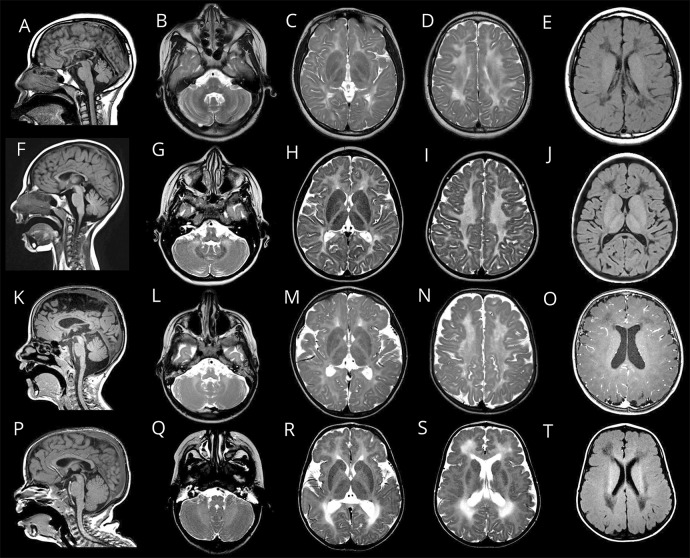
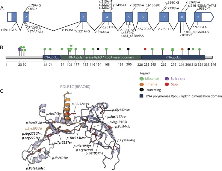
Results: Fourteen female and 9 male patients aged 7 days to 23 years were included in the study. Most participants presented early in life (birth to 6 years), and motor deterioration was seen during childhood. A notable proportion of patients required a wheelchair before adolescence, suggesting a more severe phenotype than previously described in POLR3-HLD. Dental, ocular, and endocrine features were not invariably present (70%, 50%, and 50%, respectively). Five patients (22%) had a combination of hypomyelinating leukodystrophy and abnormal craniofacial development, including 1 individual with clear Treacher Collins syndrome (TCS) features. Brain MRI revealed hypomyelination in all cases, often with areas of pronounced T2 hyperintensity corresponding to T1 hypointensity of the white matter. Twenty-nine different pathogenic variants (including 12 new disease-causing variants) in POLR1C were identified.

Conclusions: This study provides a comprehensive description of POLR3-HLD caused by biallelic POLR1C pathogenic variants based on the largest cohort of patients to date. These results suggest distinct characteristics of POLR1C-related disorder, with a spectrum of clinical involvement characterized by hypomyelinating leukodystrophy with or without abnormal craniofacial development reminiscent of TCS.


Figures

Figure 1
Figure 1. Photograph of patient 19 showing facial features compatible with Treacher Collins syndrome (TCS)
Photograph of patient 19 at age 10 years. She had facial features in keeping with TCS, including downslanted palpebral fissures, strabismus, bitemporal narrowing, external ear abnormalities, cleft palate, and prominent micrognathia.
Figure 2
Figure 2. Brain MRI characteristics of 4 patients with POLR3-HLD caused by biallelic POLR1C pathogenic variants
Sagittal T1 (A, F, K, and P), axial T2 (B–D, G–I, L–N, and Q–S) and axial T1 (E, J, O, and T) images. (A–E) MRI of patient 18 obtained at age 11 years showing diffuse hypomyelination with superimposed areas of pronounced T2 hyperintensity (C and D) and corresponding T1 hypointensity (E). Thinning of the corpus callosum and mild superior vermis atrophy are also seen (A), as well as preserved myelination of the dentate nucleus (B), globus pallidus, anterolateral nucleus of the thalamus, and optic radiation (C). (F–J) MRI of patient 4 obtained at age 5 years showing diffuse hypomyelination with preservation of the dentate nucleus (G), anterolateral nucleus of the thalamus, and optic radiation (H). There is also thinning of the corpus callosum and mild vermis atrophy (F). Areas of marked T2 hyperintensity of the white matter are seen (H and I), with corresponding pronounced T1 hypointensity (J). (K–O), MRI of patient 1 obtained at age 5 years showing a thin corpus callosum (K), relative preservation of myelination of the dentate nucleus (L), and absent T2 hypointensity of the corticospinal tracts in the posterior limb of the internal capsule (M). (P–T), MRI of patient 20.1 obtained at age 3 years showing areas of prominent T2 hyperintensity of the white matter (R and S) with corresponding T1 hypointensity (T), especially in the deep white matter. There is also bilateral frontal polymicrogyria (R, S, and T). POLR3-HLD = RNA polymerase III-related leukodystrophy.
Figure 3
Figure 3. Pathogenic variants identified in POLR1C associated with POLR3-HLD
(A–B) All reported pathogenic variants and their positions within the POLR1C gDNA (A), with missense variants represented in green, in frame in orange, truncating in black, splice site in purple, and stop in red (B). (C) Missense variants displayed on the structure of the yeast ortholog of POLR1C (RPAC40). Variants previously identified in POLR3-HLD are represented in italic, whereas newly identified variants are shown in bold. The p.Lys295del is shown in orange. The p.Thr26Ile, p.Thr27Ala, and p.Pro30Ser variants have not been represented because they are not visible in the crystal structure of RPAC40 (PDB 5M5W).,, POLR3-HLD = RNA polymerase III-related leukodystrophy.

References

    1. Kevelam SH, Steenweg ME, Srivastava S, et al. . Update on leukodystrophies: a historical perspective and adapted definition. Neuropediatrics 2016;47:349–354. - PubMed
    1. Vanderver A, Prust M, Tonduti D, et al. . Case definition and classification of leukodystrophies and leukoencephalopathies. Mol Genet Metab 2015;114:494–500. - PMC - PubMed
    1. Parikh S, Bernard G, Leventer RJ, et al. . A clinical approach to the diagnosis of patients with leukodystrophies and genetic leukoencephelopathies. Mol Genet Metab 2015;114:501–515. - PMC - PubMed
    1. Steenweg ME, Vanderver A, Blaser S, et al. . Magnetic resonance imaging pattern recognition in hypomyelinating disorders. Brain 2010;133:2971–2982. - PMC - PubMed
    1. Schiffmann R, van der Knaap MS. Invited article: an MRI-based approach to the diagnosis of white matter disorders. Neurology 2009;72:750–759. - PMC - PubMed