

Stabilizing Peri-Stent Restenosis Using a Novel Therapeutic Carrier
- PMID: 32043017
- PMCID: PMC7000871
- DOI: 10.1016/j.jacbts.2019.09.005
Stabilizing Peri-Stent Restenosis Using a Novel Therapeutic Carrier
Abstract
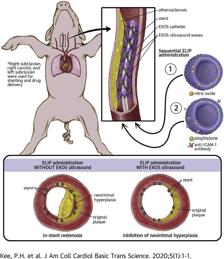
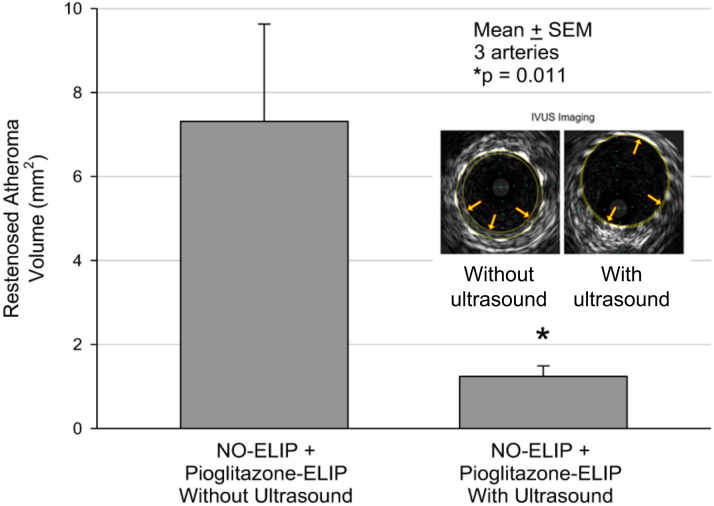
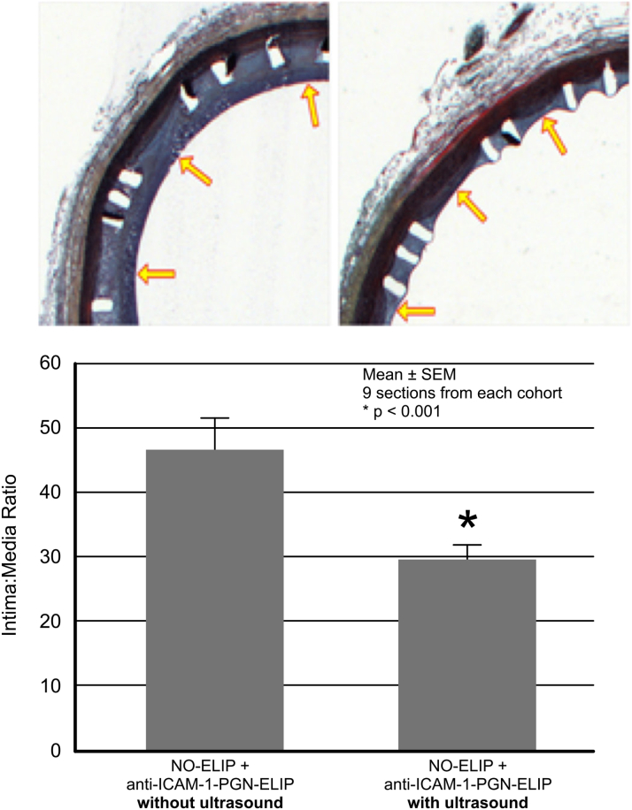
Late in-stent restenosis remains a significant problem. Bare-metal stents were implanted into peripheral arteries in miniature swine, followed by direct intra-arterial infusion of nitric oxide-loaded echogenic liposomes (ELIPs) and anti-intercellular adhesion molecule-1 conjugated ELIPs loaded with pioglitazone exposed to an endovascular catheter with an ultrasonic core. Ultrasound-facilitated delivery of ELIP formulations into stented peripheral arteries attenuated neointimal growth. Local atheroma-targeted, ultrasound-triggered delivery of nitric oxide and pioglitazone, an anti-inflammatory peroxisome proliferator-activated receptor-γ agonist, into stented arteries has the potential to stabilize stent-induced neointimal growth and obviate the need for long-term antiplatelet therapy.
Keywords: ELIP, echogenic liposome; ICAM, intercellular adhesion molecule; IVUS, intravascular ultrasound; NO, nitric oxide; PGN, pioglitazone; SPDP, 3-(2-pyridyldithio propionic acid)-N-hydroxysuccinimide ester; atherosclerosis; in-stent restenosis; nitric oxide; pioglitazone; ultrasound contrast agent.
© 2020 The Authors.
Figures
Comment in
-
In-Stent Restenosis: Chasing Our Tails in Search of a Solution.JACC Basic Transl Sci. 2020 Jan 27;5(1):12-14. doi: 10.1016/j.jacbts.2019.12.002. eCollection 2020 Jan. JACC Basic Transl Sci. 2020. PMID: 32043490 Free PMC article.
References
-
- Abdulhannan P., Russell D.A., Homer-Vanniasinkam S. Peripheral arterial disease: a literature review. Br Med Bull. 2012;104:21–39. - PubMed
-
- Brower V. Stents and biology combination for restenosis. Nat Biotechnol. 1996;14:422. - PubMed
-
- Farb A., Weber D.K., Kolodgie F.D., Burke A.P., Virmani R. Morphological predictors of restenosis after coronary stenting in humans. Circulation. 2002;105:2974–2980. - PubMed
-
- Finn A.V., Nakazawa G., Joner M. Vascular responses to drug eluting stents: importance of delayed healing. Arterioscler Thromb Vasc Biol. 2007;27:1500–1510. - PubMed
-
- Nakazawa G., Vorpahl M., Finn A.V., Narula J., Virmani R. One step forward and two steps back with drug-eluting-stents: from preventing restenosis to causing late thrombosis and nouveau atherosclerosis. J Am Coll Cardiol Img. 2009;2:625–628. - PubMed
Grants and funding
LinkOut - more resources
Full Text Sources
