

Sleep and Cellular Stress
- PMID: 32043041
- PMCID: PMC7008961
- DOI: 10.1016/j.cophys.2019.12.011
Sleep and Cellular Stress
Abstract
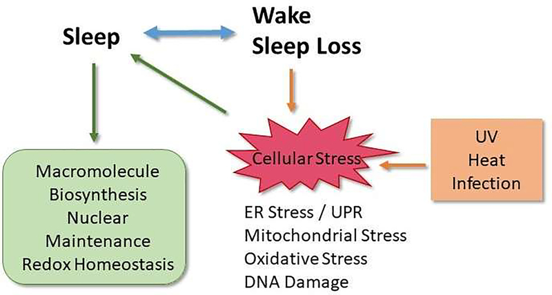
Sleep is a universal phenomenon occurring in all species studied thus far. Sleep loss results in adverse physiological effects at both the organismal and cellular levels suggesting an adaptive role for sleep in the maintenance of overall health. This review examines the bidirectional relationship between sleep and cellular stress. Cellular stress in this review refers to a shift in cellular homeostasis in response to an external stressor. Studies that illustrate the fact that sleep loss induces cellular stress and those that provide evidence that cellular stress in turn promotes sleep will be discussed.
Keywords: DNA repair; ER stress; Epidermal Growth Factor; NFκB; Unfolded protein response; immune; inflammation.
Conflict of interest statement
Conflict of Interest Statement The authors declare no conflict of interest
Figures
References
-
- Cirelli C, Gutierrez CM, and Tononi G, Extensive and divergent effects of sleep and wakefulness on brain gene expression. Neuron, 2004. 41(1): p. 35–43. - PubMed
-
- Naidoo N, et al., Sleep deprivation induces the unfolded protein response in mouse cerebral cortex. J Neurochem, 2005. 92(5): p. 1150–7. - PubMed
-
- Mackiewicz M, et al., Macromolecule biosynthesis: a key function of sleep. Physiol Genomics, 2007. 31(3): p. 441–57. - PubMed
-
- Ramm P and Smith CT, Rates of cerebral protein synthesis are linked to slow wave sleep in the rat. Physiology and Behavior, 1990. 48(5): p. 749–53. - PubMed
-
- Nakanishi H, et al., Positive correlations between cerebral protein synthesis rates and deep sleep in Macaca mulatta. European Journal of Neuroscience, 1997. 9(2): p. 271–9. - PubMed
Grants and funding
LinkOut - more resources
Full Text Sources