

Age-related differences in functional brain network segregation are consistent with a cascade of cerebrovascular, structural, and cognitive effects
- PMID: 32043045
- PMCID: PMC7006874
- DOI: 10.1162/netn_a_00110
Age-related differences in functional brain network segregation are consistent with a cascade of cerebrovascular, structural, and cognitive effects
Abstract
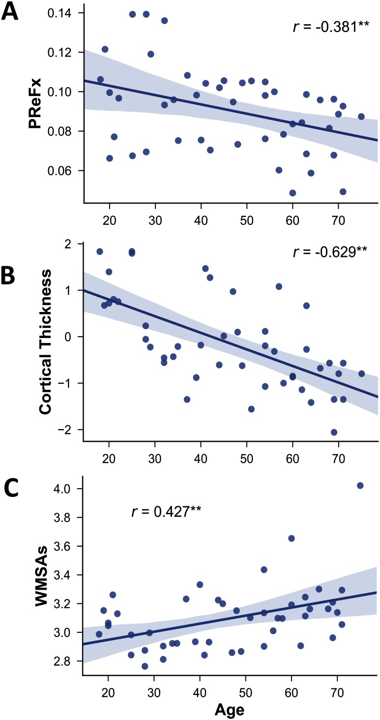
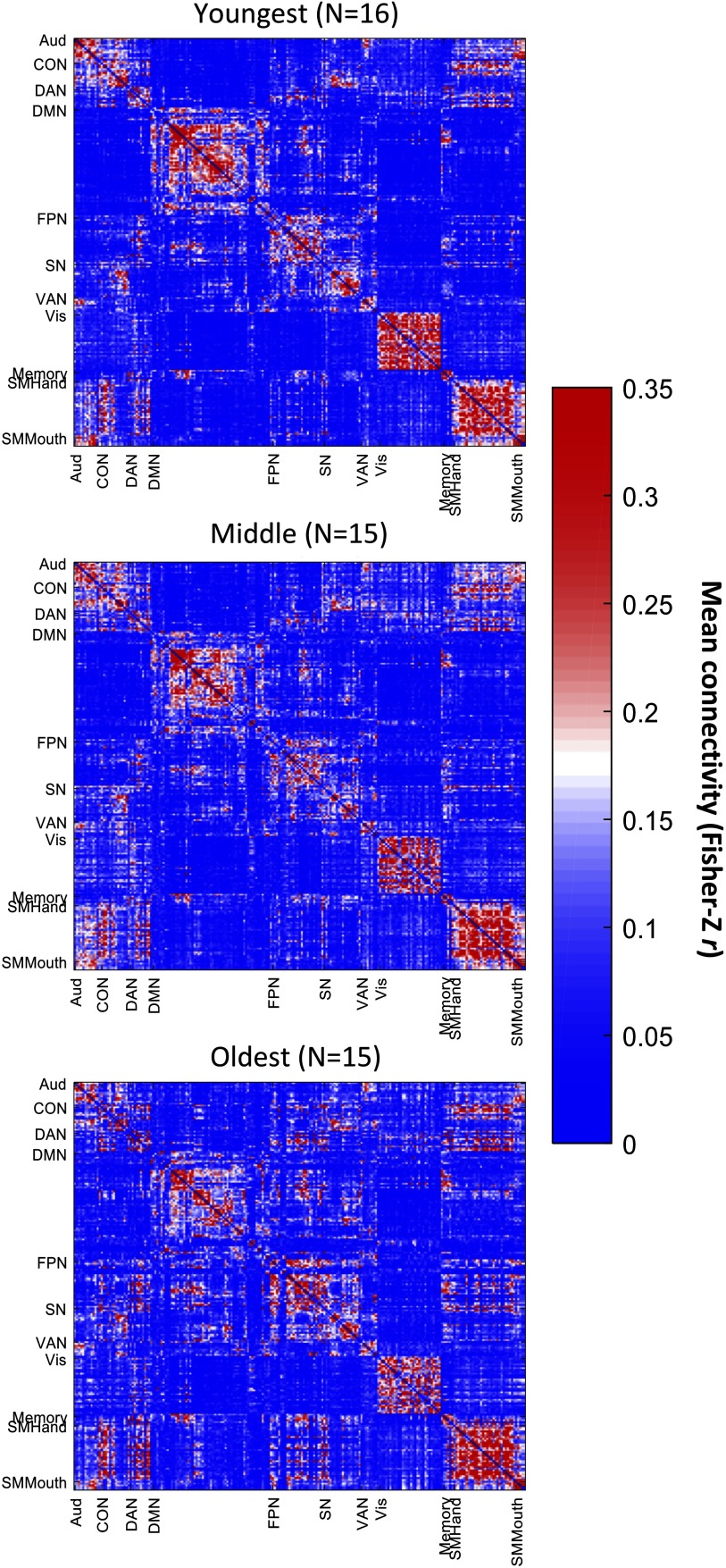
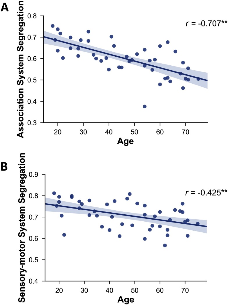
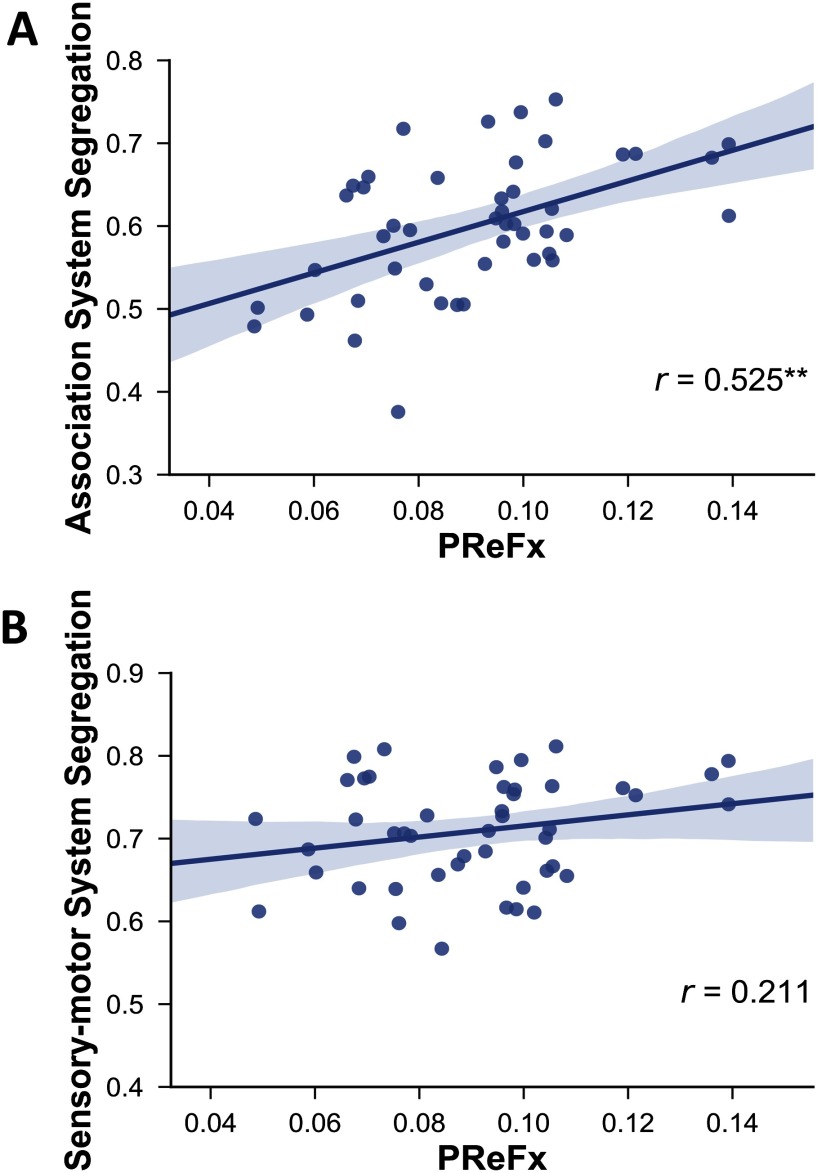
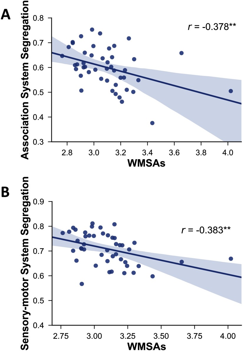
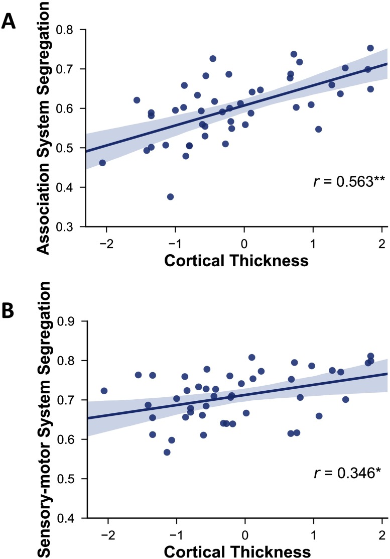
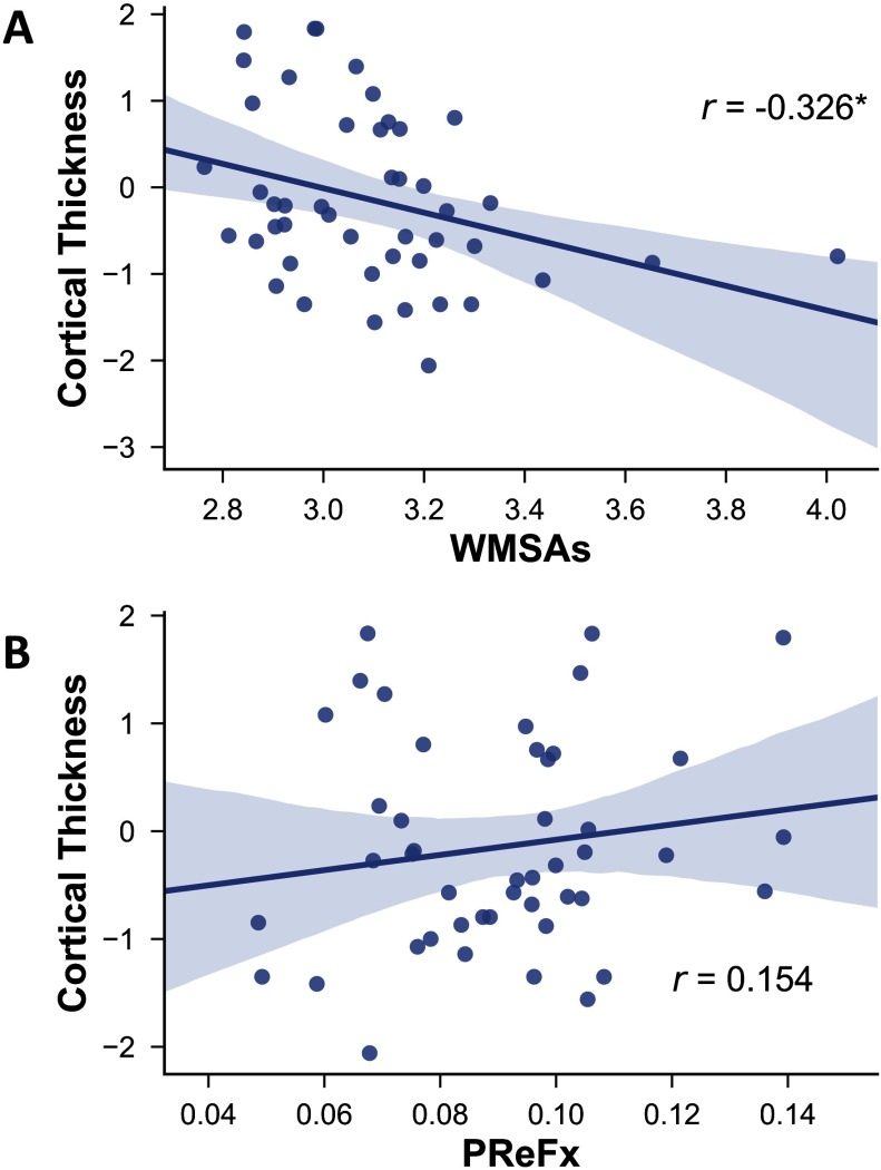
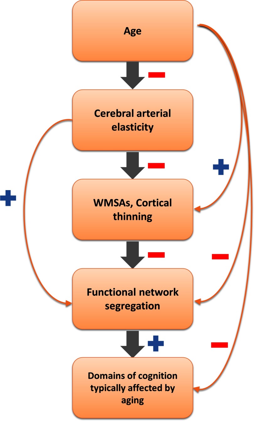
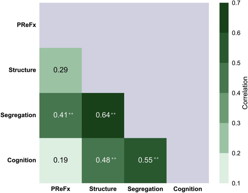
Age-related declines in cognition are associated with widespread structural and functional brain changes, including changes in resting-state functional connectivity and gray and white matter status. Recently we have shown that the elasticity of cerebral arteries also explains some of the variance in cognitive and brain health in aging. Here, we investigated how network segregation, cerebral arterial elasticity (measured with pulse-DOT-the arterial pulse based on diffuse optical tomography) and gray and white matter status jointly account for age-related differences in cognitive performance. We hypothesized that at least some of the variance in brain and cognitive aging is linked to reduced cerebrovascular elasticity, leading to increased cortical atrophy and white matter abnormalities, which, in turn, are linked to reduced network segregation and decreases in cognitive performance. Pairwise comparisons between these variables are consistent with an exploratory hierarchical model linking them, especially when focusing on association network segregation (compared with segregation in sensorimotor networks). These findings suggest that preventing or slowing age-related changes in one or more of these factors may induce a neurophysiological cascade beneficial for preserving cognition in aging.
Keywords: Aging; Cerebrovascular health; Cortical thickness; Optical brain arterial pulse (pulse-DOT); Resting-state functional connectivity (rsFC); White matter signal abnormalities (WMSAs).
© 2019 Massachusetts Institute of Technology.
Conflict of interest statement
Competing Interests: The authors have declared that no competing interests exist.
Figures
Similar articles
-
Optical measures of cerebral arterial stiffness are associated with white matter signal abnormalities and cognitive performance in normal aging.Neurobiol Aging. 2019 Dec;84:200-207. doi: 10.1016/j.neurobiolaging.2019.08.004. Epub 2019 Aug 10. Neurobiol Aging. 2019. PMID: 31500910 Free PMC article.
-
When functional blurring becomes deleterious: Reduced system segregation is associated with less white matter integrity and cognitive decline in aging.Neuroimage. 2021 Nov 15;242:118449. doi: 10.1016/j.neuroimage.2021.118449. Epub 2021 Aug 3. Neuroimage. 2021. PMID: 34358662
-
Individual differences in regional cortical volumes across the life span are associated with regional optical measures of arterial elasticity.Neuroimage. 2017 Nov 15;162:199-213. doi: 10.1016/j.neuroimage.2017.08.064. Epub 2017 Sep 1. Neuroimage. 2017. PMID: 28866349 Free PMC article.
-
Impact of pulse pressure on cerebrovascular events leading to age-related cognitive decline.Am J Physiol Heart Circ Physiol. 2018 Jun 1;314(6):H1214-H1224. doi: 10.1152/ajpheart.00637.2017. Epub 2018 Feb 16. Am J Physiol Heart Circ Physiol. 2018. PMID: 29451817 Free PMC article. Review.
-
Healthy aging by staying selectively connected: a mini-review.Gerontology. 2014;60(1):3-9. doi: 10.1159/000354376. Epub 2013 Sep 28. Gerontology. 2014. PMID: 24080587 Review.
Cited by
-
Cerebral Arterial Pulsatility and Global White Matter Microstructure Impact Spatial Working Memory in Older Adults With and Without Cardiovascular Risk Factors.Front Aging Neurosci. 2020 Aug 7;12:245. doi: 10.3389/fnagi.2020.00245. eCollection 2020. Front Aging Neurosci. 2020. PMID: 32848715 Free PMC article.
-
Age-related differences in functional network segregation in the context of sex and reproductive stage.Hum Brain Mapp. 2023 Apr 1;44(5):1949-1963. doi: 10.1002/hbm.26184. Epub 2022 Dec 21. Hum Brain Mapp. 2023. PMID: 36541480 Free PMC article.
-
Functional and structural cerebellar-behavior relationships in aging.Brain Struct Funct. 2024 Dec 18;230(1):10. doi: 10.1007/s00429-024-02862-9. Brain Struct Funct. 2024. PMID: 39692877
-
Age-related changes in cerebrovascular health and their effects on neural function and cognition: A comprehensive review.Psychophysiology. 2021 Jul;58(7):e13796. doi: 10.1111/psyp.13796. Epub 2021 Mar 16. Psychophysiology. 2021. PMID: 33728712 Free PMC article. Review.
-
Low neighborhood deprivation buffers against hippocampal neurodegeneration, white matter hyperintensities, and poorer cognition.Geroscience. 2023 Jun;45(3):2027-2036. doi: 10.1007/s11357-023-00780-y. Epub 2023 Apr 1. Geroscience. 2023. PMID: 37004594 Free PMC article.
References
-
- Badji A., Sabra D., Bherer L., Cohen-Adad J., Girouard H., & Gauthier C. J. (2019). Arterial Stiffness and Brain Integrity: A review of MRI findings. Ageing research reviews. . - PubMed
-
- Baltes P. B., Lindenberger U., Schubert G., Stober A., & Weilandt M. (1997). Emergence of a powerful connection between sensory and cognitive functions across the adult life span: A new window to the study of cognitive aging? Psychology and Aging,12(1), 12–21. - PubMed
Grants and funding
LinkOut - more resources
Full Text Sources