

Prevalence of NRAS Mutation, PD-L1 Expression and Amplification, and Overall Survival Analysis in 36 Primary Vaginal Melanomas
- PMID: 32043781
- PMCID: PMC7011659
- DOI: 10.1634/theoncologist.2019-0148
Prevalence of NRAS Mutation, PD-L1 Expression and Amplification, and Overall Survival Analysis in 36 Primary Vaginal Melanomas
Abstract
Background: Primary vaginal melanomas are uncommon and aggressive tumors with poor prognosis, and the development of new targeted therapies is essential. This study aimed to identify the molecular markers occurring in these patients and potentially improve treatment strategies.
Materials and methods: The clinicopathological characteristics of 36 patients with primary vaginal melanomas were reviewed. Oncogenic mutations in BRAF, KIT, NRAS, GNAQ and GNA11 and the promoter region of telomerase reverse transcriptase (TERT) were investigated using the Sanger sequencing. The expression and copy number of programmed death-ligand 1 (PD-L1) were also assessed.
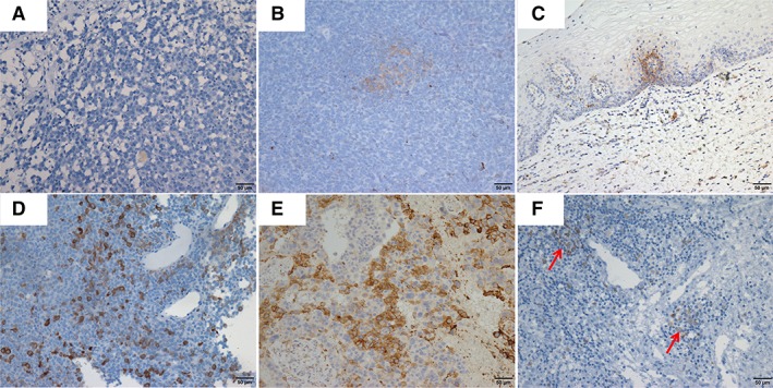
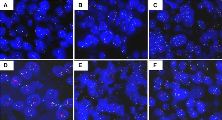
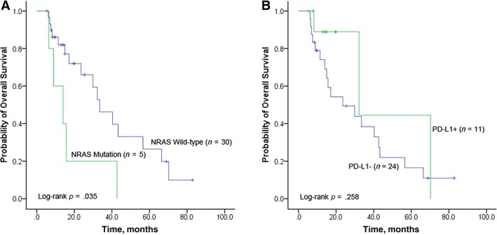
Results: Mutations in NRAS, KIT, and TERT promoter were identified in 13.9% (5/36), 2.9% (1/34), and 5.6% (2/36) of the primary vaginal melanomas, respectively. PD-L1 expression and amplification were observed in 27.8% (10/36) and 5.6% (2/36) of cases, respectively. PD-L1 positive expression and/or amplification was associated with older patients (p = .008). Patients who had NRAS mutations had a poorer overall survival compared with those with a wild-type NRAS (33.5 vs. 14.0 months; hazard ratio [HR], 3.09; 95% CI, 1.08-8.83). Strikingly, two patients with/without PD-L1 expression receiving immune checkpoint inhibitors had a satisfying outcome. Multivariate analysis demonstrated that >10 mitoses per mm2 (HR, 2.96; 95% CI, 1.03-8.51) was an independent prognostic factor.
Conclusions: NRAS mutations and PD-L1 expression were most prevalent in our cohort of primary vaginal melanomas and can be potentially considered as therapeutic targets.
Implications for practice: This study used the Sanger sequencing, immunohistochemistry, and fluorescence in situ hybridization methods to detect common genetic mutations and PD-L1 expression and copy number in 36 primary vaginal melanomas. NRAS mutations and PD-L1 expression were the most prevalent, but KIT and TERT mutations occurred at a lower occurrence in this rare malignancy. Two patients receiving immune checkpoint inhibitors had a satisfying outcome, signifying that the PD-L1 expression and amplification can be a possible predictive marker of clinical response. This study highlights the possible prospects of biomarkers that can be used for patient selection in clinical trials involving treatments with novel targeted therapies based on these molecular aberrations.
Keywords: NRAS; Ooncogenic mutations; PD-L1 expression; Primary vaginal melanoma.
© 2019 The Authors. The Oncologist published by Wiley Periodicals, Inc. on behalf of AlphaMed Press.
Conflict of interest statement
Figures
References
-
- Erdmann F, Lortet‐Tieulent J, Schüz J et al. International trends in the incidence of malignant melanoma 1953‐2008–are recent generations at higher or lower risk? Int J Cancer 2013;132:385–400. - PubMed
-
- Chang AE, Karnell LH, Menck HR. The national cancer data base report on cutaneous and noncutaneous melanoma: A summary of 84,836 cases from the past decade. The American College of Surgeons Commission on Cancer and the American Cancer Society. Cancer 1998;83:1664–1678. - PubMed
-
- Frumovitz M, Etchepareborda M, Sun CC et al. Primary malignant melanoma of the vagina. Obstet Gynecol 2010;116:1358–1365. - PubMed
-
- Cobellis L, Calabrese E, Stefanon B et al. Malignant melanoma of the vagina. A report of 15 cases. Eur J Gynaecol Oncol 2000;21:295–297. - PubMed
-
- Reid GC, Schmidt RW, Roberts JA et al. Primary melanoma of the vagina: A clinicopathologic analysis. Obstet Gynecol 1989;74:190–199. - PubMed
Publication types
MeSH terms
Substances
LinkOut - more resources
Full Text Sources
Medical
Research Materials
Miscellaneous
