

Lifetime Transfusion Burden and Transfusion-Related Iron Overload in Adult Survivors of Solid Malignancies
- PMID: 32043782
- PMCID: PMC7011667
- DOI: 10.1634/theoncologist.2019-0222
Lifetime Transfusion Burden and Transfusion-Related Iron Overload in Adult Survivors of Solid Malignancies
Abstract
Background: Limited data exist on transfusion burden and transfusion-related iron overload in adult survivors of solid malignancies.
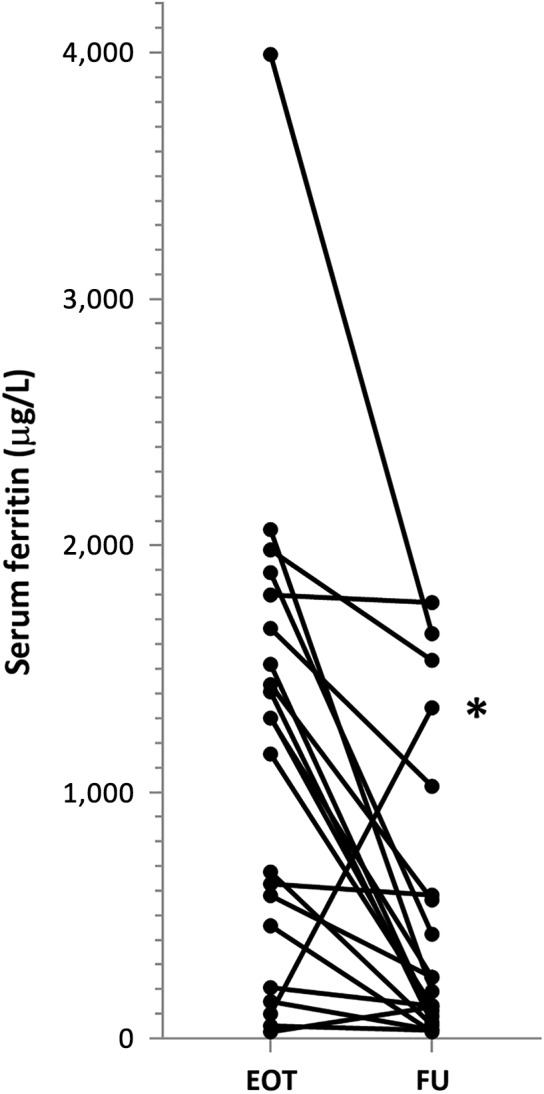
Methods: Hospital-specific cancer registry data of patients with solid tumor receiving systemic anticancer treatment between January 2008 and September 2009 at the Oncology Department of the Leiden University Medical Center (The Netherlands) were retrieved and cross-referenced with red blood cell (RBC) transfusion records. Individual lifetime transfusion burden was captured in April 2015. Multitransfused long-term survivors with serum ferritin >500 μg/L were subsequently screened for hepatic and cardiac iron overload using 1.5 Tesla magnetic resonance imaging.
Results: The study population consisted of 775 adult patients with solid cancer (45.2% male; median age, 58 years; >75% chemotherapy-treated), 423 (54.6%) of whom were transfused with a median of 6.0 RBC units (range 1-67). Transfusion triggers were symptomatic anemia or hemoglobin <8.1-8.9 g/dL prior to each myelosuppressive chemotherapy cycle. We identified 123 (15.9%) patients across all tumor types with a lifetime transfusion burden of ≥10 RBC units. In the absence of a hemovigilance program, none of these multitransfused patients was screened for iron overload despite a median survival of 4.6 years. In 2015 at disclosure of transfusion burden, 26 multitransfused patients were alive. Six (23.1%) had hepatic iron overload: 3.9-11.2 mg Fe/g dry weight. No cardiac iron depositions were found.
Conclusion: Patients with solid malignancies are at risk for multitransfusion and iron overload even when adhering to restrictive RBC transfusion policies. With improved long-term cancer survivorship, increased awareness of iatrogenic side effects of supportive therapy and development of evidence-based guidelines are essential.
Implications for practice: In the presence of a restrictive transfusion policy, ∼30% of transfused adult patients with solid cancer are multitransfused and ∼50% become long-term survivors, underscoring the need for evidence-based guidelines for the detection and management of transfusion-related iron overload in this group of patients. In each institution, a hemovigilance program should be implemented that captures the lifetime cumulative transfusion burden in all patients with cancer, irrespective of tumor type. This instrument will allow timely assessment and treatment of iron overload in cancer survivors, thus preventing organ dysfunction and decreased quality of life.
Keywords: Bone neoplasms; Erythrocyte transfusion; Iron overload; Long-term survivors; Neoplasms; Solid tumor; Testicular neoplasms; Toxicity.
© AlphaMed Press 2019.
Conflict of interest statement
Figures

 ). A relatively large proportion of patients with bone tumors (
 ) was multitransfused.
References
-
- Andrews NC. Disorders of iron metabolism. N Engl J Med 1999;341:1986–1995. - PubMed
-
- Buja LM, Roberts WC. Iron in the heart. Etiology and clinical significance. Am J Med 1971;51:209–221. - PubMed
-
- Wood JC. Guidelines for quantifying iron overload. Hematology Am Soc Hematol Educ Program 2014;2014:210–215. - PubMed
-
- Gilreath JA, Stenehjem DD, Rodgers GM. Diagnosis and treatment of cancer‐related anemia. Am J Hematol 2014;89:203–212. - PubMed
Publication types
MeSH terms
LinkOut - more resources
Full Text Sources
Medical
Miscellaneous
