. 2020 Feb 11:9:e51915.
doi: 10.7554/eLife.51915.

Etv transcription factors functionally diverge from their upstream FGF signaling in lens development

Affiliations

Etv transcription factors functionally diverge from their upstream FGF signaling in lens development

Ankur Garg et al. Elife. .

Abstract

The signal regulated transcription factors (SRTFs) control the ultimate transcriptional output of signaling pathways. Here, we examined a family of FGF-induced SRTFs - Etv1, Etv 4, and Etv 5 - in murine lens development. Contrary to FGF receptor mutants that displayed loss of ERK signaling and defective cell differentiation, Etv deficiency augmented ERK phosphorylation without disrupting the normal lens fiber gene expression. Instead, the transitional zone for lens differentiation was shifted anteriorly as a result of reduced Jag1-Notch signaling. We also showed that Etv proteins suppresses mTOR activity by promoting Tsc2 expression, which is necessary for the nuclei clearance in mature lens. These results revealed the functional divergence between Etv and FGF in lens development, demonstrating that these SRTFs can operate outside the confine of their upstream signaling.

Keywords: ETS; FGF; Notch; PEA3; developmental biology; lens development; mTOR; mouse.

Plain language summary

Many cells contain proteins known as signal-induced transcription factors, which are poised to receive messages from the environment and then react by activating genes required for the cell to respond appropriately. It is commonly thought that these transcription factors faithfully follow the instructions they receive from the external signal: for instance, if the message was to encourage the cell to grow, the transcription factors would switch on growth-related genes. As the eyes of mice and other mammals develop, a signal known as FGF is required for certain cells to specialize into lens fiber cells: these long, thin, transparent cells form the bulk of the lens, the structure that allows focused vision. Previous studies suggest that FGF activates three transcription factors known as Etv1, Etv4 and Etv5, but their precise roles in the development of the lens has remained unclear. Here, Garg, Hannan, Wang et al. confirm that FGF signaling does indeed activate all three proteins. However, mutant mice that lacked Etv1, Etv4 and Etv5 still created lens fiber cells, suggesting that the transcription factors are largely unnecessary for lens fiber cells formation. Instead, the Etv proteins participated in a cascade of molecular events involving a protein called Notch; as a result, if the transcription factors were absent, the lens fiber cells formed prematurely. In addition, deactivating Etv1, Etv4 and Etv5 also promoted the activity of a protein which interfered with the removal of internal cell compartments, a process required for lens fiber cells to mature properly. These findings reveal that the roles of Etv1, Etv4 and Etv5 deviate from and even oppose FGF signaling in the lenses of mice. Transcription factors control the ultimate fate of a cell, and there is therefore increased interest in targeting them for therapy. The work by Garg, Hannan, Wang et al. reveals an unexpected complexity in how these proteins respond to upstream signals, highlighting the importance of further dissecting these relationships.


Conflict of interest statement

AG, AH, QW, NM, JZ, HL, SY, YM, XZ No competing interests declared

Figures
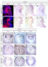
Figure 1.
Figure 1.. Etv transcription factors are controlled by FGF-ERK signaling in the lens.
(A–D) At E10.5, the invaginating lens ectoderm displays ERK phosphorylation and expression of Etv1, 4 and 5 (arrows). (E–H) Genetic ablation of Fgfr1/2 in Fgfr CKO mutants prevented lens vesicle formation and abrogated phospho-ERK and Etv expression (arrows). The lens ectoderms are marked by dotted lines. (I–K) At E14.5, Etv1, 4 and 5 are predominantly expressed in the transitional zone of the lens (arrows). (L–Q) Deletion of either Erk1/2 (Erk CKO) or Mek1/2 (Mek CKO) abolished expression of Etv genes. The lenses are circled in dotted lines.
Figure 2.
Figure 2.. Lens development requires Etv transcription factors.
(A–H) Hematoxylin and eosin (H and E) staining reveal reduced lens size in Etv1/4/5 deletion (Etv TKO) mutants (A and E). Etv null lens exhibited increased cell apoptosis shown by TUNEL staining (B and F, arrows) and reduced cell proliferation as indicated by EdU staining (C and G, arrowheads). In contrast, ectopic expression of Cyclin D1 (G, arrows) and proliferation markers EdU and pHH3 (C, D, G and H, inserts) were detected in the posterior lens. (I) Quantification of EdU and TUNEL staining.
Figure 3.
Figure 3.. Transcriptomic analysis shows ERK signaling dysregulation in Etv mutant lens.
(A) The transitional zone of the lens was isolated by laser capture microscope (LCM) for RNA sequencing analysis. (B) Cluster analysis of the top differentially expressed genes in the RNA sequencing data. (C) Heat map of the Etv regulated genes. (D and F) Spry2 is significantly down-regulated in Etv TKO mutants. (H) Gene set enrichment analysis (GSEA) indicates the MAPK pathway is elevated.
Figure 3—figure supplement 1.
Figure 3—figure supplement 1.. RNA sequencing analysis identified Etv-regulated genes.
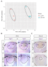
(A) Principle component analysis (PCA) showed that control and Etv TKO mutant lenses were well separated in their transcriptomes. (B) Fgfr1 and Fgfr3 were strongly expressed in the transitional zones (arrows) of the control lenses, but down regulated in Etv TKO mutant lenses. (C) Genetic deletion of Erk1/2 abolished Spry2 expression in Erk CKO mutants.
Figure 4.
Figure 4.. Lens fiber differentiation proceeds in the absence of Etv and ERK.
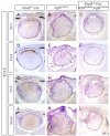
(A–J) Expression of fiber cell markers Maf, α- and γ-crystallin is unaffected in Etv TKO lenses. (K–T) Maf, α- and γ-crystallin are still expressed in Erk and Mek CKO lens despite the severely reduced lens size.
Figure 5.
Figure 5.. Etv deletion prevents FGF from inducing aberrant differentiation of the lens epithelium.
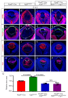
(A–P) Overexpression of Fgf3 in Fgf3OVE391 lens induced expansion of ERK phosphorylation into the anterior lens epithelium (B), which lost the epithelial marker E-cadherin (F, arrows) but expressed the fiber cell markers Maf and γ-crystallin (J and N). The expression of dominant negative EtvEnR restored normal lens differentiation pattern in Pax6Le-Cre; Fgf3OVE391;R26EtvEnR lenses (H, L and P). (Q) The lens size was enlarged in Fgf3OVE391 embryos, but reduced in both Pax6Le-Cre;R26EtvEnR and Pax6Le-Cre; Fgf3OVE391;R26EtvEnR embryos.
Figure 5—figure supplement 1.
Figure 5—figure supplement 1.. Fgf3 overexpression stimulates expressions of Etv and Spry in the lens.
(A–L) Overexpression of Fgf3 in Fgf3OVE391 led to upregulation of Etv1, 4, five and Spry2 in the anterior lens epithelium, which were greatly reduced by expression of dominant negative EtvEnR.
Figure 6.
Figure 6.. Etv induces Jag1 expression to control Notch signaling.
(A–H) The transitional zone marked by the boundaries of Prox1, Foxe3, Pax6 and E-cadherin expressions are shifted anteriorly in Etv TKO mutant lenses (A, B, E and F, arrows). This was caused by reduced expression of Jag1 in the nascent lens fibers (C and G, arrowheads) and down regulation of Notch signaling as indicated by Notch1-ICD staining in the lens epithelium (D and H, arrows). (I) Quantification of the anterior and posterior perimeters of the lens shows the relative shortening of the lens epithelium. (J) FGF2 induced Jag1 expression in the lens culture after 5 hr, which was blocked by Mek inhibitor U1206, but not by PI3K inhibitor LY294002. (K) Bioinformatic analysis identifies two Etv binding sites within the intron 2 and 5 of the Jag1 locus. The bars indicate the open chromatin regions obtained from the ATAC-seq analysis of lens epithelium and fibers. (L) Chromatin immunoprecipitation experiment in lens cultures showed that both the introns 2 and 5 sites were pulled down by Etv5 antibody but not IgG control. (M–R) The endogenous Jag1 expression in lens fiber cells (M, arrowheads) is abolished by deletion of either Erk or Mek (N and O). Overexpression of Fgf3 induces ectopic Jag1 expression in the lens epithelium (P, arrow), which is suppressed by dominant negative EtvEnR (Q and R, arrowheads).
Figure 6—figure supplement 1.
Figure 6—figure supplement 1.. FGF induces Jag1-Notch pathway in an ERK-dependent manner.
FGF2 induced expression of Jag1 and Notch1 cleavage in lens cells, which were blocked by Mek inhibitors U0126 (U) and PD0325901 (PD).
Figure 7.
Figure 7.. Activation of mTOR signaling disrupts nuclei degradation in the Etv mutant.
(A–J) The center of the wild type lens was an organelle free zone (OFZ) at P5 (F), but it still contains nuclei in Etv mutants (F, arrowheads). This is associated with increased mTOR signaling as indicated by the elevated phosphorylation of mTOR, S6 and 4EBP1 (B-D, F-H, arrowheads). (I–L) The aberrant retention of nuclei and activation of mTOR signaling are reproduced in Tsc1 knockout lens.
Figure 7—figure supplement 1.
Figure 7—figure supplement 1.. Persistent nuclei in Etv and Tsc1 mutant lenses.
(A–C, A–C’) At postnatal day 3 (P3), the lens nuclei can be visualized with strong staining of DAPI and weak staining of Lamin A/C. Whereas the nuclei in control lens were restricted to the periphery (A and A’), both Etv and Tsc1 mutants displayed persistent nuclei in the lens center (B and B’, C and C’). (D) The number of DAPI-positive nuclei within the central half of lenses were quantified.

Similar articles

Cited by

References

    1. Ashery-Padan R, Marquardt T, Zhou X, Gruss P. Pax6 activity in the Lens primordium is required for Lens formation and for correct placement of a single retina in the eye. Genes & Development. 2000;14:2701–2711. doi: 10.1101/gad.184000. - DOI - PMC - PubMed
    1. Bassnett S, Shi Y, Vrensen GFJM. Biological glass: structural determinants of eye Lens transparency. Philosophical Transactions of the Royal Society B: Biological Sciences. 2011;366:1250–1264. doi: 10.1098/rstb.2010.0302. - DOI - PMC - PubMed
    1. Basu S, Rajakaruna S, Reyes B, Van Bockstaele E, Menko AS. Suppression of MAPK/JNK-MTORC1 signaling leads to premature loss of organelles and nuclei by autophagy during terminal differentiation of Lens fiber cells. Autophagy. 2014;10:1193–1211. doi: 10.4161/auto.28768. - DOI - PMC - PubMed
    1. Carbe C, Hertzler-Schaefer K, Zhang X. The functional role of the meis/Prep-binding elements in Pax6 locus during pancreas and eye development. Developmental Biology. 2012;363:320–329. doi: 10.1016/j.ydbio.2011.12.038. - DOI - PMC - PubMed
    1. Carbe C, Garg A, Cai Z, Li H, Powers A, Zhang X. An allelic series at the paired box gene 6 (Pax6) locus reveals the functional specificity of Pax genes. The Journal of Biological Chemistry. 2013;288:12130–12141. doi: 10.1074/jbc.M112.436865. - DOI - PMC - PubMed

Publication types

MeSH terms

Associated data