

Comment
doi: 10.7554/eLife.54984.
Looking below the surface in plants
Affiliations
- PMID: 32043974
- PMCID: PMC7012594
- DOI: 10.7554/eLife.54984
Item in Clipboard
Comment
Looking below the surface in plants
Elife.
.
Abstract
A new way to culture and image flowers is uncovering the processes that take place in reproductive cells buried deep in plants.
Keywords: A. thaliana; cell biology; flower; germline; light sheet microscopy; live cell imaging; meiosis; plant biology.
© 2020, Wang and Dobritsa.
Conflict of interest statement
RW, AD No competing interests declared
Figures
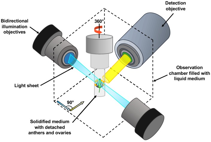
A detached flower bud with sepals and petals removed is submerged in a sugary agarose gel within a sealed capillary (grey cylinder). For long-term imaging, a closed cultivation system was created to allow the detached buds to grow under the microscope without any contamination. Light sheet fluorescent microscopy focuses a thin sheet of laser light (blue) on the specimen: this section overlaps with the focal plane of the detection pathway (in yellow). The light sheet better penetrates the sample, making the imaging of large specimens possible. Only the fluorescent protein tags within the thin sheet of laser light are excited and emit light. This eliminates the out-of-focus excitation and light emission, reducing photodamage in the rest of the sample, and therefore allowing long-term imaging. By moving the sample through the light sheet, the whole volume of the specimen can be imaged plane-by-plane. Samples can also be rotated freely, so the adjustments required by the growth of the specimen can be performed.
Comment on
-
Imaging plant germline differentiation within Arabidopsis flowers by light sheet microscopy.Elife. 2020 Feb 11;9:e52546. doi: 10.7554/eLife.52546. Elife. 2020. PMID: 32041682 Free PMC article.
References
Publication types
MeSH terms
LinkOut - more resources
Full Text Sources
