Review
. 2020 May;158(7):1913-1928.
doi: 10.1053/j.gastro.2019.11.311. Epub 2020 Feb 8.

Mechanisms of Fibrosis Development in Nonalcoholic Steatohepatitis

Affiliations
Review

Mechanisms of Fibrosis Development in Nonalcoholic Steatohepatitis

Robert F Schwabe et al. Gastroenterology. 2020 May.

Abstract

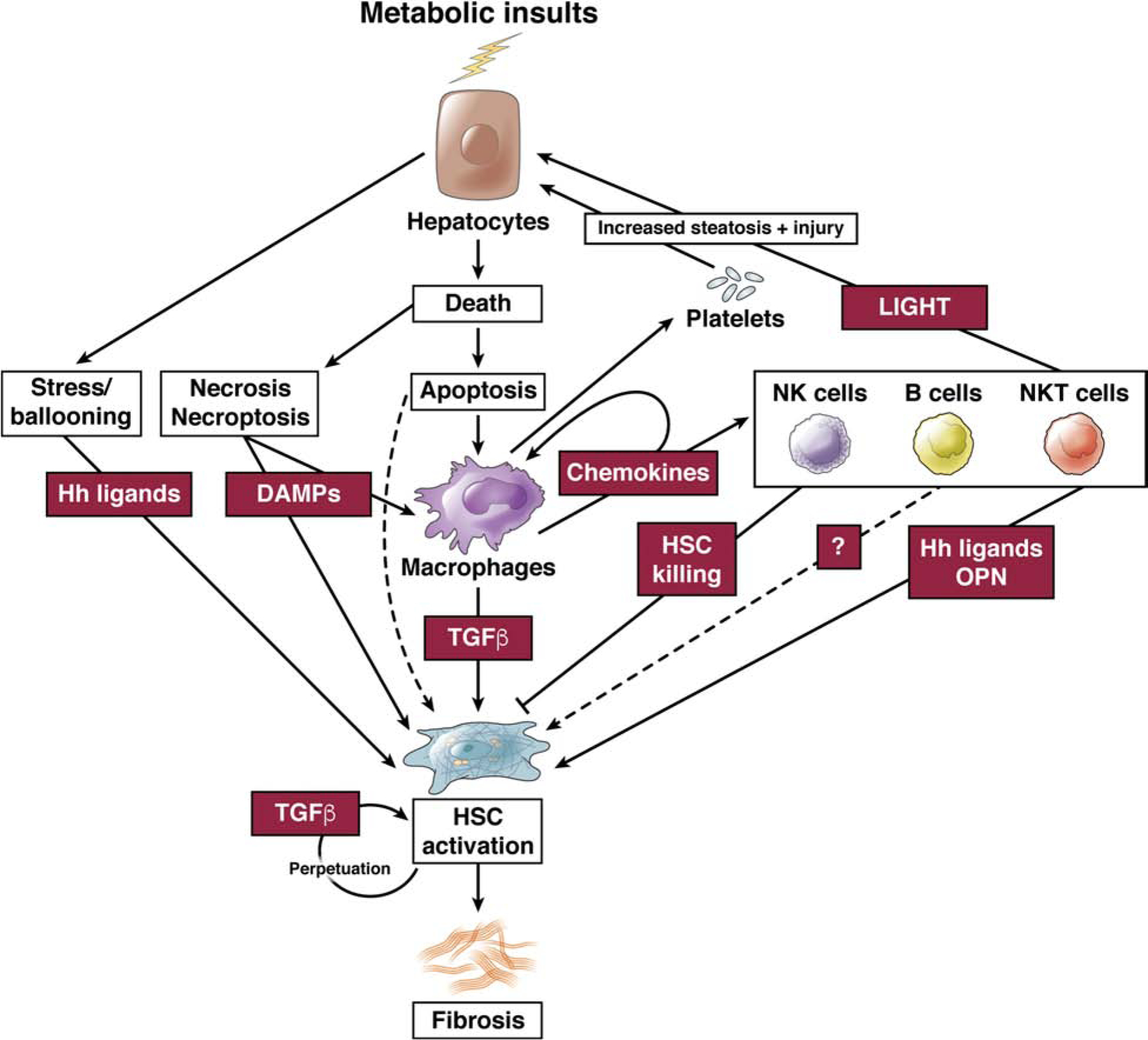
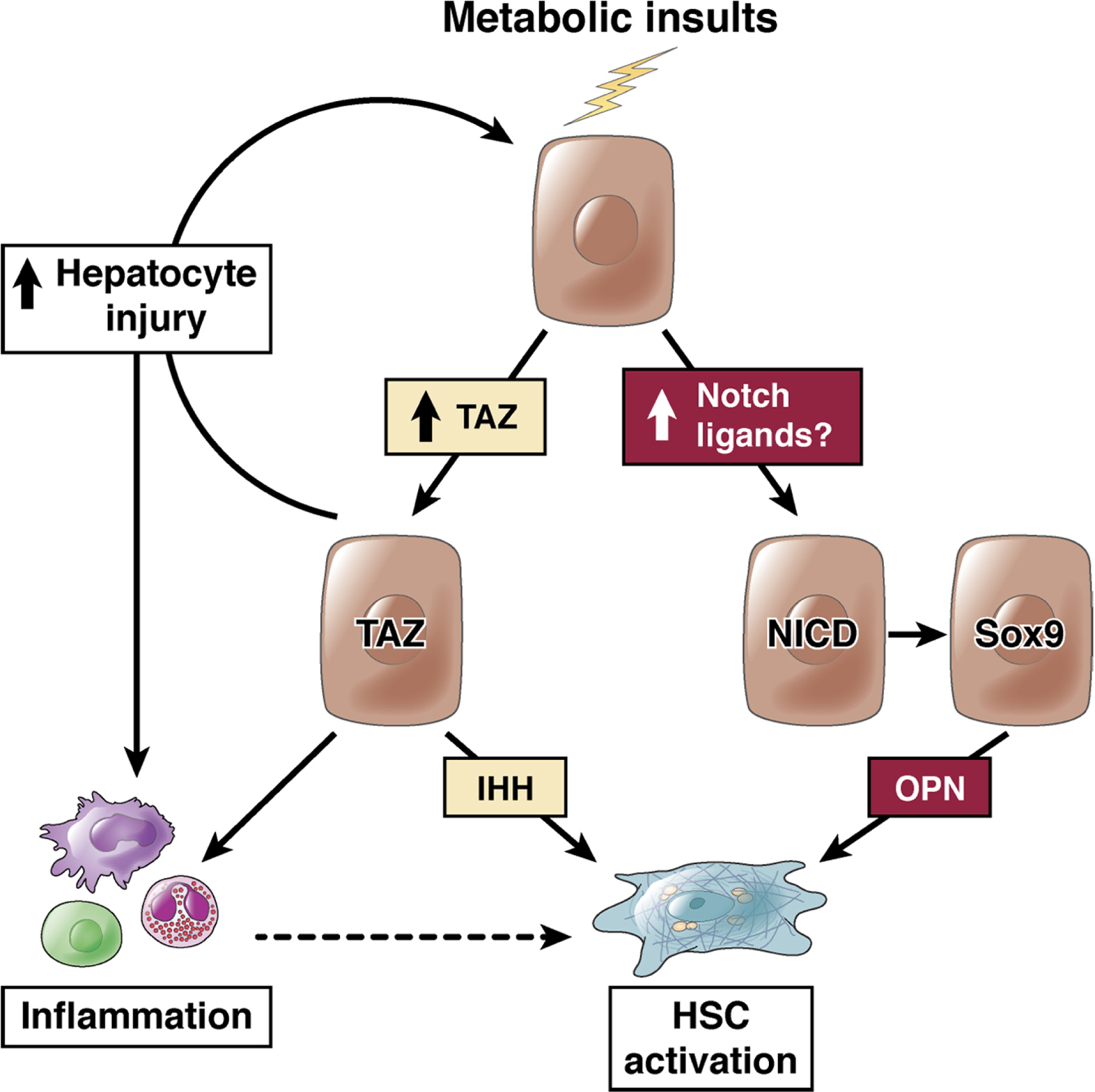
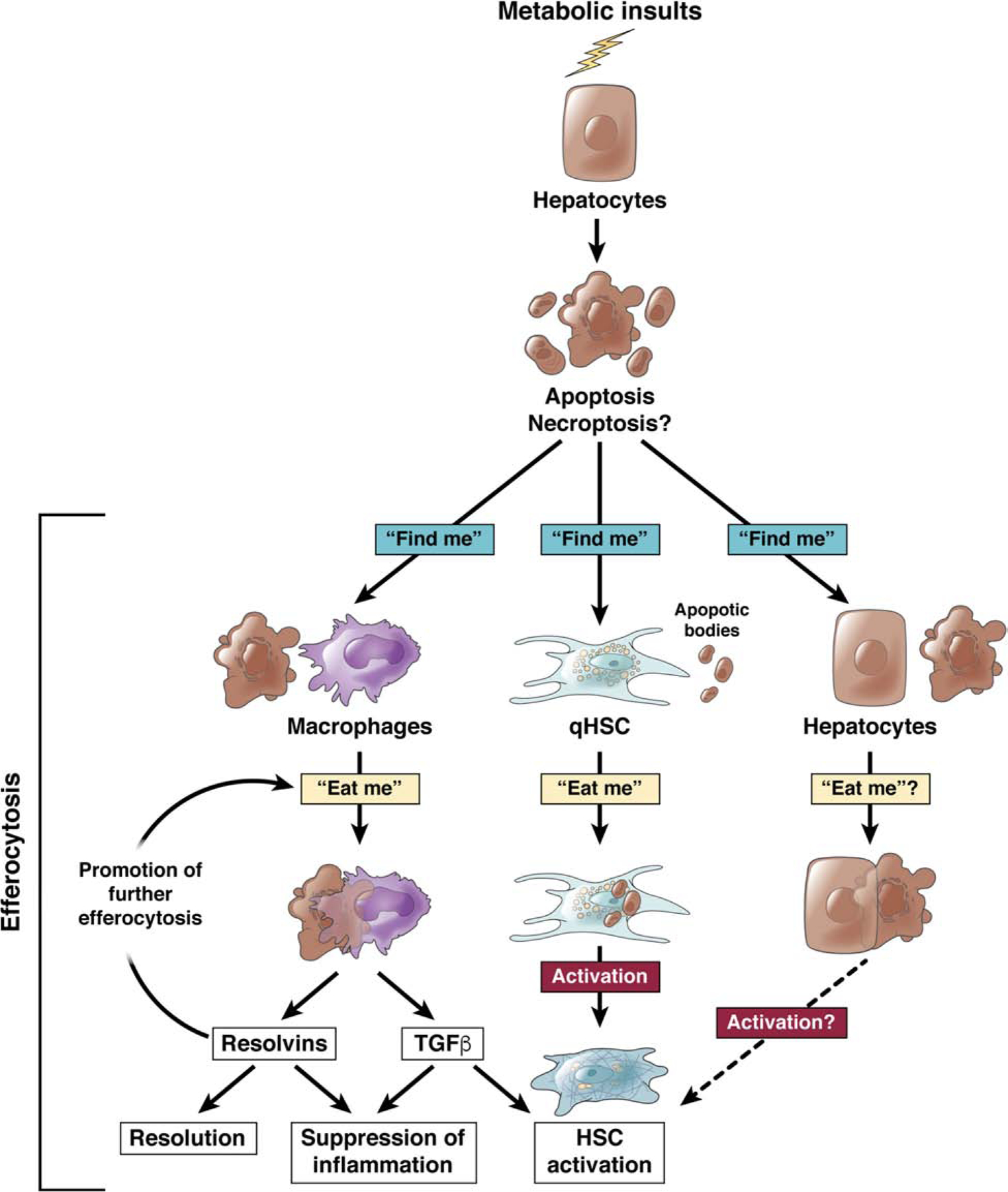
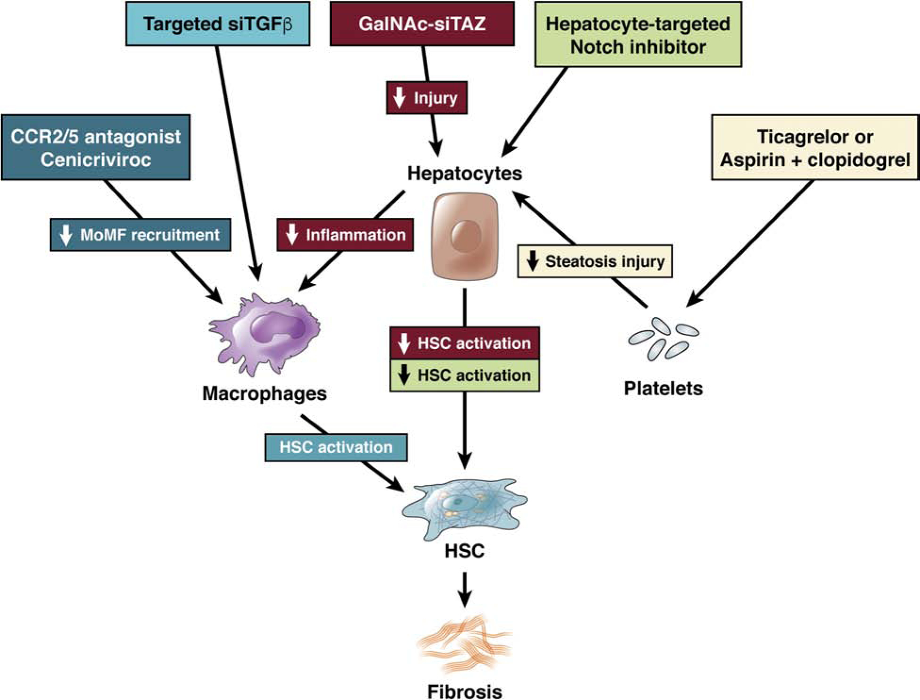
Nonalcoholic fatty liver disease is the most prevalent liver disease worldwide, affecting 20%-25% of the adult population. In 25% of patients, nonalcoholic fatty liver disease progresses to nonalcoholic steatohepatitis (NASH), which increases the risk for the development of cirrhosis, liver failure, and hepatocellular carcinoma. In patients with NASH, liver fibrosis is the main determinant of mortality. Here, we review how interactions between different liver cells culminate in fibrosis development in NASH, focusing on triggers and consequences of hepatocyte-macrophage-hepatic stellate cell (HSC) crosstalk. We discuss pathways through which stressed and dead hepatocytes instigate the profibrogenic crosstalk with HSC and macrophages, including the reactivation of developmental pathways such as TAZ, Notch, and hedgehog; how clearance of dead cells in NASH via efferocytosis may affect inflammation and fibrogenesis; and insights into HSC and macrophage heterogeneity revealed by single-cell RNA sequencing. Finally, we summarize options to therapeutically interrupt this profibrogenic hepatocyte-macrophage-HSC network in NASH.

Keywords: Drug Development; Metabolic Syndrome; Noninvasive Biomarkers; Pediatric Obesity.


Figures

Fig.1.
Fig.1.. The cellular network regulating HSC activation and fibrosis in NASH.
Metabolic insults promote hepatocyte steatosis and injury, activating a multi-cellular network consisting of macrophages, NKT cells, NK cells, B cells and NK cells that control HSC activation and the development of fibrosis.
Fig.2.
Fig.2.. The role of hepatocyte TAZ and Notch in NASH fibrosis.
Metabolic insults lead to the activation of TAZ and Notch in hepatocytes. Increased hepatocytes expression of TAZ in NASH (but not simple steatosis) directly leads to HSC activation via the release of IHH, and additionally promotes hepatocyte injury and inflammation, which may indirectly promote HSC activation. Notch activity, driven by cell-surface ligands on a neighboring cell, leads to Sox9-dependent increase in osteopontin secretion, to activate HSC.
Fig.3.
Fig.3.. Efferocytosis of dead hepatocytes as promoter of HSC activation and fibrosis.
Apoptotic hepatocytes may be detected and engulfed by macrophages in response to “find me” and “eat me” signal in a process termed efferocytosis. Efferocytosis is most commonly exerted by professional phagocytes such as macrophages, where it leads to the release of resolvins – suppressing inflammation and promoting resolution - and TGFβ - suppressing inflammation and promoting HSC activation. Efferocytosis has also been suggested to occur in HSC and promote their activation. It is conceivable that efferocytosis could occur in hepatocytes, directly or indirectly affecting HSC activation.
Fig.4.
Fig.4.. Therapeutic inhibition of NASH by targeting intercellular networks.
Targeting metabolic pathways, either in hepatocytes (ACC, SCD1, FXR/FGF19) or upstream (PPARα/δ or PPARα/γ) will improve hepatocyte metabolism and health and thereby indirectly reduce the fibrosis-promoting crosstalk with macrophages and HSC. Pathways that more directly initiate fibrosis-promoting crosstalk may be targeted at different levels.. Hepatocyte-specific TAZ silencing via GalNAc-coupled siRNA may lead to a reduction of IHH-mediated HSC activation as well as reduced inflammation and hepatocyte injury in NASH. Hepatocyte-targeted Notch inhibitors may decrease HSC activation in NASH fibrosis. Targeting the recruitment of monocyte-derived macrophages (MoMF) via CCR2/5 antagonist Cenicriviroc or the release of TGFβ from macrophages may reduce HSC activation in NASH. Targeting of the platelet-hepatocyte crosstalk via anti-platelet therapies such as Ticagrelor or aspirin+clopidogrel may reduce hepatocyte steatosis and injury, and a subsequent secondary reduction of HSC activation in NASH.

Similar articles

Cited by

References

    1. Younossi Z, Anstee QM, Marietti M, Hardy T, Henry L, Eslam M, George J, Bugianesi E. Global burden of NAFLD and NASH: trends, predictions, risk factors and prevention. Nat Rev Gastroenterol Hepatol 2018;15:11–20. - PubMed
    1. Wong RJ, Aguilar M, Cheung R, Perumpail RB, Harrison SA, Younossi ZM, Ahmed A. Nonalcoholic steatohepatitis is the second leading etiology of liver disease among adults awaiting liver transplantation in the United States. Gastroenterology 2015;148:547–55. - PubMed
    1. Diehl AM, Day C. Cause, Pathogenesis, and Treatment of Nonalcoholic Steatohepatitis. N Engl J Med 2017;377:2063–2072. - PubMed
    1. Schuppan D, Pinzani M. Anti-fibrotic therapy: lost in translation? J Hepatol 2012;56 Suppl 1:S66–74. - PubMed
    1. Friedman SL, Neuschwander-Tetri BA, Rinella M, Sanyal AJ. Mechanisms of NAFLD development and therapeutic strategies. Nat Med 2018;24:908–922. - PMC - PubMed

Publication types

MeSH terms