Review
. 2020 May;1867(5):118675.
doi: 10.1016/j.bbamcr.2020.118675. Epub 2020 Feb 8.

The diversity of ACBD proteins - From lipid binding to protein modulators and organelle tethers

Affiliations
Review

The diversity of ACBD proteins - From lipid binding to protein modulators and organelle tethers

Markus Islinger et al. Biochim Biophys Acta Mol Cell Res. 2020 May.

Abstract

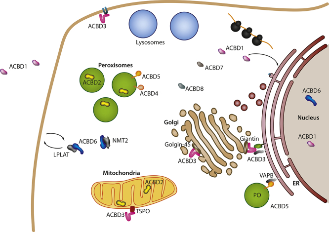
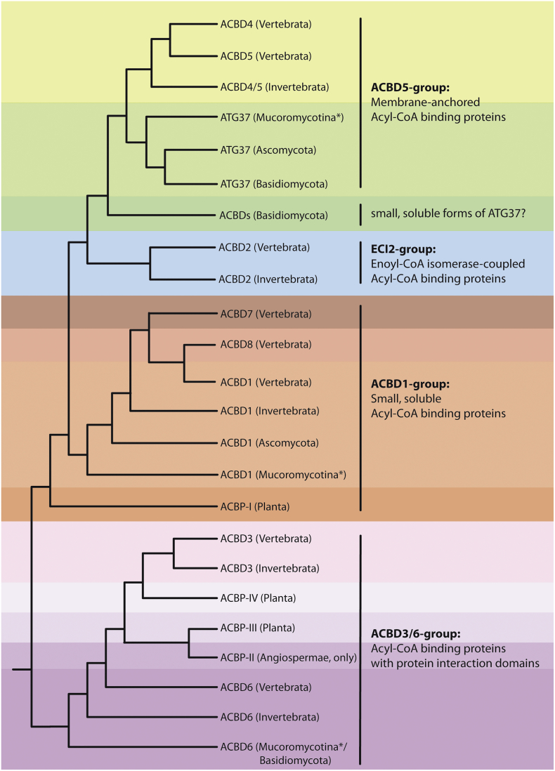
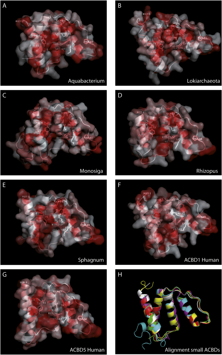
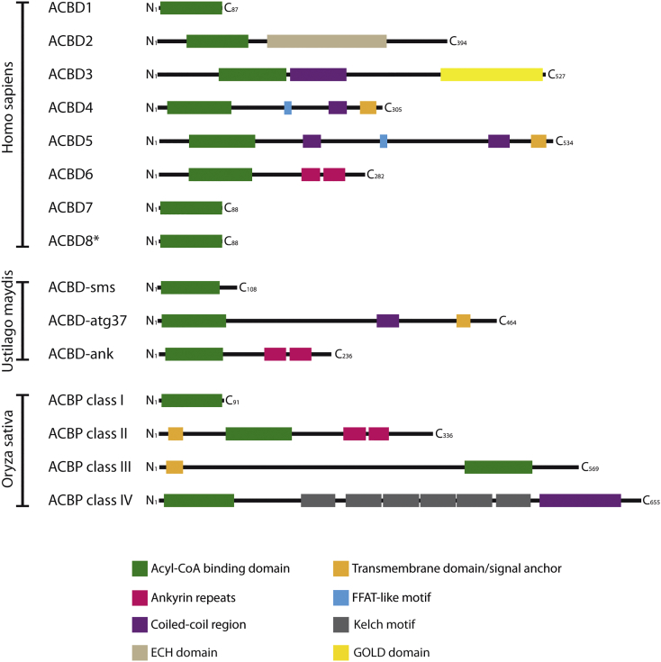
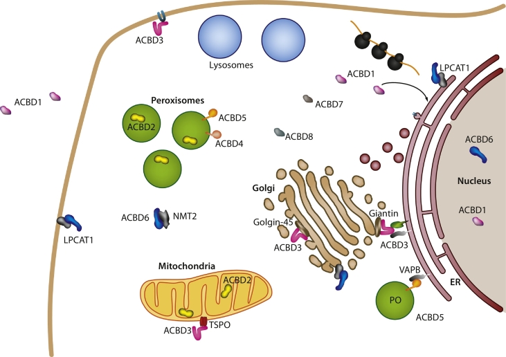
Members of the large multigene family of acyl-CoA binding domain containing proteins (ACBDs) share a conserved motif required for binding of Coenzyme A esterified fatty acids of various chain length. These proteins are present in the three kingdoms of life, and despite their predicted roles in cellular lipid metabolism, knowledge about the precise functions of many ACBD proteins remains scarce. Interestingly, several ACBD proteins are now suggested to function at organelle contact sites, and are recognized as host interaction proteins for different pathogens including viruses and bacteria. Here, we present a thorough phylogenetic analysis of the ACBD family and discuss their structure and evolution. We summarize recent findings on the various functions of animal and fungal ACBDs with particular focus on peroxisomes, the role of ACBD proteins at organelle membranes, and their increasing recognition as targets for pathogens.

Keywords: Acyl-CoA binding domain containing protein; FFAT motif; Lipid metabolism; Membrane contact sites; Pathogen host interaction; Peroxisomes.


Conflict of interest statement

Declaration of competing interest The authors declare that they have no known competing financial interests or personal relationships that could have appeared to influence the work reported in this paper.

Figures

Unlabelled Image
Graphical abstract
Fig. 1
Fig. 1
Evolution of Acyl-CoA binding domain containing proteins (ACBDs) among metazoans. The scheme summarizes data from a phylogenetic reconstruction including 449 sequences from animals, fungi and plants (for details see Fig. S1). After alignment by the ClustalW 2.0 algorithm included in the Seaview software package and manual correction of misaligned sequences, phylogenetic reconstructions were performed with PhyML3.0 using the aLRT algorithm for branch support, BioNJ for tree topology optimization, 4 rate categories for RHAS (Rate Heterogeneity Among Sites) modelling and including NNI and SPR tree searching operations. Furthermore, we used Marcoil1.0 online prediction tool [181] to screen the sequences for coiled-coil motifs and TMHMM2.0 and TMPred for TMD prediction [182,183]. Colours highlight four major categories of ACBDs – the C-terminally membrane-anchored ACBD4/5 (green), the enoyl-CoA isomerase containing ACBD2 (blue), the small soluble ACBD1 (red) and the extended forms with protein interaction domains ACBD3/6 and extended plant forms (purple; ACBP class II–IV). Taxonomic categories shown name the minimum organism group in which a distinct protein subfamily was detected. E.g. while ACBD2 was only found in vertebrate and invertebrate animals, ACBD4/5 occur in both animals and fungi, more specifically in basidiomycota and several early branching fungal divisions of incertae sedis (*symbolized with Mucoromycota in the figure). By contrast, the ankyrin repeat-containing ACBD6/ACBP II is distributed among all metazoans indicating its early evolution from the ACBD1 form. Evolution of Acyl-CoA binding domain containing proteins (ACBDs) among metazoans. The scheme summarizes data from a phylogenetic reconstruction including 449 sequences from animals, fungi and plants (for details see Fig. S1). After alignment by the ClustalW 2.0 algorithm included in the Seaview software package and manual correction of misaligned sequences, phylogenetic reconstructions were performed with PhyML3.0 using the aLRT algorithm for branch support, BioNJ for tree topology optimization, 4 rate categories for RHAS (Rate Heterogeneity Among Sites) modelling and including NNI and SPR tree searching operations. Furthermore, we used Marcoil1.0 online prediction tool [181] to screen the sequences for coiled-coil motifs and TMHMM2.0 and TMPred for TMD prediction [182,183]. Colours highlight four major categories of ACBDs – the C-terminally membrane-anchored ACBD4/5 (green), the enoyl-CoA isomerase containing ACBD2 (blue), the small soluble ACBD1 (red) and the extended forms with protein interaction domains ACBD3/6 and extended plant forms (purple; ACBP class II–IV). Taxonomic categories shown name the minimum organism group in which a distinct protein subfamily was detected. E.g. while ACBD2 was only found in vertebrate and invertebrate animals, ACBD4/5 occur in both animals and fungi, more specifically in basidiomycota and several early branching fungal divisions of incertae sedis (*symbolized with Mucoromycota in the figure). By contrast, the ankyrin repeat-containing ACBD6/ACBP II is distributed among all metazoans indicating its early evolution from the ACBD1 form.
Fig. 2
Fig. 2
3D predictions of selected acyl-CoA binding proteins/domains. The predictions were accomplished with Phyre2 using entire cDNA sequences from ACBD1 of Homo sapiens (F) and homologous small ACBDs from the β-proteobacterium Aquabacterium parvum beta (A), an hitherto undefined ACBD sequence (TFG05618) of Lokiarchaeota origin (B), the choanoflagellate Monosiga brevicollis (C), the Mucoromycota fungus Rhizopus oryzae (D), the moss Sphagnum fallax (E); for comparison the ACB domain of human ACBD5 was added subjected to 3D prediction (G). Hydrophobic regions in the protein surface structure are highlighted in red and residues conserved among all species are shown. (H) Tertiary structure reconstructions from (A)–(F) are additionally shown as sequence overlay in ribbon cartoon format to illustrate the significant structural homology in the distantly related species (depicted in different colours). Conversion of the PDB files into graphical illustrations was performed with PyMOL 2.3.2.
Fig. 3
Fig. 3
Domain structures of animal, fungal and plant acyl-CoA binding proteins. The schemes represent ACBDs from Homo sapiens (Mammalia, Animalia), Ustilago maydis (Basidiomycota, Fungi) and Oryza sativa (Angiospermae, Planta); sequence and domain lengths as well as position are proportional to the original protein structure. For some ACBDs, several isoforms have been reported or predicted. In such cases, only the currently best characterized isoform is presented. Note the structural similarity between the mammalian, fungal and plant ankyrin repeat-containing ACBDs, which suggests an early evolution already found in the LECA. ECH, enoyl-CoA hydratase domain; FFAT, two phenylalanines in an acidic tract motif.
Fig. 4
Fig. 4
Schematic overview of the localization of ACBD proteins. ACBD, acyl-CoA binding domain containing protein (ACBD1–8); ER, endoplasmic reticulum; LPLAT, acyl-CoA:lysophospholipid acyltransferase; NMT2, N-myristoyltransferase 2; PO, peroxisome; TSPO, translocator protein for cholesterol; VAPB, vesicle-associated membrane protein (VAMP)–associated protein B. For details see 3, 4, 5, 6, 7 as well as Table 1, Table 2.

References

    1. Neess D., Bek S., Engelsby H., Gallego S.F., Færgeman N.J. Long-chain acyl-CoA esters in metabolism and signaling: role of acyl-CoA binding proteins. Prog. Lipid Res. 2015;59:1–25. - PubMed
    1. Lung S.C., Chye M.L. Deciphering the roles of acyl-CoA-binding proteins in plant cells. Protoplasma. 2016;253:1177–1195. - PubMed
    1. Færgeman N.J., Knudsen J. Role of long-chain fatty acyl-CoA esters in the regulation of metabolism and in cell signalling. Biochem. J. 1997;323:1–12. - PMC - PubMed
    1. Costello J.L., Castro I.G., Hacker C., Schrader T.A., Metz J., Zeuschner D., Azadi A.S., Godinho L.F., Costina V., Findeisen P., Manner A., Islinger M., Schrader M. ACBD5 and VAPB mediate membrane associations between peroxisomes and the ER. J. Cell Biol. 2017;216:331–342. - PMC - PubMed
    1. Hua R., Cheng D., Coyaud É., Freeman S., Di Pietro E., Wang Y., Vissa A., Yip C.M., Fairn G.D., Braverman N., Brumell J.H., Trimble W.S., Raught B., Kim P.K. VAPs and ACBD5 tether peroxisomes to the ER for peroxisome maintenance and lipid homeostasis. J. Cell Biol. 2017;216:367–377. - PMC - PubMed

Publication types