Review
. 2020 Mar;32(12):e1906423.
doi: 10.1002/adma.201906423. Epub 2020 Feb 11.

From Shape to Function: The Next Step in Bioprinting

Affiliations
Review

From Shape to Function: The Next Step in Bioprinting

Riccardo Levato et al. Adv Mater. 2020 Mar.

Abstract

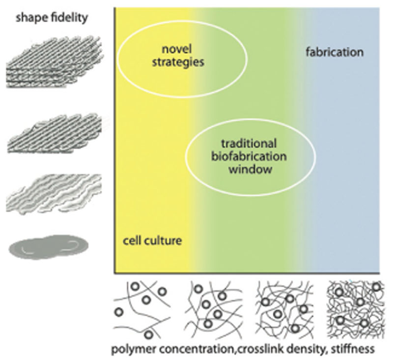
In 2013, the "biofabrication window" was introduced to reflect the processing challenge for the fields of biofabrication and bioprinting. At that time, the lack of printable materials that could serve as cell-laden bioinks, as well as the limitations of printing and assembly methods, presented a major constraint. However, recent developments have now resulted in the availability of a plethora of bioinks, new printing approaches, and the technological advancement of established techniques. Nevertheless, it remains largely unknown which materials and technical parameters are essential for the fabrication of intrinsically hierarchical cell-material constructs that truly mimic biologically functional tissue. In order to achieve this, it is urged that the field now shift its focus from materials and technologies toward the biological development of the resulting constructs. Therefore, herein, the recent material and technological advances since the introduction of the biofabrication window are briefly summarized, i.e., approaches how to generate shape, to then focus the discussion on how to acquire the biological function within this context. In particular, a vision of how biological function can evolve from the possibility to determine shape is outlined.

Keywords: biofabrication; bioinks; biological function; regenerative medicine; tissue hierarchy.


Conflict of interest statement

Conflict of Interest

The authors declare no conflict of interest.

Figures

Figure 1
Figure 1
Schematic representation of the biofabrication window as introduced in 2013. Reproduced with permission.[12] Copyright 2013, Wiley-VCH.
Figure 2
Figure 2
Overview—evolving strategies for controlling shape in bioprinting. A) To enhance chemical crosslinking strategies mainly step growth reactions and the transition from ultraviolet to visible light crosslinking have been applied more frequently. Also, technological advances like in situ photo-crosslinking can improve shape fidelity and broaden the spectrum of applicable materials based on chemical crosslinking strategies. B) In terms of physical interactions, weak bonds, like host–guest interactions and β-sheets, are applied to adjust the rheological properties of the materials. C) Rheological tuning is leading toward a two-step crosslinking utilizing a first step of crosslinking to enable printing with high shape fidelity and a second step to ensure long-term stability enabling to adjust the viscosity of the material to a level that is needed for the different fabrication steps. D) Further technological advances, like coaxial bioprinting and the application of microfluidic approaches in bioprinting, help broadening the spectrum of materials that can be applied for bioprinting. E) Also, printing into support baths is a promising strategy to enable fabrication of more sophisticated structures with less stringent demands on viscoelastic properties of bioinks.
Figure 3
Figure 3
Chemical crosslinking strategies broaden the biofabrication window of inks and improve control over their resulting physicochemical properties. A) The chain growth-based crosslinking, represented here by gelatin methacryloyl (GelMA), is susceptible to oxygen inhibition and lacks precise control over the reaction products and results in undefined oligo(methacrylates) and thus uncontrolled degradation products. In contrast, the step growth reaction, represented here by the thiol–ene reaction between allyl functionalized gelatin (GelAGE) and the crosslinker dithiothreitol (DTT), provides faster reactions and conversation rates, forms more homogeneous networks and is not prone to oxygen inhibition. Reproduced with permission.[45] Copyright 2017, Wiley-VCH. B) In situ photo-crosslinking utilizes photo-crosslinking of materials through a light permeable printing nozzle. This enables printing of low viscosity precursors and the generation of core–shell as well as filaments from different materials. Reproduced with permission.[17] Copyright 2017, Wiley-VCH. C) Visible light crosslinking is a promising strategy to improve photoreactions used in bioprinting. Shown are °CT images of GelMA/collagen constructs crosslinked with UV and Irgacure 2959 (left) or visible light and Ru/SPS (right) at the same conditions. The visible light approach resulted in more homogeneous constructs, while the UV-crosslinked construct exhibited weakly defined lattice structures with gaps and varying filament diameters. Reproduced with permission.[116] Copyright 2016, American Chemical Society.
Figure 4
Figure 4
Physical crosslinking strategies take advantage of reversible interactions to provide bioprinted filaments with structural stability, allowing to broaden the biofabrication window of low viscosity and low polymer density inks, as well as that of colloidal inks. A) From CT data to a printed cheek geometry of cm-scale using silk gelatin inks. To make the physically crosslinked, B. mori derived silk printable, it was mixed with gelatin and glycerol. The additives and the preparation enables the formation of β-sheets, increased the biological activity and helped preventing nozzle clogging. Reproduced with permission.[57] Copyright 2017, Elsevier. B) Colloidal inks from different precursors as deposited strands (scale bar 200 μm) demonstrating viscoelastic properties as proven by the intact filament spanning from the collector to the nozzle tip (i–iii). The approach shows potential to make a broader range of hydrogel systems printable due to its independency from the used crosslinking mechanism. The scale bar represents 5 mm. Reproduced under the terms of the Creative Commons CC-BY 4.0 License (https://creativecommons.org/licenses/by/4.0/).[139] Copyright 2019, The Authors, Published by Wiley-VCH. C) Extruded PEGDA without (left) and with Laponite (right). Reproduced with permission.[b] Copyright 2019, Wiley-VCH. D) Alginate methylcellulose with Laponite as viscosity enhancer, pointing out the principle of viscosity modulators and their benefits for bioprinting by significantly improving shape fidelity of the constructs. Reproduced with permission.[a] Copyright 2015, American Chemical Society.
Figure 5
Figure 5
Coaxial and microfluidic methods are versatile tools to induce rapid crosslinking of extruded filaments. Additionally, using such extrusion devices permit to print multiple materials in a single process, as well as to control the composition and physicochemical properties of the forming hydrogel strands. A) Coaxial bioprinting of an alginate-based bioink (inner compartment) and its crosslinking agent CaCl2 (outer compartment). The rapid crosslinking via calcium ions enabled in situ crosslinking ensuring high shape fidelity printing. Reproduced with permission.[149] Copyright 2016, IOPScience. B) Microfluidic approaches like the utilization of y-junctions in combination with a coaxial nozzle allowed the deposition of strands with parallel aligned multiple cell types, but has also been used to extrude different types of materials. Reproduced with permission.[18] Copyright 2017, Elsevier. C) By coaxially extruding an alginate-based ink through the outer and the crosslinking agent through the inner nozzle, perfusable inks could be produced. Reproduced with permission.[148] Copyright 2015, Elsevier. D) Microfluidic approaches can be used to generate gradients which play crucial roles in biology. Methods like flow focusing (images in the middle) can be also applied to 3D printing and help reducing filament diameters. Reproduced under the terms of the Creative Commons CC-BY 4.0 License (https://creativecommons.org/licenses/by/4.0/).[163] Copyright 2018, The Authors, Published by MDPI.
Figure 6
Figure 6
Support bath printing allows to print suspended structures and overhangs, and provides structural stability when using low viscosity bioinks, enhancing control over shape and resolution. A) Based on a CAD-model a construct from collagen I, a material that is usually not printable by itself, was printed using the support bath approach. The support bath, based on gelatin, allowed the deposition of a low viscous collagen ink, utilizing an in situ crosslinking approach, and fabrication of complex structures with high shape fidelity that closely resemble the CAD file templates. Reproduced with permission.[53] Copyright 2019, AAAS. B) The method can also be used to generate filigree multimaterial constructs which could not be printed with classical extrusion-based bioprinting. The alginate-based support bath was enzymatically degraded to gently harvest the fragile objects. Reproduced under the terms of the Creative Commons CC-BY 4.0 License (https://creativecommons.org/licenses/by/4.0/).[88] Copyright 2019, The Authors, Published by Wiley-VCH. C) Carbopol as bath material in combination with a very thin nozzle (50 μm tip) enabled printing of structures with high resolution and challenging shapes. Reproduced with permission.[168] Copyright 2015, The Authors, published by AAAS. Reproduced/modified from ref. [16]. © 2016, The Authors, some rights reserved; exclusive licensee American Association for the Advancement of Science. Distributed under a Creative Commons Attribution NonCommercial License 4.0 (CC BY-NC).
Figure 7
Figure 7
Strategies to induce anisotropy within printed constructs can be used to promote cell alignment and therefore maturation of, i.e., muscular and neural structures, as well as to generate 3D constructs with local variations of mechanical properties. A) Cardiomyocytes aligning within confined, printed hydrogel chambers. Reproduced under the terms of the Creative Commons CC-BY 3.0 License (https://creativecommons.org/licenses/by/3.0/).[38] Copyright 2018, The Authors, published by IOP Science. B) Nanocellulose particle alignment tuned by the extrusion speed and shear stress in the printing nozzle, allowing the creation of sophisticated anisotropic hydrogel patterns. Reproduced with permission.[211] Copyright 2017, Wiley-VCH. C) Fluorescently tagged fibrin-based bioinks, showing either: i) random distribution of the fibrin microfibers, when the bioink is printed at a lower printhead velocity, or ii) sub-micrometer scale alignment alongside the printing direction for higher displacement velocity, iii) which eventually drives alignment of bioprinted Schwann cells (actin-dapi staining, in green and red, respectively). Reproduced with permission.[214] Copyright 2018, IOPScience.
Figure 8
Figure 8
Bioprinted models of a cardiac ventricle, exhibiting synchronous electroconductive and contractile functions could be fabricated combining multimaterial printing and printing in a support bath, with the incorporation of high density of cardiomyocytes. A) Schematic representation of the printing process, B) construct dimensions, and C) final printed model. D) Calcium imaging of the printed structure and E) spontaneous, directional propagation of the calcium wave, indicating transmission of the action potential across the cardiomyocytes, also shown from a top view of the construct (F,G). H,I) Calcium signal propagation can be observed also after point stimulation, as also J) measured recording transient calcium waves during both spontaneous contraction or induced contractions with stimulation at 1 and 2 Hz. Reproduced with permission.[53] Copyright 2019, AAAS.
Figure 9
Figure 9
The functionality of parenchymal cells is intimately dependent on the cross-talk with stromal cells and with the vasculature, and the precise patterning of these components can be used to promote tissue maturation. A) Centimeter-scale vascularized bone constructs and bioprinted vascularized bone niche. Reproduced with permission.[24] Copyright 2016, National Academy of Sciences. B) Providing a precise 3D patterning of vascular cells within bioprinted filaments was proven beneficial to mimic healthy liver lobules, and enhance expression of cytochromes in hepatocytic cells, when compared to nonorganized mixing of endothelial cells and hepatocytes within the same bioink. Reproduced with permission.[140] Copyright 2018, Wiley-VCH. C) Free-form endothelialized vessels can be obtained via printing with sacrificial gels and further studied to understand angiogenic sprouting in 3D. Reproduced under the terms of the Creative Commons CC-BY 3.0 License (https://creativecommons.org/licenses/by/3.0/).[281] Copyright 2018, The Authors, published by IOPScience.
Figure 10
Figure 10
Possible approaches toward the convergence of bioprinting and self-organization to guide the maturation of bioprinted constructs toward the generation of functional tissues. Inspired by the composition of adult, native tissues, multiple progenitor or differentiated cells can be loaded into bioinks to build tissues or organoids. In this approach, the architecture imposed by the printing process will be templating the cell-driven development of the tissue and its subsequent maturation. Alternatively, specific stem and progenitor cells that possess the ability to autonomously organize into submillimeter to millimeter organoids that exhibit salient tissue features can be used as intermediate building blocks and as bioink components. In both processes, the stimuli provided by the biomaterials, their architecture, and bioactive factors included in the bioinks play key roles for driving the acquisition of native functions.

References

    1. Lincoln J, Lange AW, Yutzey KE. Dev Biol. 2006;294:292. - PubMed
    1. Rose JC, De Laporte L. Adv Healthcare Mater. 2018;7 1701067.
    1. Moroni L, Boland T, Burdick JA, De Maria C, Derby B, Forgacs G, Groll J, Li Q, Malda J, Mironov VA, Mota C, Nakamura M, Shu W, Takeuchi S, Woodfield TBF, Xu T, Yoo JJ, Vozzi G. Trends Biotechnol. 2018;36:384. - PubMed
    1. Groll J, Boland T, Blunk T, Burdick JA, Cho DWW, Dalton PD, Derby B, Forgacs G, Li Q, Mironov VA, Moroni L, Nakamura M, Shu W, Takeuchi S, Vozzi G, Woodfield TBFF, Xu T, Yoo JJ, Malda J. Biofabrication. 2016;8 013001. - PubMed
    1. Ozbolat IT, Hospodiuk M. Biomaterials. 2016;76:321. - PubMed

LinkOut - more resources