A Fluorescent Teixobactin Analogue
- PMID: 32045203
- PMCID: PMC7814985
- DOI: 10.1021/acschembio.9b00908
A Fluorescent Teixobactin Analogue
Abstract
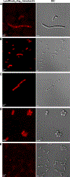
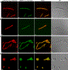
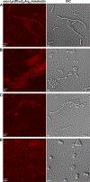
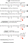
This report describes the first synthesis and application of a fluorescent teixobactin analogue that exhibits antibiotic activity and binds to the cell walls of Gram-positive bacteria. The teixobactin analogue, Lys(Rhod)9,Arg10-teixobactin, has a fluorescent tag at position 9 and an arginine in place of the natural allo-enduracididine residue at position 10. The fluorescent teixobactin analogue retains partial antibiotic activity, with minimum inhibitory concentrations of 4-8 μg/mL across a panel of Gram-positive bacteria, as compared to 1-4 μg/mL for the unlabeled Arg10-teixobactin analogue. Lys(Rhod)9,Arg10-teixobactin is prepared by a regioselective labeling strategy that labels Lys9 with an amine-reactive rhodamine fluorophore during solid-phase peptide synthesis, with the resulting conjugate tolerating subsequent solid-phase peptide synthesis reactions. Treatment of Gram-positive bacteria with Lys(Rhod)9,Arg10-teixobactin results in septal and lateral staining, which is consistent with an antibiotic targeting cell wall precursors. Concurrent treatment of Lys(Rhod)9,Arg10-teixobactin and BODIPY FL vancomycin results in septal colocalization, providing further evidence that Lys(Rhod)9,Arg10-teixobactin binds to cell wall precursors. Controls with either Gram-negative bacteria, or an inactive fluorescent homologue with Gram-positive bacteria, showed little or no staining in fluorescence micrographic studies. Lys(Rhod)9,Arg10-teixobactin can thus serve as a functional probe to study Gram-positive bacteria and their interactions with teixobactin.
Conflict of interest statement
Notes
The authors declare no competing financial interest.
ASSOCIATED CONTENT
Supporting Information
The Supporting Information is available free of charge on the ACS Publications website at DOI: Additional fluorescence micrographic images; HPLC traces, mass spectra, and excitation and emission spectra of Lys(Rhod)9,Arg10-teixobactin and
Figures


References
-
- Stone MRL, Butler MS, Phetsang W, Cooper MA, and Blaskovich MAT (2018) Fluorescent antibiotics: new research tools to fight antibiotic resistance. Trends Biotechnol. 36, 523–536. - PubMed
-
- Calloway NT, Choob M, Sanz A, Sheetz MP, Miller LW, and Cornish VW (2007) Optimized fluorescent trimethoprim derivatives for in vivo protein labeling. ChemBioChem 8, 767–774. - PubMed
-
- Akram AR, Avlonitis N, Lilienkampf A, Perez-Lopez AM, McDonald N, Chankeshwara SV, Scholefield E, Haslett C, Bradley M, Dhaliwal K (2015) A labelled-ubiquicidin antimicrobial peptide for immediate in situ optical detection of live bacteria in human alveolar lung tissue. Chem. Sci 6, 6971–6979. - PMC - PubMed
Publication types
MeSH terms
Substances
Grants and funding
LinkOut - more resources
Full Text Sources
Other Literature Sources
Medical
Molecular Biology Databases

