Review
. 2020 Jun;93(1110):20190118.
doi: 10.1259/bjr.20190118. Epub 2020 Feb 20.

The urachus revisited: multimodal imaging of benign & malignant urachal pathology

Affiliations
Review

The urachus revisited: multimodal imaging of benign & malignant urachal pathology

Jeeban Paul Das et al. Br J Radiol. 2020 Jun.

Abstract

The urachus is a fibrous tube extending from the umbilicus to the anterosuperior bladder dome that usually obliterates at week 12 of gestation, becoming the median umbilical ligament. Urachal pathology occurs when there is incomplete obliteration of this channel during foetal development, resulting in the formation of a urachal cyst, patent urachus, urachal sinus or urachal diverticulum. Patients with persistent urachal remnants may be asymptomatic or present with lower abdominal or urinary tract symptoms and can develop complications. The purpose of this review is to describe imaging features of urachal remnant pathology and potential benign and malignant complications on ultrasound, CT, positron emission tomography CT and MRI.


Figures

Figure 1.
Figure 1.
Schematic diagram (a) showing a normal urachus. Saggital (b) and axial (c) CT images following intravenous contrast demonstrating a normal urachus extending into Retzius space.
Figure 2.
Figure 2.
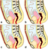
Schematic diagrams showing common urachal remnant pathology. (a) Urachal cyst; (b) Urachal sinus; (c) Patent urachus; (d) Urachal diverticulum.
Figure 3.
Figure 3.
38-year-old male with a lower urinary tract symptoms. Axial (a) and saggital (b) CT images after intravenous contrast showing a supravesical cystic structure without definite communication with the bladder or umbilicus confirmed as a urachal cyst following image-guided aspiration.
Figure 4.
Figure 4.
70-year-old male with back pain. Saggital (a) and axial (b) CT images following intravenous contrast showing a urachal diverticulum (arrows) detected as an incidental finding.
Figure 5.
Figure 5.
5-year-old child with fever and urinary retention. Longitudinal (a) and transverse (b) greyscale ultrasound images demonstrating a lesion containing internal echoes (arrows) anterosuperior to the urinary bladder (*). Surgical excision following intravenous antibiotics confirmed diagnosis of an infected urachal cyst.
Figure 6.
Figure 6.
49-year-old male with recurrent urinary tract infections. Saggital (a) and axial (b) CT images following intravenous contrast demonstrating an irregular cystic structure anterosuperior to the urinary bladder demonstrating peripheral enhancement, confirmed as an infected urachal cyst following surgical excision.
Figure 7.
Figure 7.
21-year-old male with 2-week history of fever and tender suprapubic mass. Axial CT image after intravenous contrast showing an irregular, mixed attenuation lesion anterosuperior to the bladder dome (arrow) with surrounding inflammatory change. Image-guided aspiration yielded purulent material. Subsequent surgical excision confirmed diagnosis of a urachal abscess.
Figure 8.
Figure 8.
28-year-old female with pelvic pain and fever. Axial (a) and sagittal (b) T2 weighted images showing a multiloculated heterogenously T2 hyperintense collection anterosuperior to the bladder (*), confirmed as a urachal abscess following surgical intervention.
Figure 9.
Figure 9.
32-year-old female with abdominal swelling and lower urinary tract symptoms. Axial CT image following intravenous contrast showing a solid-cystic mass (arrow) containing a focus of curvlinear calcification (arrowhead) anterosuperior to the urinary bladder (*). Histopathology confirmed the diagnosis of urachal adenocarcinoma, following surgical excision.
Figure 10.
Figure 10.
39-year-old female with abdominal bloating and mucosuria. Axial CT image demonstrating a well-defined cystic structure with mural thickening and peripheral calcification (arrowhead) anterosuperior to the urinary bladder (*). Image-guided aspiration and histopathology confirmed mucinous urachal adenocarcinoma.
Figure 11.
Figure 11.
41-year-old male with haematuria. Axial CT image showing an enhancing soft-tissue nodule (arrow) arising from left anterosuperior wall of the bladder (*). Histopathology following cystoscopy and biopsy confirmed diagnosis of urachal adenocarcinoma.
Figure 12.
Figure 12.
39-year-old female with abdominal bloating. Saggital (a) and coronal (b) T2 weighted magnetic resonance images show a paramidline cystic mass arising from the dome of the bladder (*). Histopathology confirmed mucinous urachal adenocarcinoma.
Figure 13.
Figure 13.
45-year-old female with haemturia and abdominal bloating. Axial (a) and sagittal (b) T2 weighted images demonstrate a complex, multilobulated mass involving the dome of the urinary bladder (*) with intravesical extension (arrowhead). Histopathology confirmed mucinous urachal adenocarcinoma.
Figure 14.
Figure 14.
47-year-old female with haematuria. Axial (a) and sagittal (b) 18-fluorine FDG PET CT showing an FDG-avid supravesical mass (arrows) containg punctate calcifications demonstrating a maximum SUV of 6.8. Biopsy and subsequent resection confirmed the diagnosis of urachal adenocarconoma. FDG, fludeoxyglucose; PET, positron emission tomography; SUV, standardized uptake value.

References

    1. Parada Villavicencio C, Adam SZ, Nikolaidis P, Yaghmai V, Miller FH . Imaging of the urachus: anomalies, complications, and mimics . Radiographics 2016. ; 36: 2049 – 63 . doi: 10.1148/rg.2016160062 - DOI - PubMed
    1. Ashley RA, Inman BA, Routh JC, Rohlinger AL, Husmann DA, Kramer SA . Urachal anomalies: a longitudinal study of urachal remnants in children and adults . Journal of Urology 2007. ; 178( 4S ): 1615 – 8 . doi: 10.1016/j.juro.2007.03.194 - DOI - PubMed
    1. JS Y, Kim KW, Lee HJ, Lee YJ, Yoon CS, Kim MJ . Urachal remnant diseases: spectrum of CT and US findings . RadioGraphics 2001. ; 2: 451 – 61 . - PubMed
    1. Goldman IL, Caldamone AA, Gauderer M, Hampel N, Wesselhoeft CW, Elder JS . Infected urachal cysts: a review of 10 cases . Journal of Urology 1988. ; 140: 375 – 8 . doi: 10.1016/S0022-5347(17)41612-0 - DOI - PubMed
    1. Ilica AT, Mentes O, Gur S, Kocaoglu M, Bilici A, Coban H . Abscess formation as a complication of a ruptured urachal cyst . Emerg Radiol 2007. ; 13: 333 – 5 . doi: 10.1007/s10140-006-0560-2 - DOI - PubMed