Practice Guideline
. 2020 Apr 1;38(10):1102-1111.
doi: 10.1200/JCO.19.02778. Epub 2020 Feb 11.

Best Practices Guideline for the Pathologic Diagnosis of Breast Implant-Associated Anaplastic Large-Cell Lymphoma

Affiliations
Practice Guideline

Best Practices Guideline for the Pathologic Diagnosis of Breast Implant-Associated Anaplastic Large-Cell Lymphoma

Elaine S Jaffe et al. J Clin Oncol. .

Abstract

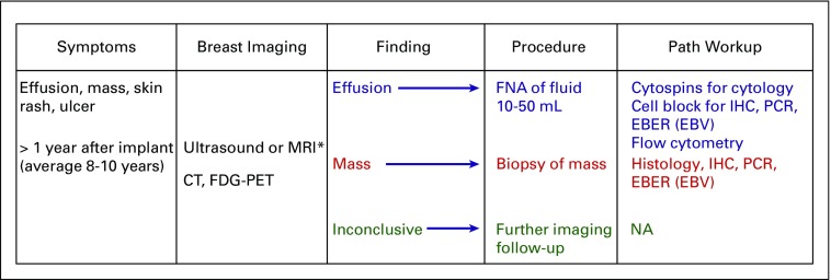
Purpose: To provide guidelines for the accurate pathologic diagnosis of breast implant-associated anaplastic large cell lymphoma (BIA-ALCL), the preoperative evaluation of the patient with suspected BIA-ALCL, and the pathologic evaluation of the capsulectomy specimen.

Methods: To better inform patients and healthcare providers about BIA-ALCL, we convened to review diagnostic procedures used in the evaluation of patients with suspected BIA-ALCL. We focused on the processing of the seroma fluid/effusion surrounding the implant, the handling of capsulectomy specimens following removal of implant(s), and the preoperative evaluation of the patient with suspected BIA-ALCL. Recommendations were based on the published literature and our experience to optimize procedures to obtain an accurate diagnosis and assess for tumor invasion and the extent of the disease.

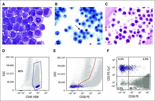
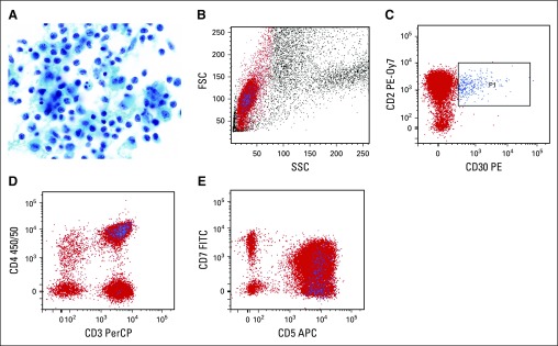
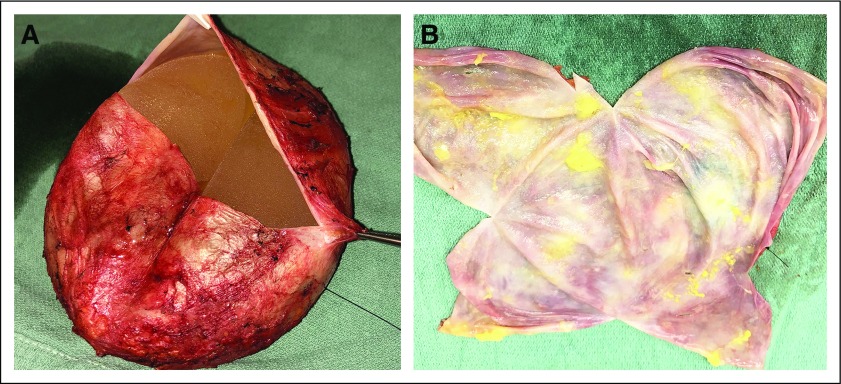
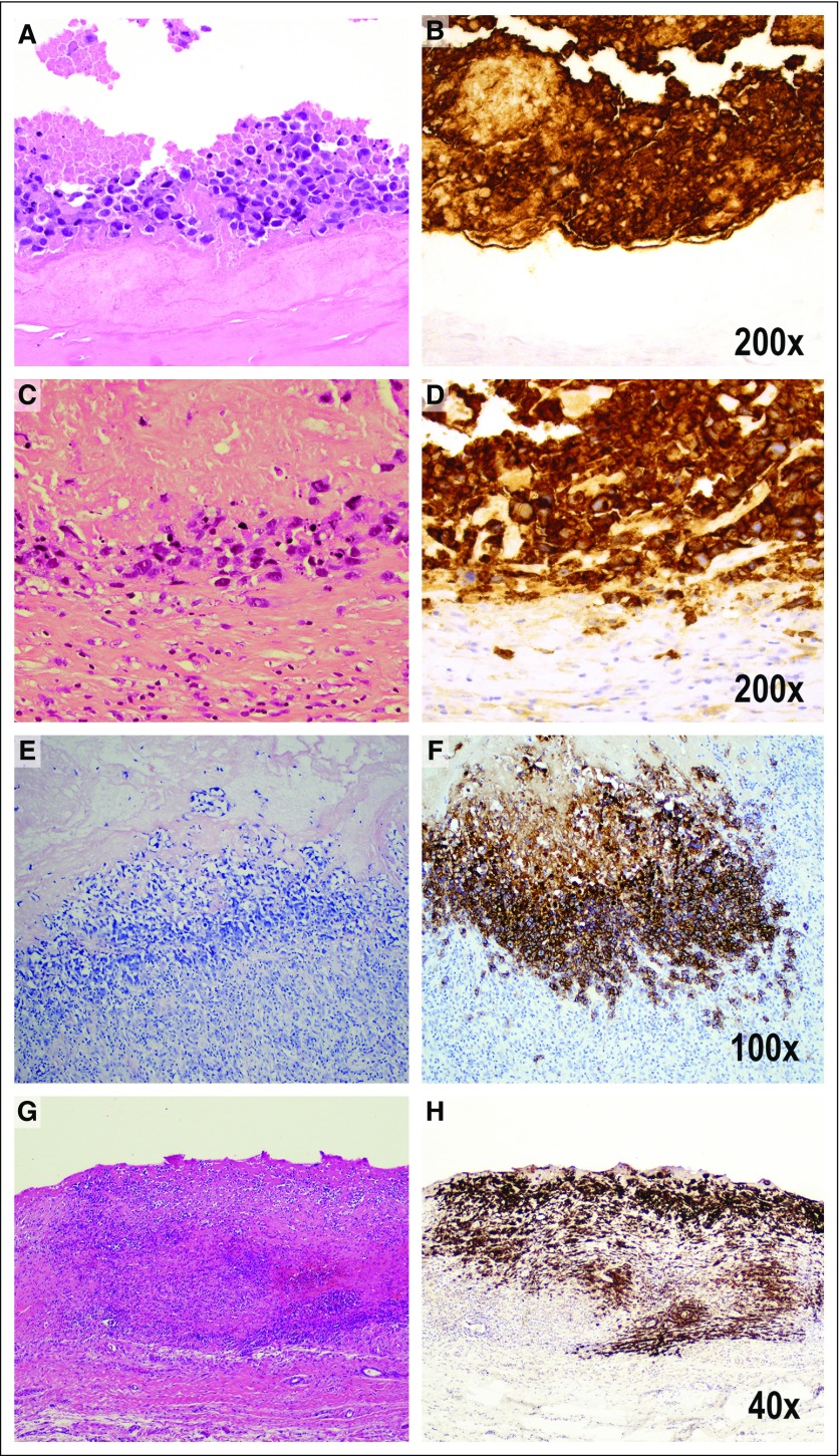
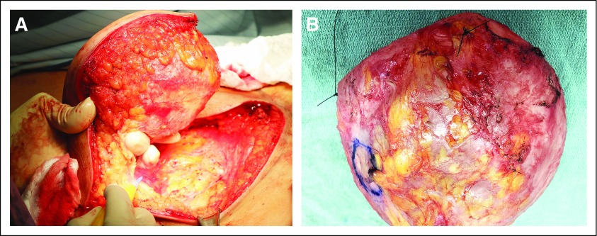
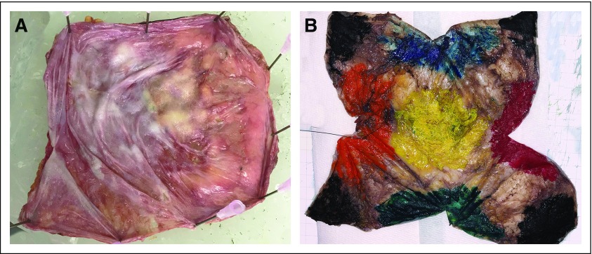
Recommendations: Early diagnosis of BIA-ALCL is important as the disease can progress and deaths have been reported. Because the most common presentation of BIA-ALCL is swelling of the breast with fluid collection, an accurate diagnosis requires cytologic evaluation of the effusion fluid surrounding the affected implant. The first priority is cytocentrifugation and filtration of fresh, unfixed effusion fluid to produce air-dried smears that are stained with Wright-Giemsa or other Romanowsky-type stains. Preparation of a cell block is desirable to allow for hematoxylin and eosin staining and immunohistochemical analysis of formalin-fixed, paraffin-embedded histologic sections. Cell block sections can be used for polymerase chain reaction-based investigation of T-cell receptor gene rearrangement to detect clonality. Fixation and mapping of the capsulectomy specimen to select multiple representative sections are advised to assess for microscopic tumor involvement and capsular invasion. It is appropriate to assess lymph node involvement by excisional biopsy material rather than fine needle aspiration, due to propensity for focal involvement.


Figures

FIG 1.
FIG 1.
Cytologic features of breast implant–associated anaplastic large-cell lymphoma (BIA-ALCL). (A) The neoplastic cells are large and pleomorphic with irregular nuclei, large prominent nucleoli, conspicuous mitotic activity, and moderate cytoplasm, which is basophilic and contains prominent vacuoles on air-dried preparations stained with a Romanowsky-type method (Giemsa, ×1,000). (B) The vesicular chromatin and nucleolar prominence and shape are better appreciated on the alcohol-fixed thin-layer preparation, which also contains some kidney-shaped hallmark cells (arrow; Papanicolaou, ×1,000). (C) Similar features are seen on the cell block preparation (hematoxylin and eosin, ×1,000). (D) Flow cytometric analysis of a BIA-ALCL effusion; in a histogram plotting CD45 versus side scatter, ALCL cells usually distribute in the lymphocyte (purple dots) and monocyte (gray dots) regions, that in this particular patient, represent 82% of cells in the specimen. (E) In a histogram plotting CD30 versus side scatter, CD30+ cells are distributed below the highlighted area (gray dots), whereas most lymphocytes are small and negative for CD30 (purple dots). (F) Histogram shows the distribution of selected (gated) cells in the histogram at top. It shows that 89.1% of gated cells express CD30, whereas 5.3% of cells are CD3+ lymphocytes but CD30−. It can be concluded that the aberrant cells (gray dots) are CD45+, CD30+, and CD3−, whereas the normal small T lymphocytes (purple dots) are CD45+, CD30−, and CD3+. PE-Cy7, phycoerythrin cyanine 7; SSC, side scatter; V500-A, Becton-Dickinson Horizon V500 (San Jose, CA) for flow cytometry.
FIG 2.
FIG 2.
Benign seroma aspirate containing nonspecific peri-implant inflammatory reaction. (A) There is a predominance of histiocytes (macrophages) and small lymphocytes. Some of the histiocytes show reactive features of curved, irregular nuclei, small nucleoli, and moderately abundant granular cytoplasm. However, their cell size is less than half that of ALCL cells, and the chromatin features are more delicate, with smaller nucleoli (Papanicolaou, ×1,000). (B-E) Flow cytometry was performed for T-cell markers and CD30. (B) On light scatter analysis, gating is performed on lymphocytes, highlighted in red, based on their low forward scatter (FSC) and side scatter (SSC) properties. (C) CD30-positive cells, highlighted in blue (P1), are small based on light scatter properties (B) and have a normal T-cell phenotype with preserved expression of CD2(C), CD3 (D) and CD5 (E), normal variable expression of CD7 (E), and predominant staining for CD4 (D), consistent with a reactive T-cell population. APC, allophycocyanin; FITC, fluorescein isothiocyanate; PE, phycoerythrin; PE-0y7, phycoerythrin-cyanine 7; PerCP, peridinin chlorophyll protein.
FIG 3.
FIG 3.
(A) The pathologist acknowledges the specimen is intact and contains landmark sutures denoting lateral (long-stitch) and superior (short-stitch) margins. After aspiration of any fluid/effusion, open the specimen through a transverse incision of the posterior aspect of the specimen. (B) After the transverse incision, a superior-to-inferior longitudinal incision allows exposure of the luminal side of the capsule, as well as assessment of the integrity of the implant.
FIG 4.
FIG 4.
Pathologic staging that reflects tumor progression from the luminal surface to the outer boundaries of the capsule. Pathologic stage T0 is not illustrated because it indicates the presence of tumor cells in the effusion, but not detected in the capsule. Pathologic stage T1 indicates (A) the lymphoma cells lie on the luminal side of the capsule, and CD30 highlights both viable and necrotic tumor cells, many of which are (B) ghosts in a background of granular material indicating cellular remnants. The tumor cells are insulated from the rest of the capsule by a dense acellular collagenous layer. Therefore, CD30 is much more extensive than the amount of identifiable viable tumor cells (A, hematoxylin and eosin [H&E]; B, CD30 immunohistochemistry, ×200). Pathologic stage T2 indicates few inflammatory cells admixed with deeper lymphoma cells (C, H&E; D, CD30 immunohistochemistry, ×200). Stage T3 indicates that numerous inflammatory cells surround lymphoma cells (E, H&E; F, CD30 immunohistochemistry, ×100). Stage 4 indicates that the tumor cells are beyond the boundaries of the fibrous capsule (G, H&E; H, CD30 immunohistochemistry, ×40).
FIG 5.
FIG 5.
Flowchart for the diagnosis of breast implant–associated anaplastic large-cell lymphoma (BIA-ALCL). Color coding indicates the preferred diagnostic approach based on the results of imaging studies, disclosing effusion, mass, or inconclusive findings. CT, computed tomography; FDG-PET, [18F]fluorodeoxyglucose–positron emission tomography; EBER, in situ hybridization for EBV; EBV, Epstein-Barr virus; FNA, fine-needle aspiration; IHC, immunohistochemistry; MRI, magnetic resonance imaging; NA, not applicable; PCR, polymerase chain reaction for T-cell receptor gene rearrangement, or immunoglobulin rearrangement, as indicated. (*) Preferred initial imaging approach. Figure modified from the 2018 National Comprehensive Cancer Network Clinical Practice Guidelines in Oncology.
FIG A1.
FIG A1.
(A) En bloc resection of the intact capsule containing the implant, with or without effusion or tumor mass, to assess margins and the extent of disease. (B) En bloc resected specimen handled for pathologic examination. It is essential to orient the specimen; in this photo, the lateral margin is marked with a long stitch and the superior margin with a short stich.
FIG A2.
FIG A2.
(A) Once the capsule lies flat, the capsule is pinned flat on a paraffin board and submerged in neutral formalin overnight. (B) After overnight fixation, the specimen is oriented, and the outer surface of the capsule is inked with 6 different colors denoting 6 different aspects of the specimen. For each aspect/side, different colors are used. Illustrated are anterior (yellow), superior (blue), inferior (green), medial (red), lateral (orange), and posterior (black). The color pattern allows mapping any lesion that is not grossly identifiable and is discovered on microscopic examination. The ink also serves to indicate the distance between the deepest site of involvement and the margin.

Comment in

References

    1. Swerdlow SH, Campo E, Pileri SA, et al. The 2016 revision of the World Health Organization classification of lymphoid neoplasms. Blood. 2016;127:2375–2390. - PMC - PubMed
    1. McCarthy CM, Loyo-Berrios N, Qureshi AA, et al: Patient Registry and Outcomes for Breast Implants and Anaplastic Large Cell Lymphoma Etiology and Epidemiology (PROFILE): Initial report of findings, 2012-2018. Plast Reconstr Surg 143:65s-73s, 2019 (suppl) - PMC - PubMed
    1. Keech JA, Jr, Creech BJ. Anaplastic T-cell lymphoma in proximity to a saline-filled breast implant. Plast Reconstr Surg. 1997;100:554–555. - PubMed
    1. de Jong D, Vasmel WL, de Boer JP, et al. Anaplastic large-cell lymphoma in women with breast implants. JAMA. 2008;300:2030–2035. - PubMed
    1. Laurent C, Delas A, Gaulard P, et al: Breast implant-associated anaplastic large cell lymphoma: Two distinct clinicopathological variants with different outcomes. Ann Oncol 27:306-314, 2016. - PMC - PubMed

Publication types

MeSH terms