Review
. 2020 Apr 1:106:1-21.
doi: 10.1016/j.actbio.2020.02.006. Epub 2020 Feb 9.

Breast cancer models: Engineering the tumor microenvironment

Affiliations
Review

Breast cancer models: Engineering the tumor microenvironment

Gokhan Bahcecioglu et al. Acta Biomater. .

Abstract

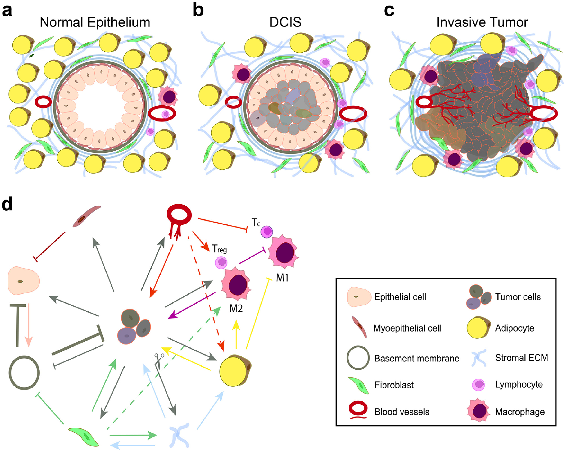
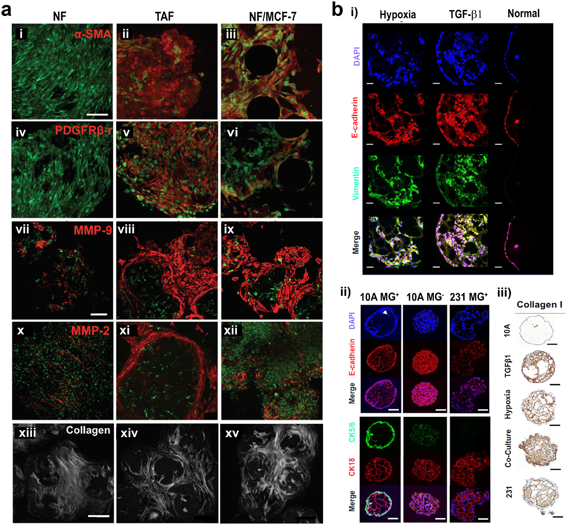
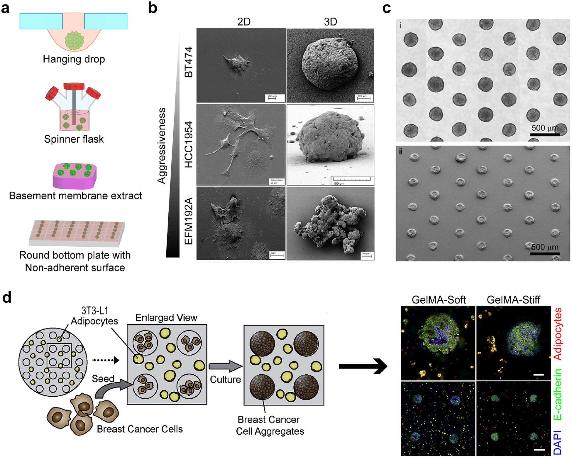
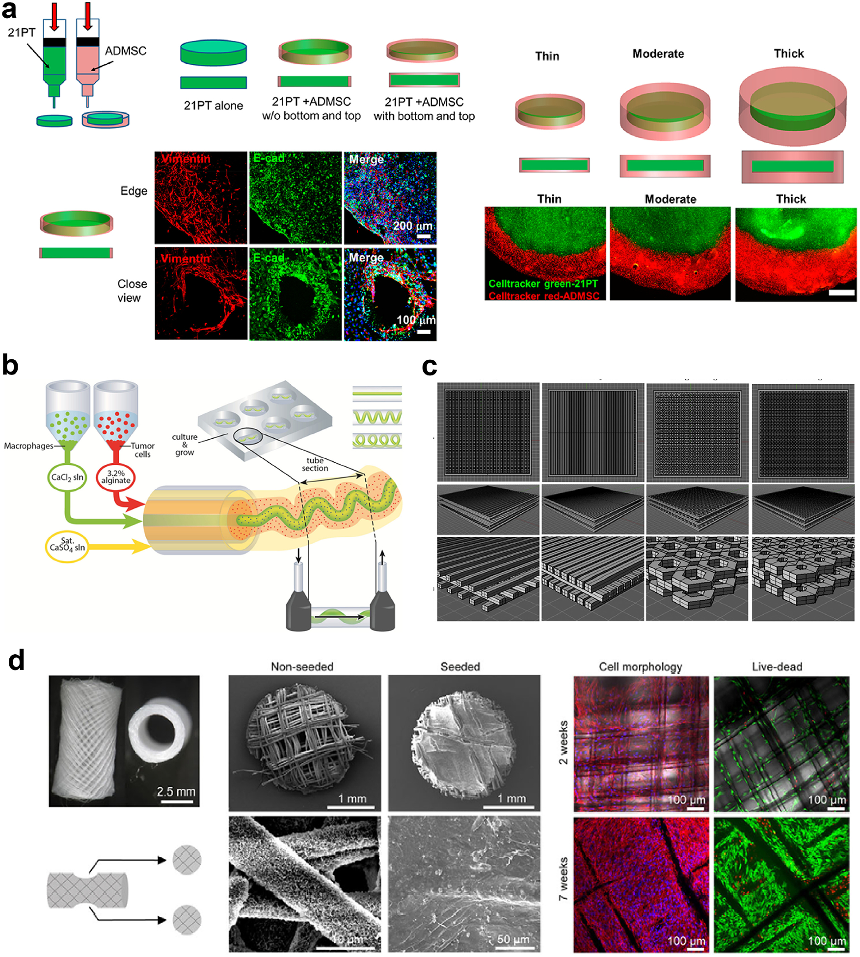
The mechanisms behind cancer initiation and progression are not clear. Therefore, development of clinically relevant models to study cancer biology and drug response in tumors is essential. In vivo models are very valuable tools for studying cancer biology and for testing drugs; however, they often suffer from not accurately representing the clinical scenario because they lack either human cells or a functional immune system. On the other hand, two-dimensional (2D) in vitro models lack the three-dimensional (3D) network of cells and extracellular matrix (ECM) and thus do not represent the tumor microenvironment (TME). As an alternative approach, 3D models have started to gain more attention, as such models offer a platform with the ability to study cell-cell and cell-material interactions parametrically, and possibly include all the components present in the TME. Here, we first give an overview of the breast cancer TME, and then discuss the current state of the pre-clinical breast cancer models, with a focus on the engineered 3D tissue models. We also highlight two engineering approaches that we think are promising in constructing models representative of human tumors: 3D printing and microfluidics. In addition to giving basic information about the TME in the breast tissue, this review article presents the state-of-the-art tissue engineered breast cancer models. STATEMENT OF SIGNIFICANCE: Involvement of biomaterials and tissue engineering fields in cancer research enables realistic mimicry of the cell-cell and cell-extracellular matrix (ECM) interactions in the tumor microenvironment (TME), and thus creation of better models that reflect the tumor response against drugs. Engineering the 3D in vitro models also requires a good understanding of the TME. Here, an overview of the breast cancer TME is given, and the current state of the pre-clinical breast cancer models, with a focus on the engineered 3D tissue models is discussed. This review article is useful not only for biomaterials scientists aiming to engineer 3D in vitro TME models, but also for cancer researchers willing to use these models for studying cancer biology and drug testing.

Keywords: 3D tumor models; Bioprinting; Breast cancer; Microfluidics; Tissue engineering; Tumor microenvironment.


Conflict of interest statement

Declaration of Competing Interest None.

Figures

Figure 1.
Figure 1.
Interactions between cells and ECM lead to alteration of normal epithelium towards the tumor. (a) Normal epithelium, (b) ductal carcinoma in situ (DCIS), and (c) invasive tumor. (d) Simplified illustration of the network of interactions between cells and the ECM. Cross-talk between the tumor cells, stromal cells (fibroblasts and adipocytes), immune cells (regulatory T cells (Treg), and type 1 (M1) and type 2 (M2) macrophages), and the endothelial cells alters the microenvironment.
Figure 2.
Figure 2.
Protein expression changes in the tumor microenvironment. (a) In TME, normal fibroblasts (NF) turn into tumor-associated fibroblasts (TAFs), with increased expression of (i-iii) α-SMA, (iv-vi) PDGF receptor, (vii-xii) MMPs and (xiii-xv) collagen. When NFs are co-cultured with MCF-7 tumor cells, expression of these factors also increase. Green: actin, red: proteins. Scale bars: 100 μm. Reproduced with permission from [38]. Copyright 2016 John Wiley and Sons. (b) Differential expression of some markers in non-tumorigenic (normal, MCF-10A) and malignant breast cancer (tumor, MDA-MB-231) cells. Expression of some markers (i) when MCF-10A cells were cultured under hypoxic conditions or stimulated with TGF-β1, and (ii, iii) when MCF-10A and MDA-MB-231 cells were incubated under various conditions. MG+: in Matrigel (normal environment), MG: Matrigel-free (DCIS environment). Scale bars: 100 μm for MG in image (ii), and 200 μm for other images. Reproduced with permission from [86]. Copyright 2018 Springer Nature.
Figure 3.
Figure 3.
Spheroids as 3D in vitro models. (a) Spheroid production methods. (b) Morphology of cells changes with respect to aggressiveness in 2D and 3D cultures. Reproduced with permission from [196]. Copyright 2016 Breslin and O’Driscoll. Creative Commons Attribution 3.0 License. (c) Several spheroids with uniform size can be produced using high throughput fabrication methods. Reproduced with permission from [17]. Copyright 2010 Elsevier Science. (d) Tumor spheroids were produced in microwells of 3T3-L1 preadipocyte-containing soft or stiff GelMA hydrogels. Adipose differentiation was hampered with the increasing stiffness of hydrogels. Red: adipocytes, green: E-cadherin, and blue: DAPI. Reproduced with permission from [226]. Copyright 2018 Elsevier Science.
Figure 4.
Figure 4.
Three dimensional printing is an efficient method in modeling the TME, as it enables spatial and morphological control on the printed materials. Multiple print heads enable (a) bioprinting of the tumor cells in the center and the mesenchymal stem cells (MSCs) in the outer region to mimic the TME (reproduced with permission from [255], Copyright 2018 American Chemical Society) or (b) co-bioprinting of the immune cells and the tumor cells, the immune cells being printed as blood vessel-like channels passing through the tumor (reproduced with permission from [260], Copyright 2015 John Wiley and Sons). (c) Scaffolds can be printed in various shapes thanks to computer aided design (CAD). Reproduced with permission from [261]. Copyright 2016 Elsevier Science. (d) 3D printing can also be used to recreate the metastasis process. Here, breast-to-bone metastasis of tumor cells is simulated using the 3D printed humanized bone. Reproduced with permission from [263]. Copyright 2014 The Company of Biologists Ltd. Creative Commons Attribution 3.0 License.
Figure 5.
Figure 5.
Microfluidic devices introduce flow to breast cancer models. (a) A microfluidic device with three channels used to study the interaction of the tumor cells, non-tumor cells and fibroblasts. Tumor cells in the vicinity of the stromal fibroblasts were more aggressive and migrated a greater distance. Reproduced with permission from [272]. Copyright 2015 Springer Nature. Creative Commons Attribution 4.0 International License. (b) Another design in which tumor cell aggregates in the center channel (mimicking the tumor) were surrounded by collagen-based gel in the side channels (mimicking the stroma) that enables the study of tumor cell invasion through the stroma in response to a chemoattractant. (i) The model. (ii, iii) Actin (red) and E-cadherin (green) expression in tumor cells. (iv) Preparation of the model. Reproduced with permission from [273]. Copyright 2018 Toh, Raja, Yu, Van Noort. Creative Commons Attribution 3.0 License. (c) A DCIS model showing the effect of flow on 3D interactions on drug sensitivity of the tumor cells. Tumor cells were more sensitive to drugs after interacting with the epithelial cells and then with stromal fibroblasts. Reproduced with permission from [274]. Copyright 2015 Royal Society of Chemistry.

References

    1. Siegel RL, Miller KD, Jemal A, Cancer Statistics, 2018, CA. Cancer J. Clin 68 (2018) 7–30. doi: 10.3322/caac.21387. - DOI - PubMed
    1. Bray F, Ferlay J, Soerjomataram I, Siegel RL, Torre LA, Jemal A, Global cancer statistics 2018: GLOBOCAN estimates of incidence and mortality worldwide for 36 cancers in 185 countries, CA. Cancer J. Clin 68 (2018) 394–424. doi: 10.3322/caac.21492. - DOI - PubMed
    1. Weigelt B, Peterse JL, Van’t Veer LJ, Breast cancer metastasis: Markers and models, Nat. Rev. Cancer (2005). doi: 10.1038/nrc1670. - DOI - PubMed
    1. Chaffer CL, Weinberg RA, A Perspective on Cancer Cell Metastasis, Science (80-. ). 331 (2011) 1559–1564. doi: 10.1126/science.1203543. - DOI - PubMed
    1. Fong ELS, Harrington DA, Farach-Carson MC, Yu H, Heralding a new paradigm in 3D tumor modeling, Biomaterials. 108 (2016) 197–213. doi: 10.1016/j.biomaterials.2016.08.052. - DOI - PMC - PubMed

Publication types