

GDNF, A Neuron-Derived Factor Upregulated in Glial Cells during Disease
- PMID: 32046031
- PMCID: PMC7073520
- DOI: 10.3390/jcm9020456
GDNF, A Neuron-Derived Factor Upregulated in Glial Cells during Disease
Abstract
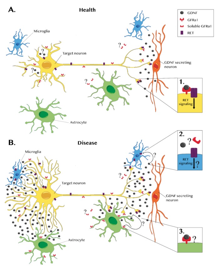
In a healthy adult brain, glial cell line-derived neurotrophic factor (GDNF) is exclusively expressed by neurons, and, in some instances, it has also been shown to derive from a single neuronal subpopulation. Secreted GDNF acts in a paracrine fashion by forming a complex with the GDNF family receptor α1 (GFRα1), which is mainly expressed by neurons and can act in cis as a membrane-bound factor or in trans as a soluble factor. The GDNF/GFRα1 complex signals through interactions with the "rearranged during transfection" (RET) receptor or via the neural cell adhesion molecule (NCAM) with a lower affinity. GDNF can also signal independently from GFRα1 by interacting with syndecan-3. RET, which is expressed by neurons involved in several pathways (nigro-striatal dopaminergic neurons, motor neurons, enteric neurons, sensory neurons, etc.), could be the main determinant of the specificity of GDNF's pro-survival effect. In an injured brain, de novo expression of GDNF occurs in glial cells. Neuroinflammation has been reported to induce GDNF expression in activated astrocytes and microglia, infiltrating macrophages, nestin-positive reactive astrocytes, and neuron/glia (NG2) positive microglia-like cells. This disease-related GDNF overexpression can be either beneficial or detrimental depending on the localization in the brain and the level and duration of glial cell activation. Some reports also describe the upregulation of RET and GFRα1 in glial cells, suggesting that GDNF could modulate neuroinflammation.
Keywords: GDNF family receptor alpha 1; Parkinson’s disease; astrocyte; gene therapy; glial-cell-line-derived neurotrophic factor; microglia; neuroinflammation; rearranged during transfection.
Conflict of interest statement
The authors declare no conflict of interest.
Figures
Similar articles
-
Lipid Rafts Are Physiologic Membrane Microdomains Necessary for the Morphogenic and Developmental Functions of Glial Cell Line-Derived Neurotrophic Factor In Vivo.J Neurosci. 2015 Sep 23;35(38):13233-43. doi: 10.1523/JNEUROSCI.2935-14.2015. J Neurosci. 2015. PMID: 26400951 Free PMC article.
-
Endogenous GDNF Is Unable to Halt Dopaminergic Injury Triggered by Microglial Activation.Cells. 2023 Dec 29;13(1):74. doi: 10.3390/cells13010074. Cells. 2023. PMID: 38201277 Free PMC article.
-
Identification of the key amino acids of glial cell line-derived neurotrophic factor family receptor alpha1 involved in its biological function.J Biol Chem. 2004 Jan 2;279(1):109-16. doi: 10.1074/jbc.M306287200. Epub 2003 Oct 16. J Biol Chem. 2004. PMID: 14563851
-
Novel functions and signalling pathways for GDNF.J Cell Sci. 2003 Oct 1;116(Pt 19):3855-62. doi: 10.1242/jcs.00786. J Cell Sci. 2003. PMID: 12953054 Review.
-
Other neurotrophic factors: glial cell line-derived neurotrophic factor (GDNF).Microsc Res Tech. 1999 May 15-Jun 1;45(4-5):292-302. doi: 10.1002/(SICI)1097-0029(19990515/01)45:4/5<292::AID-JEMT13>3.0.CO;2-8. Microsc Res Tech. 1999. PMID: 10383122 Review.
Cited by
-
Astroglial Dysfunctions in Mood Disorders and Rodent Stress Models: Consequences on Behavior and Potential as Treatment Target.Int J Mol Sci. 2024 Jun 8;25(12):6357. doi: 10.3390/ijms25126357. Int J Mol Sci. 2024. PMID: 38928062 Free PMC article. Review.
-
Cellular Localization of gdnf in Adult Zebrafish Brain.Brain Sci. 2020 May 11;10(5):286. doi: 10.3390/brainsci10050286. Brain Sci. 2020. PMID: 32403347 Free PMC article.
-
From Homeostasis to Neuroinflammation: Insights into Cellular and Molecular Interactions and Network Dynamics.Cells. 2025 Jan 5;14(1):54. doi: 10.3390/cells14010054. Cells. 2025. PMID: 39791755 Free PMC article. Review.
-
Therapeutic Potential of AAV1-Rheb(S16H) Transduction against Neurodegenerative Diseases.Int J Mol Sci. 2021 Mar 17;22(6):3064. doi: 10.3390/ijms22063064. Int J Mol Sci. 2021. PMID: 33802760 Free PMC article. Review.
-
GDNF synthesis, signaling, and retrograde transport in motor neurons.Cell Tissue Res. 2020 Oct;382(1):47-56. doi: 10.1007/s00441-020-03287-6. Epub 2020 Sep 8. Cell Tissue Res. 2020. PMID: 32897420 Free PMC article. Review.
References
-
- Kirik D., Rosenblad C., Bjorklund A., Mandel R.J. Long-term rAAV-mediated gene transfer of GDNF in the rat Parkinson’s model: Intrastriatal but not intranigral transduction promotes functional regeneration in the lesioned nigrostriatal system. J. Neurosci. 2000;20:4686–4700. doi: 10.1523/JNEUROSCI.20-12-04686.2000. - DOI - PMC - PubMed
-
- Ramaswamy S., McBride J.L., Han I., Berry-Kravis E.M., Zhou L., Herzog C.D., Gasmi M., Bartus R.T., Kordower J.H. Intrastriatal CERE-120 (AAV-Neurturin) protects striatal and cortical neurons and delays motor deficits in a transgenic mouse model of Huntington’s disease. Neurobiol. Dis. 2009;34:40–50. doi: 10.1016/j.nbd.2008.12.005. - DOI - PubMed
-
- Blits B., Carlstedt T.P., Ruitenberg M.J., de Winter F., Hermens W.T., Dijkhuizen P.A., Claasens J.W., Eggers R., van der Sluis R., Tenenbaum L., et al. Rescue and sprouting of motoneurons following ventral root avulsion and reimplantation combined with intraspinal adeno-associated viral vector-mediated expression of glial cell line-derived neurotrophic factor or brain-derived neurotrophic factor. Exp. Neurol. 2004;189:303–316. doi: 10.1016/j.expneurol.2004.05.014. - DOI - PubMed
-
- Georgievska B., Kirik D., Bjorklund A. Aberrant sprouting and downregulation of tyrosine hydroxylase in lesioned nigrostriatal dopamine neurons induced by long-lasting overexpression of glial cell line derived neurotrophic factor in the striatum by lentiviral gene transfer. Exp. Neurol. 2002;177:461–474. doi: 10.1006/exnr.2002.8006. - DOI - PubMed
Publication types
Grants and funding
LinkOut - more resources
Full Text Sources
Research Materials
Miscellaneous
