Review
. 2020 Feb 7;9(2):460.
doi: 10.3390/jcm9020460.

An Introduction to High Intensity Focused Ultrasound: Systematic Review on Principles, Devices, and Clinical Applications

Affiliations
Review

An Introduction to High Intensity Focused Ultrasound: Systematic Review on Principles, Devices, and Clinical Applications

Zahra Izadifar et al. J Clin Med. .

Abstract

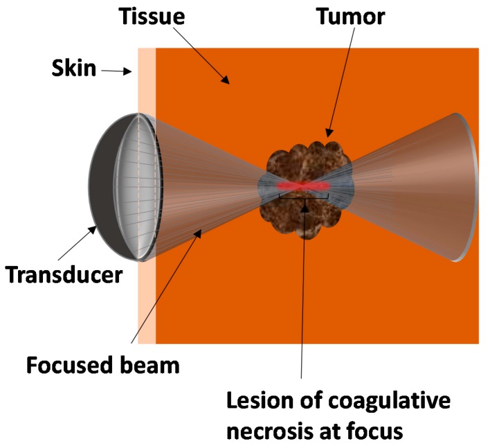
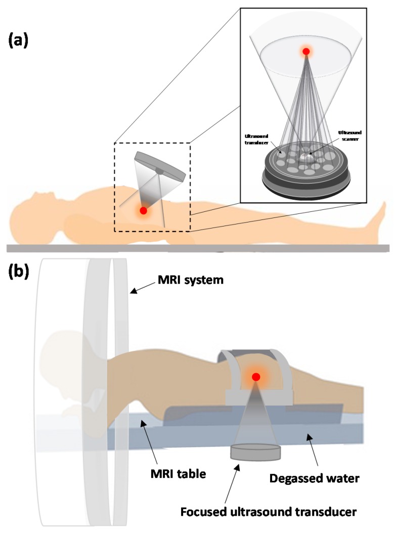
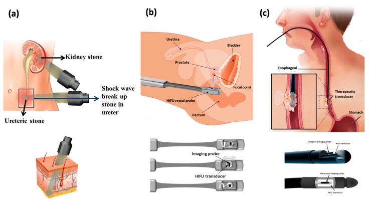
Ultrasound can penetrate deep into tissues and interact with human tissue via thermal and mechanical mechanisms. The ability to focus an ultrasound beam and its energy onto millimeter-size targets was a significant milestone in the development of therapeutic applications of focused ultrasound. Focused ultrasound can be used as a non-invasive thermal ablation technique for tumor treatment and is being developed as an option to standard oncologic therapies. High-intensity focused ultrasound has now been used for clinical treatment of a variety of solid malignant tumors, including those in the pancreas, liver, kidney, bone, prostate, and breast, as well as uterine fibroids and soft-tissue sarcomas. Magnetic resonance imaging and Ultrasound imaging can be combined with high intensity focused ultrasound to provide real-time imaging during ablation. Magnetic resonance guided focused ultrasound represents a novel non-invasive method of treatment that may play an important role as an alternative to open neurosurgical procedures for treatment of a number of brain disorders. This paper briefly reviews the underlying principles of HIFU and presents current applications, outcomes, and complications after treatment. Recent applications of Focused ultrasound for tumor treatment, drug delivery, vessel occlusion, histotripsy, movement disorders, and vascular, oncologic, and psychiatric applications are reviewed, along with clinical challenges and potential future clinical applications of HIFU.

Keywords: application; clinical device; high intensity focused ultrasound; principle.


Conflict of interest statement

The authors declare no conflict of interest.

Figures

Figure 1
Figure 1
Overview schematic of high-intensity focused ultrasound for tumor therapy.
Figure 2
Figure 2
(a) Schematic of a concave focusing transducer and (b) an arranged multiple piston transducer or the atruncated surface of a spherical bowl (c) fully populated phased array.
Figure 3
Figure 3
Schematic of (a) the structure of an extracorporeal high intensity focused ultrasound (HIFU) transducer, including both imaging and therapy probes, depicting an ultrasound-guided technique on a patient and (b) magnetic resonance-guided extracorporeal focused ultrasound system treatment technique.
Figure 4
Figure 4
Schematic of high-intensity focused ultrasound applications in (a) lithotripsy; with an extracorporeal ultrasound transducer (b) prostate cancer; with a typical transrectal ultrasound transducer for prostate cancer treatment with both therapy and imaging transducers incorporated into the head of the transducer probe (c) and esophageal cancer; the front and side view of the head of the interstitial transducer used for the treatment of esophageal tumors.
Figure 5
Figure 5
An ExAblate Neuro (InSightec, Haifa, Israel) MRgFUS transducer helmet on an MRI table (located at Sunnybrook hospital, Toronto, Canada).
Figure 6
Figure 6
(a) The patient lies on the MRI bed and the head is placed inside the phased-array ultrasound transducer helmet. (b) The patient’s head is covered with a flexible silicon membrane that is sealed to outer face of the ultrasound transducer helmet. Degassed and chilled water is circulated in the volume between the patient’s head and the transducer to cool down the surface temperature and avoid damage. This water is also used to fill the space between the patient’s head and the transducers to keep the skull bone temperature within a safe range.
Figure 7
Figure 7
Schematic of different ways of drug delivery utilizing microbubbles. (a) Free drug particles (yellow circles) are circulated along with ultrasound microbubbles (grey circles) in vessels, and the effect of ultrasound on growth and burst of microbubbles results in extravasation of drug into adjacent soft tissues. (b) Drugs encapsulated inside microbubbles are circulated in the vasculature and microbubbles are ruptured by ultrasound and the transported substances are released into the surrounding targeted tissue. (c) Drugs loaded on the external membrane of microbubbles, freely circulated in vessels and then ruptured by ultrasound with the drug being released into the target.

Similar articles

Cited by

References

    1. Lafon C., Melodelima D., Salomir R., Chapelon J.Y. Interstitial devices for minimally invasive thermal ablation by high-intensity ultrasound. Int. J.Hyperth. 2007;23:153–163. doi: 10.1080/02656730601173029. - DOI - PubMed
    1. Lehmann J.F. The biophysical basis of biologic ultrasonic reactions with special reference to ultrasonic therapy. Arch. Phys. Med. Rehabil. 1953;34:139. - PubMed
    1. Woo J. A Short History of the Development of Ultrasound in Obstetrics and Gynecology. [(accessed on 14 May 2011)];2002 Available online: http://www.ob-ultrasound.net/history1.html.
    1. Fry F., Ades H., Fry W. Production of reversible changes in the central nervous system by ultrasound. Science. 1958;127:83–84. doi: 10.1126/science.127.3289.83. - DOI - PubMed
    1. Jolesz F.A. MRI-guided focused ultrasound surgery. Annu. Rev. Med. 2009;60:417–430. doi: 10.1146/annurev.med.60.041707.170303. - DOI - PMC - PubMed

LinkOut - more resources