

New Insights into the Susceptibility of Immunocompetent Mice to Usutu Virus
- PMID: 32046265
- PMCID: PMC7077335
- DOI: 10.3390/v12020189
New Insights into the Susceptibility of Immunocompetent Mice to Usutu Virus
Abstract
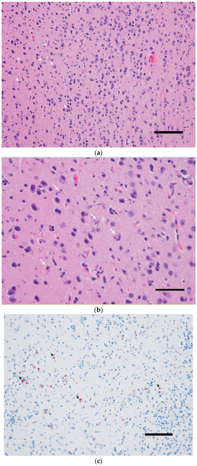
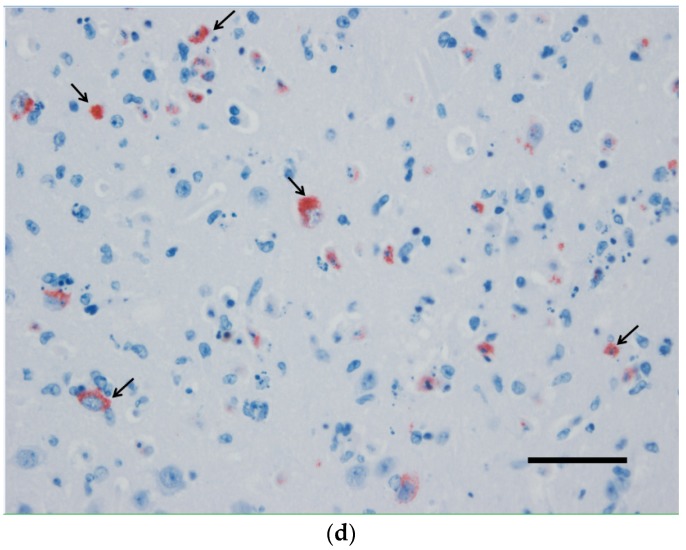
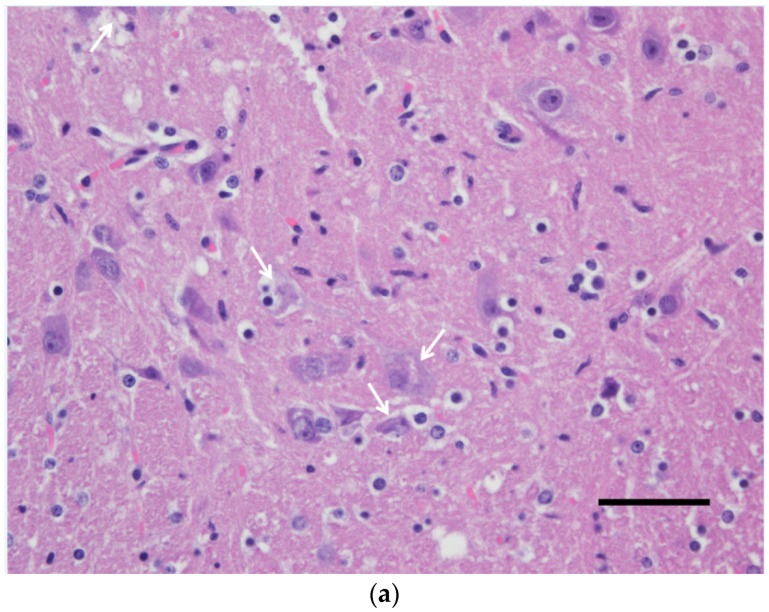
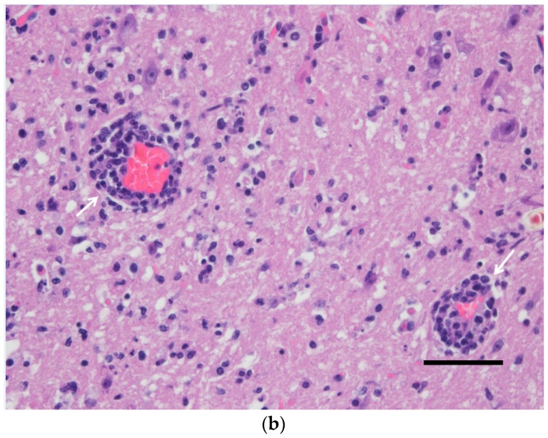
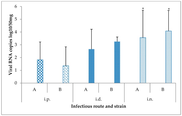
Usutu virus (USUV) is a mosquito-borne flavivirus that shares many similarities with the closely related West Nile virus (WNV) in terms of ecology and clinical manifestations. Initially distributed in Africa, USUV emerged in Italy in 1996 and managed to co-circulate with WNV in many European countries in a similar mosquito-bird life cycle. The rapid geographic spread of USUV, the seasonal mass mortalities it causes in the European avifauna, and the increasing number of infections with neurological disease both in healthy and immunocompromised humans has stimulated interest in infection studies to delineate USUV pathogenesis. Here, we assessed the pathogenicity of two USUV isolates from a recent Belgian outbreak in immunocompetent mice. The intradermal injection of USUV gave rise to disorientation and paraplegia and was associated with neuronal death in the brain and spinal cord in a single mouse. Intranasal inoculation of USUV could also establish the infection; viral RNA was detected in the brain 15 days post-infection. Overall, this pilot study probes the suitability of this murine model for the study of USUV neuroinvasiveness and the possibility of direct transmission in mammals.
Keywords: Usutu virus; encephalitis; immunocompetent; infection; mice.
Conflict of interest statement
The authors declare no conflicts of interest.
Figures
References
-
- Lindenbach B.D., Murray C.L., Thiel H.-J., Rice C.M. Flaviviridae. In: Knipe D.M., Howley P.M., editors. Fields Virology. LippincottWilliams & Wilkins; Philadelphia, PA, USA: 2013. pp. 712–746.
-
- Benzarti E., Sarlet M., Franssen M., Cadar D., Schmidt-Chanasit J., Rivas J., Linden A., Desmecht D., Garigliany M. Usutu Virus Epizootic in Belgium in 2017 and 2018: Evidence of Virus Endemization and Ongoing Introduction Events. Vector Borne Zoonotic Dis. 2019;20:43–50. doi: 10.1089/vbz.2019.2469. - DOI - PubMed
-
- García-bocanegra I., Paniagua J., Gutiérrez-guzmán A.V., Lecollinet S., Boadella M., Arenas-montes A., Cano-terriza D., Lowenski S., Gortázar C., Höfle U. Spatio-temporal trends and risk factors affecting West Nile virus and related flavivirus exposure in Spanish wild ruminants. BMC Vet. Res. 2016;12:249. doi: 10.1186/s12917-016-0876-4. - DOI - PMC - PubMed
Publication types
MeSH terms
Substances
Supplementary concepts
LinkOut - more resources
Full Text Sources
