. 2020 Feb 8;8(1):77.
doi: 10.3390/vaccines8010077.

A Novel Combined Scientific and Artistic Approach for the Advanced Characterization of Interactomes: The Akirin/Subolesin Model

Affiliations

A Novel Combined Scientific and Artistic Approach for the Advanced Characterization of Interactomes: The Akirin/Subolesin Model

Sara Artigas-Jerónimo et al. Vaccines (Basel). .

Expression of concern in

Abstract

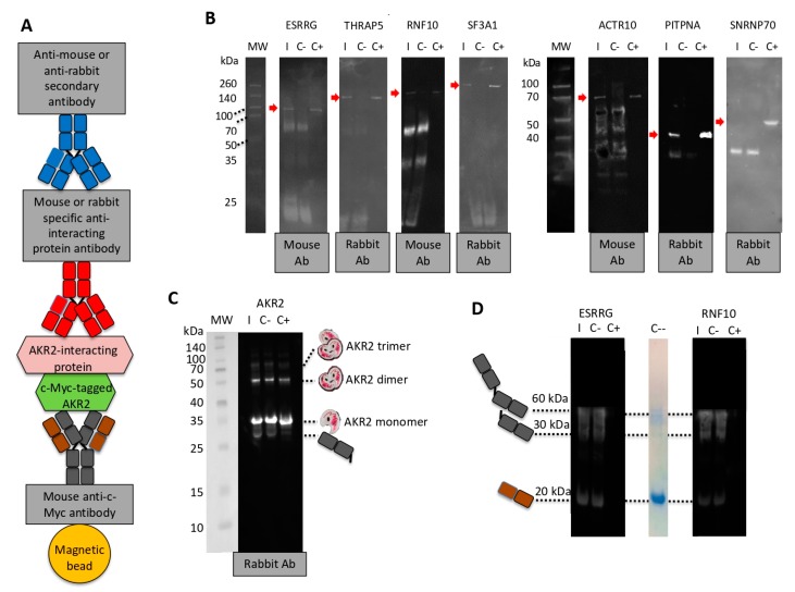
The main objective of this study was to propose a novel methodology to approach challenges in molecular biology. Akirin/Subolesin (AKR/SUB) are vaccine protective antigens and are a model for the study of the interactome due to its conserved function in the regulation of different biological processes such as immunity and development throughout the metazoan. Herein, three visual artists and a music professor collaborated with scientists for the functional characterization of the AKR2 interactome in the regulation of the NF-κB pathway in human placenta cells. The results served as a methodological proof-of-concept to advance this research area. The results showed new perspectives on unexplored characteristics of AKR2 with functional implications. These results included protein dimerization, the physical interactions with different proteins simultaneously to regulate various biological processes defined by cell type-specific AKR-protein interactions, and how these interactions positively or negatively regulate the nuclear factor kappa-light-chain-enhancer of activated B cells (NF-κB) signaling pathway in a biological context-dependent manner. These results suggested that AKR2-interacting proteins might constitute suitable secondary transcription factors for cell- and stimulus-specific regulation of NF-κB. Musical perspective supported AKR/SUB evolutionary conservation in different species and provided new mechanistic insights into the AKR2 interactome. The combined scientific and artistic perspectives resulted in a multidisciplinary approach, advancing our knowledge on AKR/SUB interactome, and provided new insights into the function of AKR2-protein interactions in the regulation of the NF-κB pathway. Additionally, herein we proposed an algorithm for quantum vaccinomics by focusing on the model proteins AKR/SUB.

Keywords: NF-κB; akirin; art; evolution; interactome; music; protective epitope; quantum vaccinomics; subolesin; vaccine; yeast two-hybrid.


Conflict of interest statement

The authors declare no conflict of interest.

Figures

Figure 1
Figure 1
Akirin (AKR) dimerization/multimerization. (A) The piece “El Beso” (The Kiss) by Israel León Viera (mixed media on canvas, 2018, 150 × 133 cm; dedicated to his daughter Sofía León Jorge; courtesy KGJ Collection, Spain) represents protein–protein interactions that play a key role in AKR function, and a new and unexplored facet of possible AKR dimerization. (B) Protein interacting regions identified by Y2H. SID (selected interaction domain) is the amino acid sequence shared by all prey fragments matching the same reference protein, which have been found to contain structural or functional domains. Only regions containing bait fragments, SIDs, or predicted functional and structural domains were considered. (C,D) In vitro characterization of AKR–AKR protein interactions using Ixodes scapularis SUB (Subolesin; Q4VRW2), human AKR2 (Q53H80), and I. scapularis B7PDL0 negative control. SUB, AKR2, and B7PDL0 were incubated alone and SUB also in combination with B7PDL0 (SUB + B7PDL0), followed by analysis in (C) polyacrylamide gels or (D) Western blot using anti-SUB polyclonal IgG antibodies. (E) Protein gel filtration calibration curve (R2 = 0.97) using conalbumin (C; 75 kDa), ovalbumin (O; 44 kDa), carbonic anhydrase (CA; 29 kDa), ribonuclease A (R; 13.7 kDa), and aprotinin (Apr; 6.5 kDa). (F) Size exclusion chromatography (SEC) analysis of AKR2 and SUB. The milli absorbance units (mAu) and elution volume (Ve) are shown. The partition coefficient (Kav) and molecular weight (MW) of AKR2/SUB proteins’ monomer and dimer were calculated using the equations derived from the calibration curve, Kav = Ve − Vo/Vc − Vo and MW = 166086e−3.377Kav. (G) Predicted model of the percentage of amino acids involved in residue–residue interactions for tick SUB–SUB and human AKR2–AKR2 interactions. Predictions were made using the iFrag server (http://sbi.imim.es/web/index.php/research/servers/iFrag?).
Figure 2
Figure 2
Human AKR2–protein interactions. (A) The piece “La Danza Molecular” (Molecular Dance) by Leandro Soto (mixed media on paper, 2018, 51 × 71 cm; courtesy KGJ Collection, Spain) also represents protein–protein interactions, and the possibility that AKR/SUB physically interacts with different proteins simultaneously to regulate various biological processes defined by tissue/cell-specific protein–protein interactions. (B) Results of the Y2H analysis of human AKR2 interactions in human placenta cells. Only proteins identified with A–D scores as potential candidates for interactions with AKR2 are shown. The full description of the proteins is shown in Table 1, Figure S1, and Data S1.
Figure 3
Figure 3
Characterization of human AKR2–protein interactions with highest confidence. (A) Representation of the percentage of total interactions after Y2H by proteins with very high and high confidence of interactions with human AKR2 in human placenta cells. (B) Subcellular localization of AKR2-interacting proteins. (C) Biological processes associated with AKR2-interacting proteins. (D) Prediction of specific interacting residues of human AKR2 with THRAP5 (Q9Y2X0) and RNF10 (Q8N5U6) proteins. The interacting residues formed by AKR2 dimer (amino acids 59-159) are not shown. (E) Predicted model of the percentage of amino acids involved in residue–residue interactions for human AKR2–interferon alpha 1 (IFN-a1) (negative control), AKR2–RNF10, and AKR2–THRAP5. Predictions in (D,E) were made using the iFrag server (http://sbi.imim.es/web/index.php/research/servers/iFrag?), and protein structure models were obtained from The Protein Model Portal.
Figure 4
Figure 4
Corroboration of human AKR2–protein interactions with highest confidence. (A) Representation of the components of the protein pull-down experiment using c-Myc magnetic beads with Myc-tagged human AKR2. (B) Western blot analysis of AKR2–protein interactions. Interacting proteins were incubated with AKR2 and immunopresipitated with c-Myc magnetic beads specific for the AKR2 protein tag (I) or with the c-Myc magnetic beads only as negative control (C-). Recombinant interacting proteins were included as positive control (C+). Mouse or rabbit antibodies specific for each protein were used as primary antibodies and then anti-mouse or anti-rabbit secondary antibodies were used to identify the presence of the interacting proteins (red arrow). The origin of the primary antibody is shown. (C) Corroboration of human AKR2–AKR2 interaction using protein pull-down with c-Myc magnetic beads with human Myc-tagged AKR2. The experiment was conducted as described in (B). The origin of the primary antibody and predicted size for AKR2 monomer, dimer, and trimer together with bands corresponding to fragments of the mouse anti-c-Myc antibody attached to magnetic beads are shown. (D) In order to identify unrelated proteins eluted from the c-Myc magnetic beads, a similar experiment was conducted with selected proteins but incubating only with the secondary antibody. A lane of SDS-PAGE stained with Bio-Safe Coomassie Stain corresponding to c-Myc magnetic beads alone eluted using Laemmli sample buffer was included (C--). The bands corresponding to fragments of the mouse anti-c-Myc antibody attached to magnetic beads are shown.
Figure 5
Figure 5
The sound of AKR evolution and protein interactions. (A) Examples of regularities observed when comparing AKR/SUB musical scores between different species (Caenorhabditis elegans akr NM_058903.6, Drosophila melanogaster akr NM_139856.4, I. scapularis akr/subolesin AY652654.1, Xenopus laevis akr2 NM_001092015.1, Salvelinus alpinus akr2 GQ247760.1, and Homo sapiens akr2 NM_018064.3; Figure S2). (B) Examples of the findings when comparing the six species in a polyphonic context (Figure S3). (C) The AKR2-RNF10 and AKR2-THRAP5 but not AKR2-IFN-α1 interactions are manifested in the higher and repeated presence of unison between both sequences (e.g., AKR2-RNF10 bars 232–235, AKR2-THRAP5 bars 83–85), as well as a higher presence of fifth and fourth consonances (e.g., AKR2–RNF10 bars 210–219, AKR2–THRAP5 bars 67–78) (Figure S4). Audio files were uploaded and can be found in Figure S4 (78998 to 479009).
Figure 6
Figure 6
Function of AKR2 interactome in the regulation of NF-κB. (A) The piece “Nothing wants to say something” by Raúl Cordero (acrylic, polyester, oil and metallic pigments on canvas, 210 × 190 cm; courtesy of the artist) challenges the view of random AKR–protein interactions and suggests that these interactions are functionally relevant in the regulation of different biological processes. (B) Western blot analysis of AKR2–IRF6 and AKR2–WNT2 interactions. Interacting proteins were incubated with AKR2 and immunoprecipitated with c-Myc magnetic beads specific for the AKR2 protein tag (I) or with the c-Myc magnetic beads only as negative control (C-). Recombinant interacting proteins were included as positive control (C+). Antibodies specific for each protein were used to identify the presence of the interacting proteins (red arrow). The origin of the primary antibody is shown. (C,D) Regulation of NF-κB in response to AKR2 and interacting proteins. (C) The NF-κB reporter was used for monitoring the activity of the NF-κB signaling pathway in human placenta-cultured cells after gene knockdown by RNAi. The ratio of firefly luminescence from the NF-κB reporter to Renilla luciferase vector control was represented as average + SD and compared between siRNA-treated groups and the siRNA negative control (C-) (black asterisks), and between combined siRNA-treated groups and the AKR2 siRNA (red asterisks) by Student’s t-test with unequal variance and one-way ANOVA with similar results (* p < 0.05, ** p < 0.005; n = 6 biological replicates). (D) The NF-κB reporter was used for monitoring the activity of the NF-κB signaling pathway in human placenta cells after transfection of recombinant proteins AKR2, RNF10, WNT2, IRF6, and combinations with AKR2. As a positive control, a FITC-antibody was transfected. Negative control cells were transfected with the ESRRG protein. The firefly to Renilla normalized luciferase activity for NF-κB reporter was represented as average + SD and compared between protein-treated groups and the ESRRG control by Student’s t-test with unequal variance (* p < 0.05, ** p < 0.005; n = 4 biological replicates).
Figure 7
Figure 7
AKR2 interactome differentially regulated the NF-κB pathway. (A) Protein transfection was confirmed in FITC-antibody positive control-transfected cells and untreated negative control cells by fluorescence microscopy. Host cell nucleus was stained with DAPI (blue). Image of negative control cells was also collected by phase contrast microscopy and superimposed to FITC/DAPI merged image. Bar: 20 µm. (B) The expression of IFN-β was characterized in human placenta cells after gene knockdown by qRT-PCR. The IFN-β mRNA levels were normalized against human β-actin, and normalized Ct values were compared between test siRNA-treated placenta cells and controls treated with non-targeting siRNA (C-) by Student’s t-test with unequal variance (* p < 0.05; n = 6 biological replicates). (C) The expression of AKR2; genes coding for interacting proteins AKR1, ESRRG, RNF10, THRAP5, IRF6, and WNT2; and regulated genes IFN-β and IL-6 was characterized in human placenta cells treated with lipopolysaccharides (LPS) by qRT-PCR. The mRNA levels were normalized against human β-actin, the normalized LPS to PBS-treated control (C-) ratio (LPS: C-) Ct values were calculated, and normalized Ct values were compared between LPS-treated and C-cells by chi2 test (* p < 0.001; n = 6 biological replicates).
Figure 8
Figure 8
SUB/AKR interactome and possibilities for quantum vaccinomics. (A) Pipeline for quantum vaccinomics by focusing on protective epitopes in peptide sequences involved in protein–protein interactions or SID that are particularly relevant for proteins such as SUB/AKR that function through these interactions. (B) Alignment of SID amino acid sequences for human AKR2 and AKR1, and I. scapularis tick SUB with the corresponding region in the SUB/AKR chimeric Q38 and Q41 protective antigens. The Q38- and Q41-identified and/or predicted protective epitopes are shown.
Figure 9
Figure 9
Model for NF-κB regulation by human AKR2 interactome in human placenta cells after (A) gene knockdown by RNAi, (B) protein transfection, (C) IFN-β and IL-6 expression after treatment with LPS, and (D) IFN-β expression after gene knockdown by RNAi. Predictions are based on results from experiments shown in Figure 6C,D and Figure 7B,C.

References

    1. Jeffries S. When two tribes meet: Collaborations between artists and scientists. [(accessed on 1 August 2019)];The Guardian. 2011 Aug 21; Available online: https://www.theguardian.com/artanddesign/2011/aug/21/collaborations-betw....
    1. Tayag Y., Wells B. Art and evolution: A work in progress. [(accessed on 1 August 2019)];Scientific American. 2014 May 6; Available online: https://blogs.scientificamerican.com/guest-blog/art-and-evolution-a-work...
    1. Schulkin J., Raglan G.B. The evolution of music and human social capability. Front. Neurosci. 2014;8:292. doi: 10.3389/fnins.2014.00292. - DOI - PMC - PubMed
    1. de la Fuente J., Estrada-Peña A., Cabezas-Cruz A., Brey R. Flying ticks: Anciently evolved associations that constitute a risk of infectious disease spread. Parasit. Vectors. 2015;8:538. doi: 10.1186/s13071-015-1154-1. - DOI - PMC - PubMed
    1. Veis N. Four takes in the evolution of art. Nature. 2017;543:490. doi: 10.1038/543490a. - DOI

LinkOut - more resources