. 2020 Feb 8;12(2):397.
doi: 10.3390/cancers12020397.

Predictors of Postoperative Seizure Outcome in Low Grade Glioma: From Volumetric Analysis to Molecular Stratification

Affiliations

Predictors of Postoperative Seizure Outcome in Low Grade Glioma: From Volumetric Analysis to Molecular Stratification

Tamara Ius et al. Cancers (Basel). .

Abstract

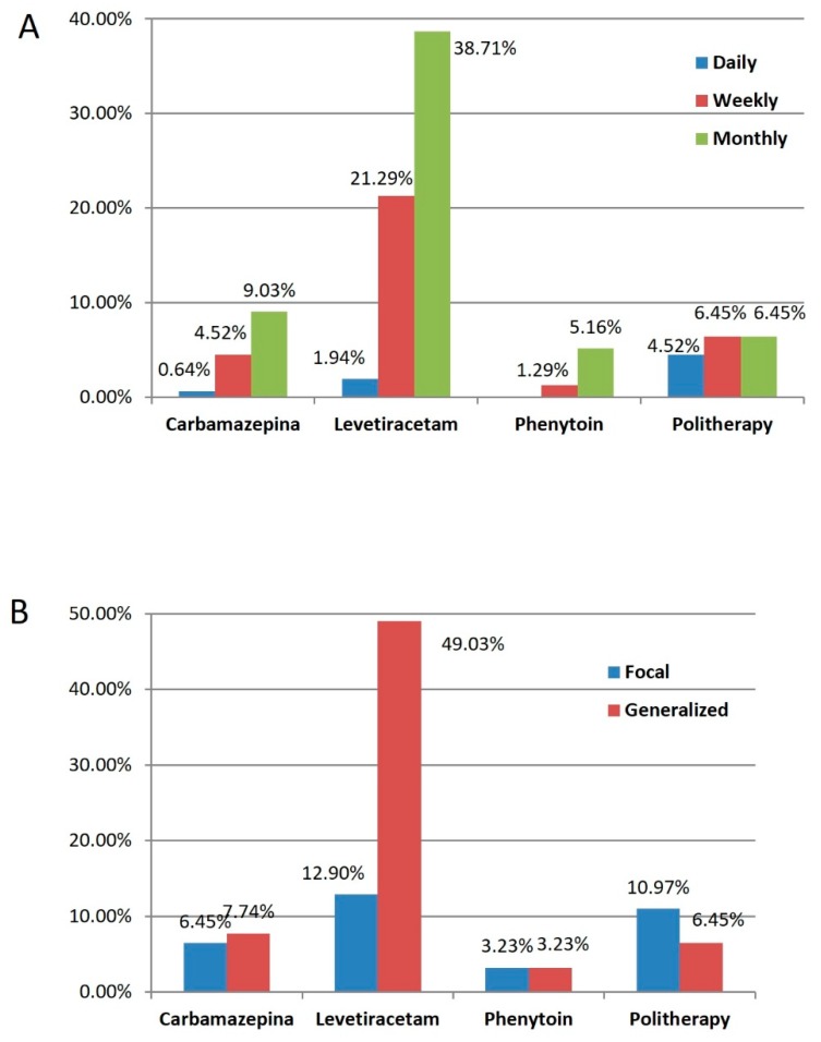
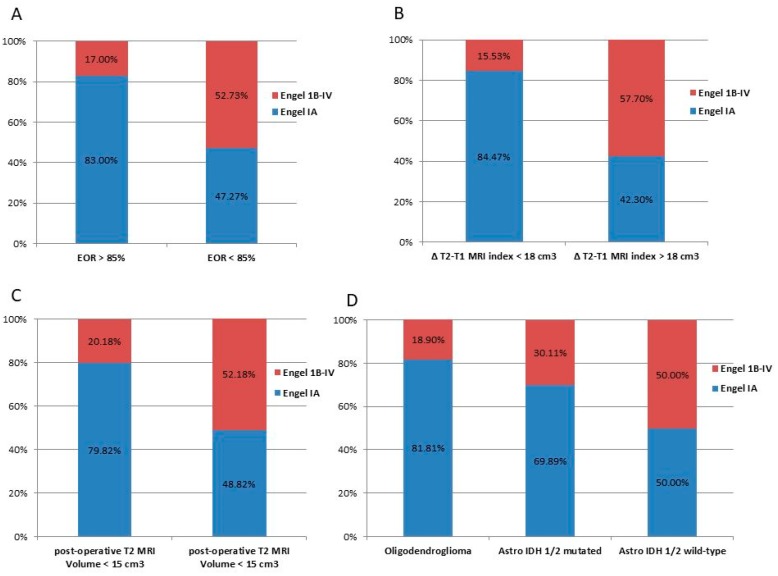
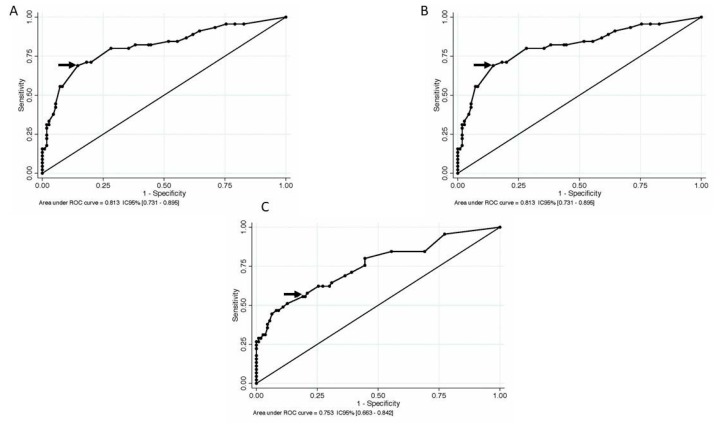
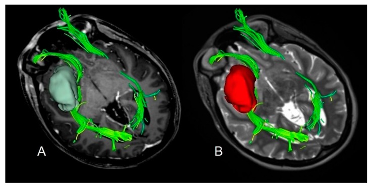
The importance of the extent of resection (EOR) has been widely demonstrated as the main predictor for survival, nevertheless its effect on tumor related epilepsy is less investigated. A total of 155 patients were enrolled after a first-line surgery for supratentorial Diffuse Low Grade Gliomas (DLGGs). Postoperative seizure outcome was analyzed stratifying the results by tumor volumetric data and molecular markers according to 2016 WHO classification. Receiver operating characteristic (ROC) curves were computed to asses EOR, residual tumor volume, and ΔT2T1 MRI index (expressing the tumor growing pattern) corresponding to optimal seizure outcome. A total of 70.97% of patients were seizure-free 18 months after surgery. Better seizure outcome was observed in IDH1/2 mutated and 1p/19q codeleted subgroup. At multivariate analysis, age (p = 0.014), EOR (p = 0.030), ΔT2T1 MRI index (p = 0.016) resulted as independent predictors of postoperative seizure control. Optimal parameters to improve postoperative seizure outcome were EOR ≥ 85%, ΔT2T1 MRI index ≤ 18 cm3, residual tumor volume ≤ 15 cm3. This study confirms the role of EOR and tumor growing pattern on postoperative seizure outcome independently from the molecular class. Higher ΔT2T1 MRI index, representing the infiltrative component of the tumor, is associated with worse seizure outcome and strengthens the evidence of common pathogenic mechanisms underlying tumor growth and postoperative seizure outcome.

Keywords: ROC curves; extent of resection; low grade glioma; molecular markers; seizure outcome; tumor growth pattern.


Conflict of interest statement

The authors declare no conflict of interest.

Figures

Figure 1
Figure 1
Graph illustrating the preoperative Anti-Epileptic Drugs (AESs) stratified by seizure frequency (A) and by onset seizure type (B).
Figure 2
Figure 2
Graph illustrating 18-months postoperative seizure outcome stratified by the significant variables at univariate analysis. Blue bars indicate patients in Engel Class IA; yellow bars indicate those in Engel Class IB or above. Individual bar totals are the total number of patients with postoperative seizures within each category. (A) Distribution of patients stratified by the EOR; (B) Distribution of patients stratified by the preoperative tumor growing pattern expressed by the ΔT2T1 MRI index; (C) distribution of patients stratified by the residual tumor computed on T2 weighted images; (D) distribution of patients stratified by the molecular class according to the 2016 WHO classification.
Figure 3
Figure 3
A receiver operating characteristic (ROC) curve for EOR, preoperative ΔT2T1 MRI index, and postoperative residua tumor on T2-weighted images, to predict seizures relapse after surgery. The optimal diagnostic point is the one with maximal sensitivity and specificity. It is the point closest to the top left corner of the graph, indicated by the arrow. (A) The optimal threshold corresponded to an EOR of 85%, which was the point with the highest sensitivity (0.764) and specificity (0.644), with a resulting area under the curve of 0.783 (CI 95% 0.700–0.865) and a predictive accuracy of 72.90%; (B) for the preoperative ΔT2T1 MRI index, the threshold of 18 cm3 corresponded to the point with the highest sensitivity (0.689) and specificity (0.855), with a resulting area under the curve of 0.813 (CI 95% 0.731–0.895) and a predictive accuracy of 82.65%; (C) regarding the residual tumor, the optimal threshold corresponded to 15 cm3, which was the point with the highest sensitivity (0.556) and specificity (0.809), with a resulting area under the curve of 0.753 (CI 95% 0.663–0.842) and a predictive accuracy of 73.55%.
Figure 4
Figure 4
A case of insular diffuse low grade gliomas (DLGG) with a regular shape, determining similar tumor volume in both post-contrast T1-weighted MRI and T2-weighted MRI sequences and displacing the Fronto-Occipital Longitudinal Fasciculus. The preoperative tumor volume computed on post-contrast T1-weighted magnetic resonance imaging (MRI) was 32 cm3 (axial slices A). The preoperative tumor volume computed on T2-weighted MRI was 34 cm3 (axial slices B). The preoperative ΔT2T1 MRI index was 2 cm3, showing the prevalence of the proliferative tumor growing pattern. The patient was in Engel Class IA 18 months after surgery.
Figure 5
Figure 5
A case of insular DLGG infiltrating the Cortico-Spinal tract. The preoperative tumor volume computed on post-contrast T1-weighted magnetic resonance imaging (MRI) was 58 cm3 (axial slices (A)). The preoperative tumor volume computed on T2-weighted MRI was 83 cm3 (axial slices (B)). The preoperative ΔT2T1 MRI index was 25 cm3. The prevalence of the diffusive and infiltrative growth generates the tumor diffusion along the white matter, resulting in a complex shape with digitations more visible on T2-weighted images. The patient was in Engel Class IB 18 months after surgery.

References

    1. Aronica E., Leenstra S., van Veelen C.W., van Rijen P.C., Hulsebos T.J., Tersmette A.C., Yankaya B., Troost D. Glioneuronal tumors and medically intractable epilepsy: A clinical study with long-term follow-up of seizure outcome after surgery. Epilepsy. Res. 2001;43:179–191. doi: 10.1016/S0920-1211(00)00208-4. - DOI - PubMed
    1. Rudà R., Trevisan E., Soffietti R. Epilepsy and brain tumors. Curr. Opin. Oncol. 2010;22:611–620. doi: 10.1097/CCO.0b013e32833de99d. - DOI - PubMed
    1. Rossetti A.O., Stupp R. Epilepsy in brain tumor patients. Curr. Opin. Neurol. 2010;23:603–609. doi: 10.1097/WCO.0b013e32833e996c. - DOI - PubMed
    1. Rudà R., Bello L., Duffau H., Soffietti R. Seizures in low-grade gliomas: Natural history, pathogenesis, and outcome after treatments. Neuro. Oncol. 2012;14(Suppl. 4):iv55–iv64. doi: 10.1093/neuonc/nos199. - DOI - PMC - PubMed
    1. Vecht C.J., Wagner G.L., Wilms E.B. Treating seizures in patients with brain tumors: Drug interactions between antiepileptic and chemotherapeutic agents. Semin. Oncol. 2003;30(Suppl. 19):49–52. doi: 10.1053/j.seminoncol.2003.11.030. - DOI - PubMed

LinkOut - more resources