Comparative Study
. 2020 Feb 8;9(2):393.
doi: 10.3390/cells9020393.

Myelin Measurement Using Quantitative Magnetic Resonance Imaging: A Correlation Study Comparing Various Imaging Techniques in Patients with Multiple Sclerosis

Affiliations
Comparative Study

Myelin Measurement Using Quantitative Magnetic Resonance Imaging: A Correlation Study Comparing Various Imaging Techniques in Patients with Multiple Sclerosis

Laetitia Saccenti et al. Cells. .

Abstract

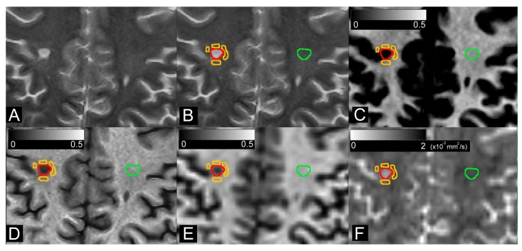
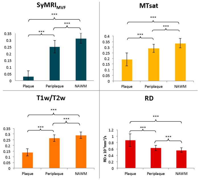
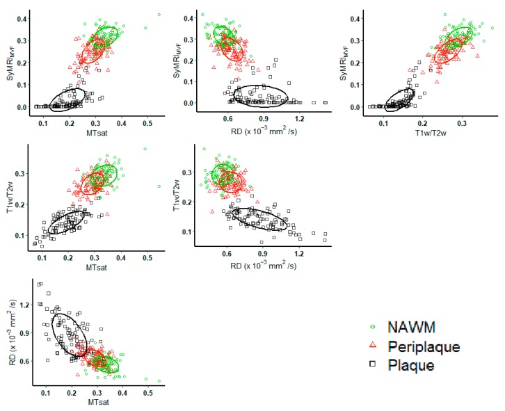
Evaluation of myelin by magnetic resonance imaging (MRI) is a difficult challenge, but holds promise in demyelinating diseases, such as multiple sclerosis (MS). Although multiple techniques have been developed, no gold standard has been established. This study aims to evaluate the correlation between synthetic MRI myelin volume fraction (SyMRIMVF) and myelin fraction estimated by other techniques, i.e., magnetization transfer saturation (MTsat), T1-weighted images divided by T2-weighted images (T1w/T2w), and radial diffusivity (RD) in patients with MS. We also compared the sensitivities of these techniques for detecting MS-related myelin damage. SyMRIMVF, MTsat, T1w/T2w, and RD were averaged on plaque, periplaque white matter, and normal-appearing white matter (NAWM). Pairwise correlation was calculated using Spearman's correlation analysis. For all segmented regions, strong correlations were found between SyMRIMVF and T1w/T2w (Rho = 0.89), MTsat (Rho = 0.82), or RD (Rho = -0.75). For each technique, the average estimated myelin differed significantly among regions, but the percentage change of NAWM from both periplaque white matter and plaque were highest in SyMRIMVF. SyMRIMVF might be suitable for myelin evaluation in MS patients, with relevant results as compared to other well-studied techniques. Moreover, it presented better sensitivity for the detection of the difference between plaque or periplaque white matter and NAWM.

Keywords: multiple sclerosis; myelin; synthetic magnetic resonance imaging.


Conflict of interest statement

The authors declare no conflicts of interest.

Figures

Figure 1
Figure 1
Synthetic T2-weighted images (A) were used for semi-automated segmentation of plaque (red), periplaque (orange), and contralateral normal-appearing white matter (green) (B). Regions-of-interest were copied and pasted onto SyMRIMVF (C), T1w/T2w (D), MTsat (E), and RD (F) maps.
Figure 2
Figure 2
Estimation of myelin with 4 different techniques (SyMRIMVF, MTsat, T1w/T2w, and RD). Each technique can differentiate plaque, periplaque, and NAWM regions. These metrics were the lowest in plaque for SyMRIMVF, MTsat, and T1w/T2w and the highest in plaque for RD. *** p < 0.001.
Figure 3
Figure 3
Percentage changes in the metrics (SyMRIMVF, T1w/T2w, MTsat, and RD) for plaque and periplaque compared to NAWM regions. SyMRIMVF showed the highest contrast between NAWM and both plaque and periplaque regions. * p < 0.05. ** p < 0.01. *** p < 0.001.
Figure 4
Figure 4
Scatterplots of SyMRIMVF, MTsat, T1w/T2w, and RD, plotted by subgroup of segmented regions-of-interest: black square = plaque, red triangle = periplaque, green circle = NAWM. Plot concentration ellipses are also drawn, with concentration levels of 0.5.

References

    1. de Hoz L., Simons M. The emerging functions of oligodendrocytes in regulating neuronal network behaviour. BioEssays. 2015;37:60–69. doi: 10.1002/bies.201400127. - DOI - PubMed
    1. Duval T., Stikov N., Cohen-Adad J. Modeling white matter microstructure. Funct. Neurol. 2016;31:217–228. doi: 10.11138/FNeur/2016.31.4.217. - DOI - PMC - PubMed
    1. Heath F., Hurley S.A., Johansen-Berg H., Sampaio-Baptista C. Advances in noninvasive myelin imaging. Dev. Neurobiol. 2018;78:136–151. doi: 10.1002/dneu.22552. - DOI - PMC - PubMed
    1. King E., Sabatier M., Hoque M., Kesar T., Backus D., Borich M. Myelin status is associated with change in functional mobility following slope walking in people with multiple sclerosis. Mult. Scler. J. Exp. Transl. Clin. 2018;4 doi: 10.1177/2055217318773540. - DOI - PMC - PubMed
    1. Prasloski T., Rauscher A., MacKay A.L., Hodgson M., Vavasour I.M., Laule C., Mädler B. Rapid whole cerebrum myelin water imaging using a 3D GRASE sequence. NeuroImage. 2012;63:533–539. doi: 10.1016/j.neuroimage.2012.06.064. - DOI - PubMed

Publication types