. 2020 Feb 10;64(1):3092.
doi: 10.4081/ejh.2020.3092.

Expression, localization and synthesis of small leucine-rich proteoglycans in developing mouse molar tooth germ

Affiliations

Expression, localization and synthesis of small leucine-rich proteoglycans in developing mouse molar tooth germ

Angammana Randilini et al. Eur J Histochem. .

Abstract

The gene expression and protein synthesis of small leucine-rich proteoglycans (SLRPs), including decorin, biglycan, fibromodulin, and lumican, was analyzed in the context of the hypothesis that they are closely related to tooth formation. In situ hybridization, immunohistochemistry, and organ culture with metabolic labeling of [35S] were carried out in mouse first molar tooth germs of different developmental stages using ICR mice at embryonic day (E) 13.5 to postnatal day (P) 7.0. At the bud and cap stage, decorin mRNA was expressed only in the surrounding mesenchyme, but not within the tooth germ. Biglycan mRNA was then expressed in the condensing mesenchyme and the dental papilla of the tooth germ. At the apposition stage (late bell stage), both decorin and biglycan mRNA were expressed in odontoblasts, resulting in a switch of the pattern of expression within the different stages of odontoblast differentiation. Decorin mRNA was expressed earlier in newly differentiating odontoblasts than biglycan. With odontoblast maturation and dentin formation, decorin mRNA expression was diminished and localized to the newly differentiating odontoblasts at the cervical region. Simultaneously, biglycan mRNA took over and extended its expression throughout the new and mature odontoblasts. Both mRNAs were expressed in the dental pulp underlying the respective odontoblasts. At P7.0, both mRNAs were weakly expressed but maintained their spatial expression patterns. Immunostaining showed that biglycan was localized in the dental papillae and pulp. In addition, all four SLRPs showed clear immunostaining in predentin, although the expressions of fibromodulin and lumican mRNAs were not identified in the tooth germs examined. The organ culture data obtained supported the histological findings that biglycan is more predominant than decorin at the apposition stage. These results were used to identify biglycan as the principal molecule among the SLRPs investigated. Our findings indicate that decorin and biglycan show spatial and temporal differential expressions and play their own tissue-specific roles in tooth development.


Conflict of interest statement

Conflict of interest: The authors declare that they have no competing interests, and all authors confirm accuracy.

Figures
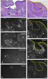
Figure 1.
Figure 1.
Frontally cut mouse head (a-e) and enlargement of lower molar tooth germ (TG) (f-i) at E13.5. TB staining (a,f ) and in situ hybridization for decorin mRNA (b,g), biglycan mRNA (c,h), fibromodulin mRNA (d,i), and lumican mRNA (e). a,f ) Tooth germ at the bud stage, developing tongue muscle (T), and mandibular bone (MB) with Meckel’s cartilage (MC) were seen. b,g) Decorin mRNA was expressed in tongue muscles, surrounding mesenchyme of the submandibular gland, developing mandibular bone (asterisks), and mesenchyme around tooth germ (arrowheads). c,h) Biglycan mRNAs was expressed in the lingual septum, perichondrium of Meckel’s cartilage, developing mandibular bone (asterisks), and condensing mesenchyme of the tooth germ (arrowheads). d,i) Fibromodulin mRNA was weakly expressed in the perichondrium of Meckel’s cartilage and the mandibular bone (asterisks), but not in the tooth germ. e) Lumican mRNA did not show significant expression. Scale bars: a-e) 200 mm; f-i) 80 mm.
Figure 2.
Figure 2.
Frontally cut mouse head (a-d) and magnification of lower molar tooth germ (TG) (e-h) at E16.0. TB staining (a,e) and in situ hybridization for decorin (b,f ), biglycan (c,g), and fibromodulin mRNA (d,h). a,e) Masseter muscle (MM) was attached to developing mandibular bone (MB); developing tooth germ was in the late cap stage. a-h) Decorin mRNA was expressed in the periosteum of the mandibular bone (asterisk in b), and the mesenchyme around the tooth germ (arrowheads in b and f ). Biglycan mRNA was expressed in the periosteum of the mandibular bone (asterisk in c), and additionally in dental papillae (arrowheads in c and g). Fibromodulin mRNA was not expressed in tooth germ. Note that three mRNAs were expressed in the epimysium of the masseter muscle (arrows in b-d). Scale bars: 50 mm.
Figure 3.
Figure 3.
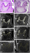
Frontally cut mouse lower molar tooth germs at E18.5 (ad) and at P1.0 (e-h). TB staining (a,e) and in situ hybridization for decorin (b,f ), biglycan (c,g) and fibromodulin mRNA (d, h). a e) Odontoblasts (OB) with formation of predentin (PD) and/or dentin (D) were evident at cusp tips. b-d) Decorin mRNA was well expressed in differentiating odontoblasts at cup tips (arrowhead in b) and dental papillae beneath them (asterisks in b); biglycan mRNA was expressed at low intensity in differentiating odontoblasts (arrowheads in c); fibromodulin mRNA was not expressed within the tooth germ. f-h) Decorin mRNA expression was reduced in mature odontoblasts (arrowheads in f ), but extended to newly differentiating odontoblasts in the cervical region (arrows in f ) and also in the dental pulp beneath odontoblasts (asterisks in f ); biglycan mRNA expression was enhanced in mature odontoblasts (arrowheads in g) and underlying dental pulp (asterisks in g), but still weak in newly differentiating odontoblasts (arrows in g); fibromodulin mRNA was not expressed within the tooth germ. All mRNAs were expressed in the epimysium of masseter muscles (large arrows in b-d, f-h). Scale bars: 100 mm.
Figure 4.
Figure 4.
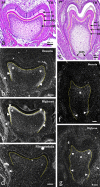
Frontally cut mouse lower molar tooth germs at P3.0 (ad) and P7.0 (e-g). TB staining (a,e) and in situ hybridization for decorin (b,f ), biglycan (c,g), and fibromodulin mRNA (d). a,e) Dentin (D) and enamel (E) formation were evident, and Hertwig's epithelial root sheath (HERS) had started to form. bd) Decorin mRNA expression was localized in the newly differentiating odontoblasts at the cervical region (arrowheads in b) and underlying pulp (asterisks in b); biglycan mRNA was expressed strongly throughout the odontoblast layer (arrowheads in c) and underlying pulp (asterisks in c); fibromodulin mRNA was not expressed within the tooth germ (d). f,g) Decorin mRNA expression only remained in odontoblasts at the cervical tip (arrowheads in f ) and weakly in the coronal pulp (asterisk in f ); biglycan mRNA expression was also reduced, but remained weak throughout the odontoblast layer (arrowheads in g) and in the coronal pulp (asterisk in g). Epimysium of masseter muscles expressed all three mRNAs (arrows in b-d). OB, odontoblasts; PD, predentin. Scale bars: 100 mm.
Figure 5.
Figure 5.
Sagittally cut mouse incisor tooth germ at P7.0 (a-c) and sagittally cut mouse lower limb at P1.0 (d-g). TB staining (a,d) and in situ hybridization for decorin (b,e), biglycan (c,f ), and fibromodulin mRNA (g). a) All differential stages of odontoblasts can be identified (arrowheads in a). b, c) Decorin mRNA was expressed in newly differentiating odontoblasts and reduced towards the coronal region (arrowheads in c); biglycan mRNA was also expressed in odontoblasts and maintained its reactivity more towards the tip region (arrowheads in c). d) Tendons of the quadriceps femoris muscle (QFt) were continuous to the epimysium (em) of the tibialis anterior muscle (TA). e-g) Three mRNAs were expressed in the tendons (arrows), the continuous epimysium (arrowheads), and in the perimysium (asterisks), although decorin was weakly expressed in the epimysium (arrowhead in e). Scale bars: 200 mm.
Figure 6.
Figure 6.
Immunohistochemistry in frontally cut mouse molar tooth germs at E16.0 (a-d), E18.5 (e-h), and P3.0 (i-l) for decorin (a,e,i), biglycan (b,f,j), fibromodulin (c,g,k), and lumican (d,h,l) proteins. a-d) Decorin immunoreactivity was detected in the lamina propria of oral epithelium (arrowhead in a); biglycan immunoreactivity was detected in the dental papillae (asterisk in b). Lumican immunoreactivity was detected in the osteoid (arrowhead in d). e-l) Immunoreactivity for all SLRPs was detected in the predentin (arrows) and osteoid (arrowheads); biglycan immunoreactivity was additionally detected in the central region of the dental pulp (asterisks in f and j), but not in the peripheral region (black circles in f and j). Scale bars: 100 mm.
Figure 7.
Figure 7.
a) [35S]-labeled macromolecules released into explants at E16.0, 18.0, and P3.0. From E16.0 to E18.0, the amount of macromolecules was significantly increased, and was further increased at P3.0 compared with E18.0; data are presented as means ± SD (n = 4); *P<0.01, significantly different between indicated groups; **P<0.05, significantly different between indicated groups. b) Superose 6 elution profiles of explants from E16.0, 18.0, and P3.0 with or without enzymatic treatments; peaks 1 and 2 in E16.0 samples were slightly susceptible to chondroitinase ABC, and remaining peaks at 1 and 2 regions were not susceptible to digestion of heparitinase and keratanase. In E18.0 samples, remaining peaks at 1 and 2 regions after chondroitinase ABC digestion, were susceptible to heparitinase (~10%), but were not susceptible to subsequent digestion by keratanase. Peak 3 was not susceptible to either heparitinase or keratanase; in the P3.0 sample, peak 1 became small and peaks 1 and 2 were largely susceptible to chondroitinase ABC, and remaining peaks were further susceptible to heparitinase, but not susceptible to keratanase; peak 4 and 5 were also recognized, but were not susceptible to any enzymatic treatment. c) SDS-PAGE analysis; a band at the top of the gel (band 1), and two broad bands of ~250 kDa (band 2) and ~100kDa (band 3), were recognized for all three ages. Compared to E16.0 and E18.0, band 2 of P3.0 was significantly thicker than band 3.

Similar articles

Cited by

References

    1. McEwan P, Scott P, Bishop P, Bella J. Structural correlations in the family of small leucine-rich repeat proteins and proteoglycans. J Struct Biol 2006;155:294-305. - PubMed
    1. Schaefer L, Iozzo R. Biological functions of the small leucinerich proteoglycans: from genetics to signal transduction. J Biol Chem 2008;283:21305-9. - PMC - PubMed
    1. Nikitovic D, Aggelidakis J, Young M, Iozzo R, Karamanos N, Tzanakakis G. The biology of small leucine-rich proteoglycans in bone pathophysiology. J Biol Chem 2012;287:33926-33. - PMC - PubMed
    1. Chen S, Birk D. The regulatory roles of small leucine-rich proteoglycans in extracellular matrix assembly. FEBS J 2013; 280:2120-37. - PMC - PubMed
    1. Orsini G, Ruggeri A, Mazzoni A, Nato F, Manzoli L, Putignano A, et al. A review of the nature, role, and function of dentin non-collagenous proteins. Part 1: proteoglycans and glycoproteins. Endodontic Topics 2012;21:1-18.

MeSH terms