Multicenter Study
. 2020 Jul 1;177(7):589-600.
doi: 10.1176/appi.ajp.2019.19060583. Epub 2020 Feb 12.

Mapping Subcortical Brain Alterations in 22q11.2 Deletion Syndrome: Effects of Deletion Size and Convergence With Idiopathic Neuropsychiatric Illness

Affiliations
Multicenter Study

Mapping Subcortical Brain Alterations in 22q11.2 Deletion Syndrome: Effects of Deletion Size and Convergence With Idiopathic Neuropsychiatric Illness

Christopher R K Ching et al. Am J Psychiatry. .

Abstract

Objective: 22q11.2 deletion syndrome (22q11DS) is among the strongest known genetic risk factors for schizophrenia. Previous studies have reported variable alterations in subcortical brain structures in 22q11DS. To better characterize subcortical alterations in 22q11DS, including modulating effects of clinical and genetic heterogeneity, the authors studied a large multicenter neuroimaging cohort from the ENIGMA 22q11.2 Deletion Syndrome Working Group.

Methods: Subcortical structures were measured using harmonized protocols for gross volume and subcortical shape morphometry in 533 individuals with 22q11DS and 330 matched healthy control subjects (age range, 6-56 years; 49% female).

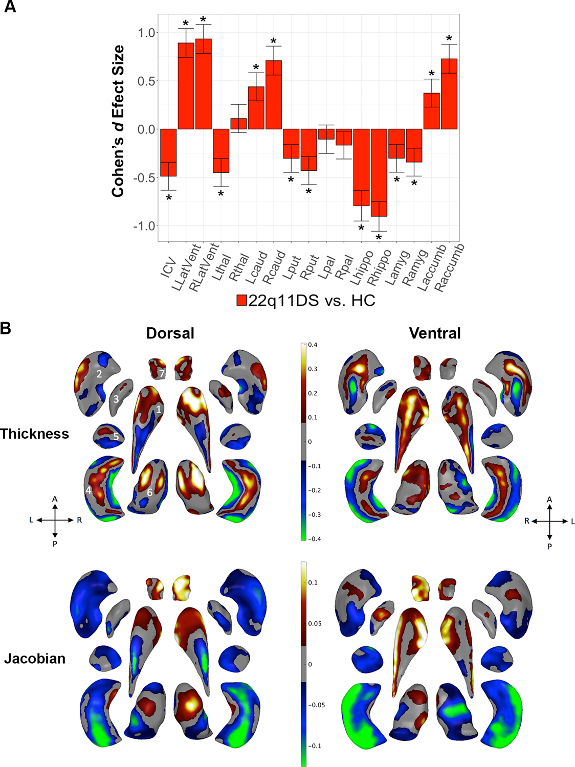
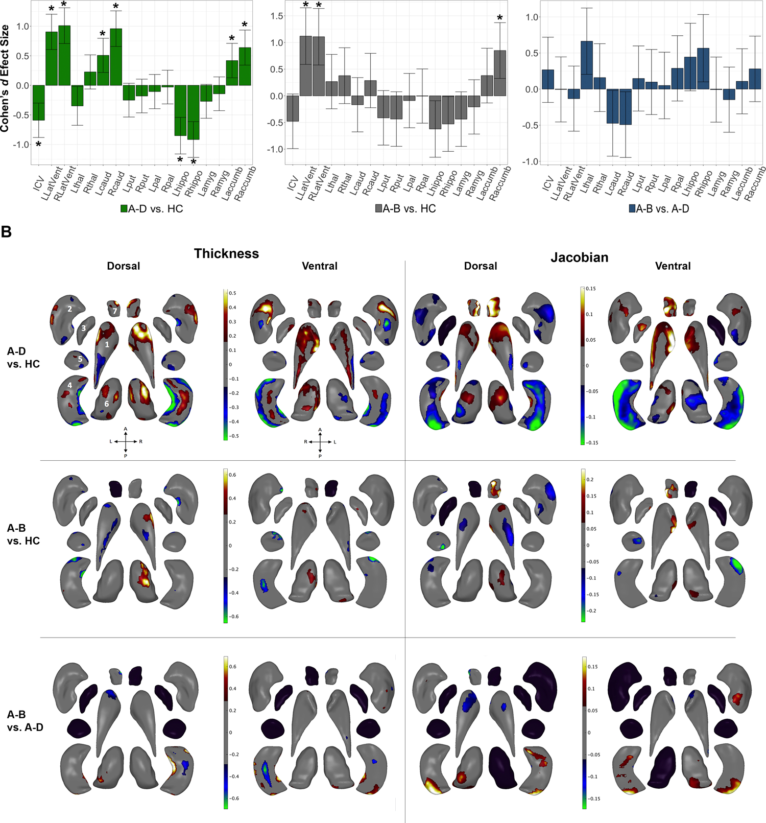
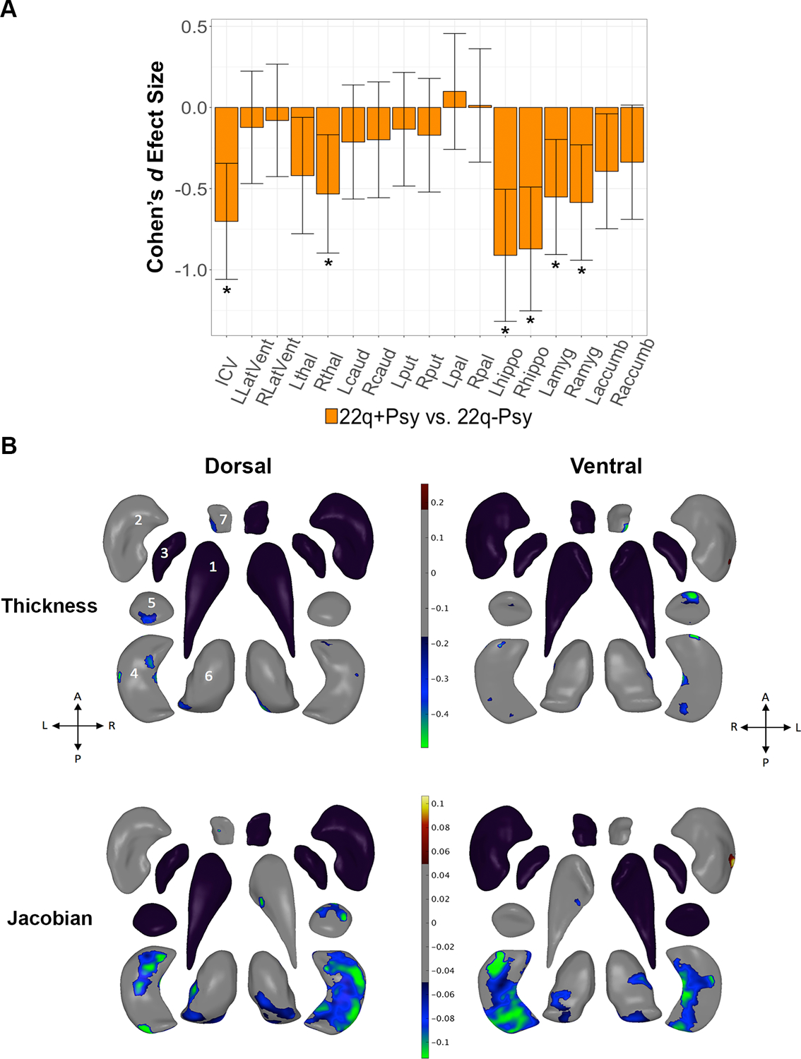
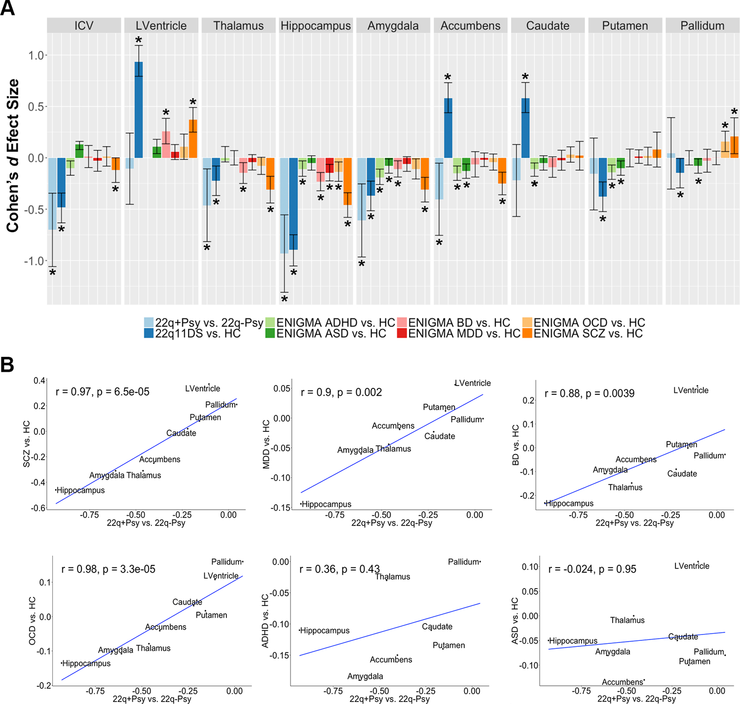
Results: Compared with the control group, the 22q11DS group showed lower intracranial volume (ICV) and thalamus, putamen, hippocampus, and amygdala volumes and greater lateral ventricle, caudate, and accumbens volumes (Cohen's d values, -0.90 to 0.93). Shape analysis revealed complex differences in the 22q11DS group across all structures. The larger A-D deletion was associated with more extensive shape alterations compared with the smaller A-B deletion. Participants with 22q11DS with psychosis showed lower ICV and hippocampus, amygdala, and thalamus volumes (Cohen's d values, -0.91 to 0.53) compared with participants with 22q11DS without psychosis. Shape analysis revealed lower thickness and surface area across subregions of these structures. Compared with subcortical findings from other neuropsychiatric disorders studied by the ENIGMA consortium, significant convergence was observed between participants with 22q11DS with psychosis and participants with schizophrenia, bipolar disorder, major depressive disorder, and obsessive-compulsive disorder.

Conclusions: In the largest neuroimaging study of 22q11DS to date, the authors found widespread alterations to subcortical brain structures, which were affected by deletion size and psychotic illness. Findings indicate significant overlap between 22q11DS-associated psychosis, idiopathic schizophrenia, and other severe neuropsychiatric illnesses.

Keywords: 22q11.2 Deletion Syndrome; Copy Number Variant; Neuroanatomy; Neurodevelopment; Psychosis; Schizophrenia.


Conflict of interest statement

Disclosures

Declan G. Murphy has received honoraria from Roche. Paul M. Thompson and Christopher R. K. Ching have received grant support from Biogen, Inc. (Boston, USA) for work unrelated to the topic of this manuscript. All other authors have no conflicts of interest to disclose.

Figures

Figure 1:
Figure 1:
22q11DS vs. HC: Gross volume and shape analysis. A. Cohen’s d effect size (with 95% confidence intervals) plotted for major pairwise gross volumetric comparisons. An asterisk (*) indicates significant group difference after correction for multiple comparisons for 22q11DS versus HC (FDR q<0.05). Positive effect sizes indicate 22q11DS>HC; negative effect sizes indicate HC>22q11DS. Models were adjusted for age, age2, sex, ICV, and scan site. Full statistical model outputs including standard error, coefficient estimates, p-values, and % difference may be found in Supplemental Table S14. Abbreviations: L/R, left/right; LatVent, lateral ventricle; thal, thalamus; caud, caudate; put, putamen; pal, pallidum; hippo, hippocampus; amyg, amygdala; accumb, accumbens; ICV, intracranial volume. B. Shape analysis with regression coefficients plotted in regions passing correction for multiple comparisons (FDR q<0.05). Blue/green colors indicate negative coefficients, or regions of lower thickness (i.e., local radial distance) or Jacobian (i.e., local surface area contraction) measures in 22q11DS versus HC. Red/yellow colors indicate positive coefficients, or regions of greater thickness or Jacobian values in 22q11DS versus HC. The top row includes thickness results; the bottom row includes Jacobian surface area results. Dorsal and ventral views of the structures are provided: A, anterior; P, posterior; L, left; R, right. 1. Caudate; 2. Putamen; 3. Globus Pallidus; 4. Hippocampus; 5. Amygdala; 6. Thalamus; 7. Nucleus Accumbens. Gray regions indicate areas of no significant difference after correction for multiple comparisons.
Figure 2:
Figure 2:
Effects of deletion size: Gross volume and shape analysis. A. Cohen’s d effect size (with 95% confidence intervals) plotted for major pairwise gross volumetric comparisons (Left panel: A-D vs. HC, middle panel: A-B vs. HC, right panel: A-B vs. A-D). An asterisk (*) indicates significant group difference after correction for multiple comparisons (FDR q<0.05). FDR corrected p-values<0.05 were considered significant. All models were adjusted for age, age2, sex, ICV, and scan site. Full statistical model outputs including standard error, coefficient estimates, p-values, and % difference may be found in Supplemental Table S14. Abbreviations: L/R, left/right; LatVent, lateral ventricle; thal, thalamus; caud, caudate; put, putamen; pal, pallidum; hippo, hippocampus; amyg, amygdala; accumb, accumbens; ICV, intracranial volume. B. Shape analysis with regression coefficients plotted in regions passing correction for multiple comparisons (FDR q<0.05). Blue/green colors indicate negative coefficients, or regions of lower thickness or Jacobian measures in cases versus controls (group listed first = case, group listed second = control). Red/yellow colors indicate positive coefficients, or regions of greater thickness or Jacobian values in cases versus controls. The left two columns include thickness results; the right two columns include Jacobian results. Thickness represents local radial distance and Jacobian represents local surface area dilation/contraction. Dorsal and ventral views of the structures are provided: A, anterior; P, posterior; L, left; R, right. 1. Caudate; 2. Putamen; 3. Globus Pallidus; 4. Hippocampus; 5. Amygdala; 6. Thalamus; 7. Nucleus Accumbens. Gray regions indicate areas of no significant difference after correction for multiple comparisons. Black structures are those for which no vertex-wise test was significant after correction for multiple comparisons.
Figure 3:
Figure 3:
Effects of psychotic illness in 22q11DS: 22q+Psy versus 22q-Psy gross volume and shape analysis. A. Cohen’s d effect size (with 95% confidence intervals), plotted for major pairwise gross volumetric comparisons. An asterisk (*) indicates significant group difference after correction for multiple comparisons (FDR q<0.05) for 22q+Psy versus 22q-Psy. Models were adjusted for age, age2, sex, and scan site. Full statistical model outputs including standard error, coefficient estimates, p-values, and % difference may be found in Supplemental Table S14. Abbreviations: L/R, left/right; LatVent, lateral ventricle; thal, thalamus; caud, caudate; put, putamen; pal, pallidum; hippo, hippocampus; amyg, amygdala; accumb, accumbens; ICV, intracranial volume. B. Shape analysis comparing 22q+Psy versus 22q-Psy with regression coefficient values plotted in regions passing correction for multiple comparisons (q<0.05). Blue/green colors indicate negative coefficients, or regions of lower thickness (i.e., local radial distance) or Jacobian (i.e., local surface area contraction) measures in 22q+Psy versus 22q-Psy. Red/yellow colors indicate positive coefficients, or regions of greater thickness or Jacobian values in 22q+Psy versus 22q-Psy. The top row includes thickness results; the bottom row includes Jacobian results. Dorsal and ventral views of the structures are provided: A, anterior; P, posterior; L, left; R, right. 1. Caudate; 2. Putamen; 3. Globus Pallidus; 4. Hippocampus; 5. Amygdala; 6. Thalamus; 7. Nucleus Accumbens. Gray regions indicate areas of no significant difference after correction for multiple comparisons. Black structures are those for which no vertex-wise test was significant after correction for multiple comparisons.
Figure 4:
Figure 4:
Cross-disorder comparisons from the ENIGMA psychiatric working group subcortical studies. A. Case-control Cohen’s d effect size estimates from the ENIGMA schizophrenia (5), major depression (30), bipolar disorder (31), obsessive-compulsive disorder (32), autism spectrum (33), and attention deficit hyperactivity disorder (34) working group studies. Asterisk (*) indicates significant group difference, including 95% confidence intervals from original study publication. Note that the ENIGMA ADHD group did not assess lateral ventricle volume in their subcortical study. B. Spearman rank correlations between 22q+Psy vs. 22q-Psy effect size estimates and those from the other ENIGMA psychiatric working groups (8 ROIs: lateral ventricle, amygdala, hippocampus, thalamus, caudate, putamen, pallidum, and nucleus accumbens). Significant correlations were found between 22q+Psy and the ENIGMA schizophrenia, major depressive disorder, bipolar disorder, and obsessive-compulsive disorder working group studies.

Comment in

Similar articles

Cited by

  • Subcortical brain alterations in carriers of genomic copy number variants.
    Kumar K, Modenato C, Moreau C, Ching CRK, Harvey A, Martin-Brevet S, Huguet G, Jean-Louis M, Douard E, Martin CO, Younis N, Tamer P, Maillard AM, Rodriguez-Herreros B, Pain A, Richetin S; 16p11.2 European Consortium, Simons Searchlight Consortium; Kushan L, Isaev D, Alpert K, Ragothaman A, Turner JA, Wang L, Ho TC, Schmaal L, Silva AI, van den Bree MBM, Linden DEJ, Owen MJ, Hall J, Lippé S, Dumas G, Draganski B, Gutman BA, Sønderby IE, Andreassen OA, Schultz L, Almasy L, Glahn DC, Bearden CE, Thompson PM, Jacquemont S. Kumar K, et al. medRxiv [Preprint]. 2023 Feb 22:2023.02.14.23285913. doi: 10.1101/2023.02.14.23285913. medRxiv. 2023. Update in: Am J Psychiatry. 2023 Sep 1;180(9):685-698. doi: 10.1176/appi.ajp.20220304. PMID: 36865328 Free PMC article. Updated. Preprint.
  • Pleiotropy and Cross-Disorder Genetics Among Psychiatric Disorders.
    Lee PH, Feng YA, Smoller JW. Lee PH, et al. Biol Psychiatry. 2021 Jan 1;89(1):20-31. doi: 10.1016/j.biopsych.2020.09.026. Epub 2020 Oct 10. Biol Psychiatry. 2021. PMID: 33131714 Free PMC article. Review.
  • Chemoarchitectural signatures of subcortical shape alterations in generalized epilepsy.
    Meng Y, Xiao J, Yang S, Li J, Xu Q, Zhang Q, Lu G, Chen H, Zhang Z, Liao W. Meng Y, et al. Commun Biol. 2024 Aug 20;7(1):1019. doi: 10.1038/s42003-024-06726-0. Commun Biol. 2024. PMID: 39164447 Free PMC article.
  • Source-based morphometry reveals structural brain pattern abnormalities in 22q11.2 deletion syndrome.
    Ge R, Ching CRK, Bassett AS, Kushan L, Antshel KM, van Amelsvoort T, Bakker G, Butcher NJ, Campbell LE, Chow EWC, Craig M, Crossley NA, Cunningham A, Daly E, Doherty JL, Durdle CA, Emanuel BS, Fiksinski A, Forsyth JK, Fremont W, Goodrich-Hunsaker NJ, Gudbrandsen M, Gur RE, Jalbrzikowski M, Kates WR, Lin A, Linden DEJ, McCabe KL, McDonald-McGinn D, Moss H, Murphy DG, Murphy KC, Owen MJ, Villalon-Reina JE, Repetto GM, Roalf DR, Ruparel K, Schmitt JE, Schuite-Koops S, Angkustsiri K, Sun D, Vajdi A, van den Bree M, Vorstman J, Thompson PM, Vila-Rodriguez F, Bearden CE. Ge R, et al. Hum Brain Mapp. 2024 Jan;45(1):e26553. doi: 10.1002/hbm.26553. Hum Brain Mapp. 2024. PMID: 38224541 Free PMC article.
  • Pathways to understanding psychosis through rare - 22q11.2DS - and common variants.
    Gur RE, Roalf DR, Alexander-Bloch A, McDonald-McGinn DM, Gur RC. Gur RE, et al. Curr Opin Genet Dev. 2021 Jun;68:35-40. doi: 10.1016/j.gde.2021.01.007. Epub 2021 Feb 8. Curr Opin Genet Dev. 2021. PMID: 33571729 Free PMC article. Review.

References

    1. McDonald-McGinn DM, Sullivan KE, Marino B, et al.: 22q11.2 deletion syndrome. Nat Rev Dis Primers 2015; 1:15071. - PMC - PubMed
    1. Schneider M, Debbané M, Bassett AS, et al.: Psychiatric disorders from childhood to adulthood in 22q11.2 deletion syndrome: results from the International Consortium on Brain and Behavior in 22q11.2 Deletion Syndrome. Am J Psychiatry 2014; 171:627–39 - PMC - PubMed
    1. Jonas RK, Montojo CA, Bearden CE: The 22q11.2 deletion syndrome as a window into complex neuropsychiatric disorders over the lifespan. Biol Psychiatry 2014; 75:351–60 - PMC - PubMed
    1. Hiroi N, Takahashi T, Hishimoto A, et al.: Copy number variation at 22q11.2: from rare variants to common mechanisms of developmental neuropsychiatric disorders. Mol Psychiatry 2013; 18:1153–65 - PMC - PubMed
    1. Bassett AS, Chow EW, Abdel Malik P, et al.: The schizophrenia phenotype in 22q11 deletion syndrome. Am J Psychiatry 2003; 160:1580–6 - PMC - PubMed

Publication types