

The effect of preconditioning with high-intensity training on tissue levels of G-CSF, its receptor and C-kit after an acute myocardial infarction in male rats
- PMID: 32046645
- PMCID: PMC7011373
- DOI: 10.1186/s12872-020-01380-w
The effect of preconditioning with high-intensity training on tissue levels of G-CSF, its receptor and C-kit after an acute myocardial infarction in male rats
Abstract
Background: Exercise training is known as a practical way to increase cardioprotection against stress, and it seems that stem cell recruitment is one of its mechanisms. The purpose of the present study was to investigate the effect of preconditioning with High-intensity interval training (HIIT) on tissue levels of G-CSF, its receptor and C-Kit following acute myocardial infarction in male rats.
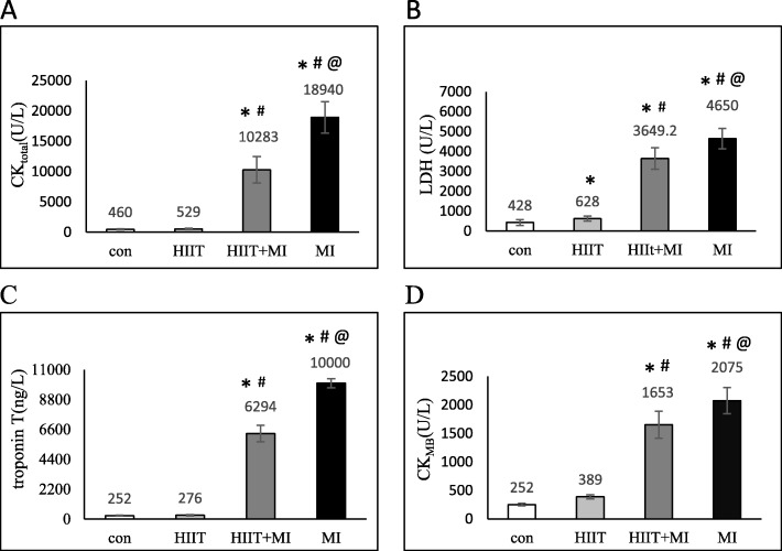
Methods: Twenty Male Wistar rats were randomly divided into 4 groups of control, MI, HIIT, and HIIT+MI. Training groups performed 2 weeks of high intensity interval training in 4 sections. The first section consisted training in 3 days and 2 sessions in each day (4 × 2 min with 35-40 m/min and 3 × 2 min with 25-30 m/min between high intervals. The second part included 2 days of training (4 × 2 min with 40 to 45 m/min and 3 × 2 min with 28 to 32 m /min). The third part was performed in 3 days with one more repetition. The fourth section consisted 2 days of training and with one more repetition compared to section 3. For induction of myocardial infarction, subcutaneous injection of isoprenaline was used. CK, total CK, LDH, and troponin T were measured in serum and G-CSF, G-CSFR and C-Kit proteins were measured by the Western Blot method in the heart tissue.
Results: The results of this study showed that enzymes of CK, total CK, LDH, troponin T had a significant increase in both MI and HIIT+MI groups compared to the other two groups (P < 0.001) and these indices in the MI group were significantly higher than the HIIT+MI group. Also, the results demonstrated that G-CSF, G-CSFR and C-Kit protein expression in the heart tissue significantly increased after MI. As well as, 2 weeks of HIIT training significantly increased G-CSF and C-kit in the training group compared to the control group, but the training caused that these proteins does not increase in HIIT+MI group as much as MI group.
Conclusions: Along with other protective pathways, high intensity interval training can increase cardioprotection and decrease heart injuries through the increase in G-CSF, G-CSFR and C-kit level.
Keywords: Acute myocardial infarction; Cardioprotection; High-intensity interval training; Preconditioning.
Conflict of interest statement
The authors declare that they have no competing interests.
Figures


Similar articles
-
The Exercise Preconditioning Effect on Cardiac Tissue Injury following Induction of Myocardial Infarction in Male Rats.Biomed Res Int. 2023 Jul 13;2023:3631458. doi: 10.1155/2023/3631458. eCollection 2023. Biomed Res Int. 2023. PMID: 37483656 Free PMC article.
-
High-intensity interval training increases myocardial levels of Klotho and protects the heart against ischaemia-reperfusion injury.Exp Physiol. 2020 Apr;105(4):652-665. doi: 10.1113/EP087994. Epub 2020 Mar 17. Exp Physiol. 2020. PMID: 32052504
-
The greater effect of high-intensity interval training versus moderate-intensity continuous training on cardioprotection against ischemia-reperfusion injury through Klotho levels and attenuate of myocardial TRPC6 expression.BMC Cardiovasc Disord. 2019 May 16;19(1):118. doi: 10.1186/s12872-019-1090-7. BMC Cardiovasc Disord. 2019. PMID: 31096903 Free PMC article.
-
High-intensity interval training increase GATA4, CITED4 and c-Kit and decreases C/EBPβ in rats after myocardial infarction.Life Sci. 2019 Mar 15;221:319-326. doi: 10.1016/j.lfs.2019.02.045. Epub 2019 Feb 22. Life Sci. 2019. PMID: 30802510
-
Pleiotropic effects of cytokines on acute myocardial infarction: G-CSF as a novel therapy for acute myocardial infarction.Curr Pharm Des. 2003;9(14):1121-7. doi: 10.2174/1381612033455008. Curr Pharm Des. 2003. PMID: 12769752 Review.
Cited by
-
The Exercise Preconditioning Effect on Cardiac Tissue Injury following Induction of Myocardial Infarction in Male Rats.Biomed Res Int. 2023 Jul 13;2023:3631458. doi: 10.1155/2023/3631458. eCollection 2023. Biomed Res Int. 2023. PMID: 37483656 Free PMC article.
-
The Molecular Signature of High-intensity Training in the Human Body.Int J Sports Med. 2022 Mar;43(3):195-205. doi: 10.1055/a-1551-9294. Epub 2021 Oct 12. Int J Sports Med. 2022. PMID: 34265857 Free PMC article. Review.
-
Post-myocardial infarction fibrosis: Pathophysiology, examination, and intervention.Front Pharmacol. 2023 Mar 28;14:1070973. doi: 10.3389/fphar.2023.1070973. eCollection 2023. Front Pharmacol. 2023. PMID: 37056987 Free PMC article. Review.
-
Effects of exercise training on left ventricular systolic and diastolic function after myocardial infarction: systematic review and meta-analysis.Front Cardiovasc Med. 2025 Mar 25;12:1526326. doi: 10.3389/fcvm.2025.1526326. eCollection 2025. Front Cardiovasc Med. 2025. PMID: 40201788 Free PMC article.
References
-
- French JP, Quindry JC, Falk DJ, Staib JL, Lee Y, Wang KK, et al. Ischemia-reperfusion-induced calpain activation and SERCA2a degradation are attenuated by exercise training and calpain inhibition. Am J Physiol Heart Circ Physiol. 2006;290(1):H128–HH36. doi: 10.1152/ajpheart.00739.2005. - DOI - PubMed
-
- Hoffman JW, Jr, Gilbert TB, Poston RS, Silldorff EP. Myocardial reperfusion injury: etiology, mechanisms, and therapies. J Extra Corpor Technol. 2004;36(4):391–411. - PubMed
Publication types
MeSH terms
Substances
Grants and funding
LinkOut - more resources
Full Text Sources
Medical
Research Materials
