. 2020 Feb 11;18(1):23.
doi: 10.1186/s12964-020-0514-4.

Immune response in piglets orally immunized with recombinant Bacillus subtilis expressing the capsid protein of porcine circovirus type 2

Affiliations

Immune response in piglets orally immunized with recombinant Bacillus subtilis expressing the capsid protein of porcine circovirus type 2

Shuai Zhang et al. Cell Commun Signal. .

Abstract

Background: Porcine circovirus type 2 (PCV2) is the causative agent of postweaning multisystemic wasting syndrome, and is associated with a number of other diseases. PCV2 is widely distributed in most developed swine industries, and is a severe economic burden. With an eye to developing an effective, safe, and convenient vaccine against PCV2-associated diseases, we have constructed a recombinant Bacillus subtilis strain (B. subtilis-Cap) that expresses the PCV2 capsid protein (Cap).

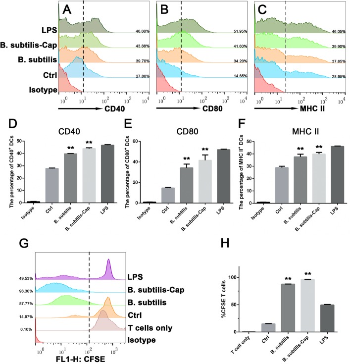
Methods: Electroporation of a plasmid shuttle vector encoding the PCV2 Cap sequence was use to transform Bacillus subtilis. Flow cytometry was used to evaluate in vitro bone marrow derived dendritic cell (BM-DC) maturation and T cell proliferation induced by B. subtilis-Cap. Orally inoculated piglets were used for in vivo experiments; ELISA and western blotting were used to evaluate B. subtilis-Cap induced PCV2-specific IgA and IgG levels, as well as the secretion of cytokines and the expression of Toll-like receptor 2 (TLR2) and Toll-like receptor 9 (TLR9).

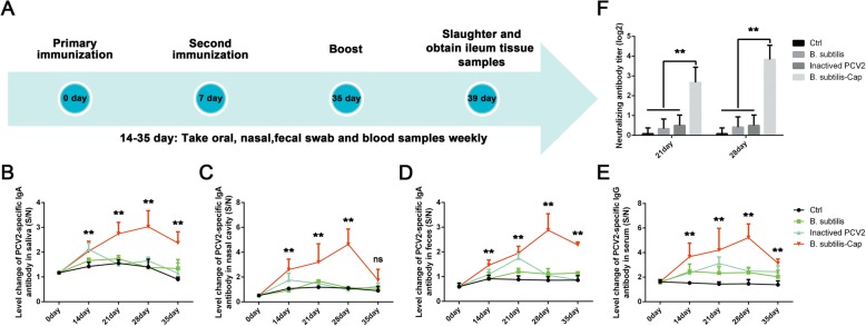
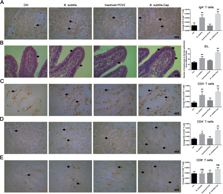
Results: We evaluated the immune response to B. subtilis-Cap in vitro using mouse BM-DCs and in vivo using neonatal piglets orally inoculated with B. subtilis-Cap. Our results showed that the recombinant B. subtilis-Cap activated BM-DCs, significantly increased co-stimulatory molecules (CD40 and CD80) and major histocompatibility complex II, and induced allogenic T cells proliferation. Piglets immunized with B. subtilis-Cap had elevated levels of PCV2-specific IgA in the mucosal tissues of the digestive and respiratory tract, and PCV2-specific IgG in serum (P < 0.05 or P < 0.01). Ileal immunocompetent cells, such as the IgA-secreting cells (P < 0.01), intestinal intraepithelial lymphocytes (IELs) (P < 0.01), CD3+ T lymphocytes (P < 0.01) and CD4+ T lymphocytes (P < 0.01) increased significantly in the B. subtilis-Cap immunized piglets. Additionally, B. subtilis-Cap inoculation resulted in increased the expression of TLR2 and TLR9 (P < 0.01), and induced the secretion of cytokines IL-1β, IL-6, interferon-γ, and β-defensin 2 (P < 0.01).

Conclusions: We constructed a prototype PCV2 vaccine that can be administered orally and elicits a more robust humoral and cellular immunity than inactivated PCV2. B. subtilis-Cap is a promising vaccine candidate that is safe, convenient, and inexpensive. Further in vivo research is needed to determine its full range of efficacy in pigs.

Keywords: Bacillus subtilis WB800N; Capsid protein; Dendritic cells; Immune responses; Neonatal piglets; Porcine circovirus type 2 (PCV2).


Conflict of interest statement

The authors declare they have no competing interests.

Figures

Fig. 1
Fig. 1
Schematic of pHT43-Cap construction and protein expression. a The Cap sequence was amplified from pMD19-T-Cap, and inserted into pHT43 between the BamHI and SmaI sites, generating plasmid pHT43-Cap. b Agarose gel of pHT43-Cap, pHT43, and Cap fragment amplified from pMD19-T-Cap in lanes 1, 2, and 3 respectively. Plasmids were digested with BamHI and SmaI before electrophoresis. Lane M is a 5000 bp DNA ladder. c pHT43 (vector control) and pHT43-Cap recombinant plasmids were transformed into B. subtilis by electroporation. d Western blotting analysis of the PCV2 Cap protein expressed in B. subtilis-Cap, lanes 1. Extracts of B. subtilis (transfected with pHT43) were run in lanes 2, and lane M is protein molecular weight ladder
Fig. 2
Fig. 2
B. subtilis-Cap stimulates phenotypic alteration of BM-DCs and promotes T cells proliferation in vitro. a-fB. subtilis, B. subtilis-Cap, and LPS, were incubated with mouse BM-DCs for 24 h. Expression of CD40, CD80, and MHCII were analyzed by flow cytometry. Untreated BM-DCs served as a negative control. g and h Mixed lymphocyte reaction (MLR) experiments. B. subtilis, B. subtilis-Cap, LPS, or PBS treated BM-DCs were co-cultured with carboxyfluorescein succinimidyl ester (CFSE)-labeled allogeneic T lymphocytes (DC:T cell ratio, 1:1) for another 5 days. Untreated BM-DCs co-cultured with T lymphocytes served as a negative control. T lymphocytes proliferation was evaluated by flow cytometry. Data are presented as means ± SD from three independent experiments. (* 0.01 < p < 0.05, ** p < 0.01)
Fig. 3
Fig. 3
Immune response in piglets orally inoculated with B. subtilis-Cap. a Flow chart of inoculation and sample collection time course. Piglets were orally inoculated with B. subtilis-Cap, B. subtilis, inactivated PCV2, and PBS on day 0 and 7. Saliva, nasal swab, feces, and serum were collected as indicated and analyzed by ELISA for PCV2-specific IgA and IgG. b Levels of PCV2-specific IgA in the oral cavity. c Levels of PCV2-specific IgA in the intranasal cavity. d Levels of PCV2-specific IgA in feces. e Levels of PCV2-specific IgG in serum. f The results of neutralizing titer activities of swine serum antibody on day 21 and day 28, expressed as the reciprocal of the highest dilution of serum causing a 50% reduction in fluorescence. Data shown are the means ± S.D. of six samples. * 0.01 < p < 0.05, ** p < 0.01 (compared to the Ctrl group). The error bars represent standard deviations
Fig. 4
Fig. 4
Effect of orally administered recombinant B. subtilis-Cap on ileal immunocompetent cells. Piglets were immunized as described above, then boosted on day 35 and sacrificed on the day 39. Ileum tissues were fixed with Bonn’s liquid, embedded in paraffin and sectioned to 5 μm thickness, the sections were subjected to hematoxylin–eosin staining or immunohistochemistry. a Distribution of IgA-secreting cells in piglet ileums (Magnification, × 40) and the average IOD of IgA-secreting cells distributed in piglet ileums. The IgA-secreting plasma cells, rounded with a deep yellow-brown nucleus (black arrows), were located mainly in the submucosa. b Distribution of IELs in ileums (Magnification, × 40) and the average of number of IELs counted per dose group. The IELs (black arrows) are located between intestinal epithelial cells, and beneath the intercellular tight junctions at the basal region of the epithelium. c, d, and e Distribution of CD3+, CD4+, and CD8+ T lymphocytes in piglet ileums (Magnification, × 40). The average IOD of CD3+, CD4+ and CD8+ T lymphocytes in the ileums are displayed in the histogram. Photomicrographs showing representative CD3+, CD4+ and CD8+ T lymphocytes which are stained a deep yellow-brown (black arrows). The average IOD was calculated from ten separate fields of ileum from each piglet, using Image-Pro Plus 6.0 software. The error bars represent standard deviations. * 0.01 < p < 0.05, ** p < 0.01 (compared to the Ctrl group)
Fig. 5
Fig. 5
The expression levels of Toll-like receptors (TLRs), IgA, and cytokines. Associated immune factors were measured in ileum homogenates prepared on day 39 of the experiment (at slaughter). a TLR2 protein expression, the ratios of TLR2 to GAPDH were normalized to the control. b TLR9 protein expression, the ratios of TLR9 to GAPDH were normalized to the control. c IgA expression. d-h Cytokine IL1β, IL-6, IL-10, TNF-α, and IFN-γ expression. i Porcine β-defensin 2 expression. The error bars represent standard deviations. * 0.01 < p < 0.05, ** p < 0.01 (compared to the Ctrl group)

Similar articles

Cited by

References

    1. Lefkowitz EJ, Dempsey DM, Hendrickson RC, Orton RJ, Siddell SG, Smith DB. Virus taxonomy: the database of the international committee on taxonomy of viruses (ICTV) Nucleic Acids Res. 2018;46:D708–D717. doi: 10.1093/nar/gkx932. - DOI - PMC - PubMed
    1. Tischer I, Gelderblom H, Vettermann AW, Koch MA. A very small porcine virus with circular single-stranded DNA. Nature. 1982;295:64. doi: 10.1038/295064a0. - DOI - PubMed
    1. Nawagitgul P, Morozov I, Bolin SR, Harms PA, Sorden SD, Paul PS. Open reading frame 2 of porcine circovirus type 2 encodes a major capsid protein. Jgenvirol. 2000;81:2281–2287. - PubMed
    1. Allan GM, Ellis JA. Porcine Circoviruses: a review. J Vet Diagn Investig. 2000;12:3–14. doi: 10.1177/104063870001200102. - DOI - PubMed
    1. Hamel AL, Lin LL, Nayar GPS. Nucleotide sequence of porcine Circovirus associated with Postweaning multisystemic wasting syndrome in pigs. J Virol. 1998;72:5262. doi: 10.1128/JVI.72.6.5262-5267.1998. - DOI - PMC - PubMed

Publication types

MeSH terms